
Article Figures & Data
Figures
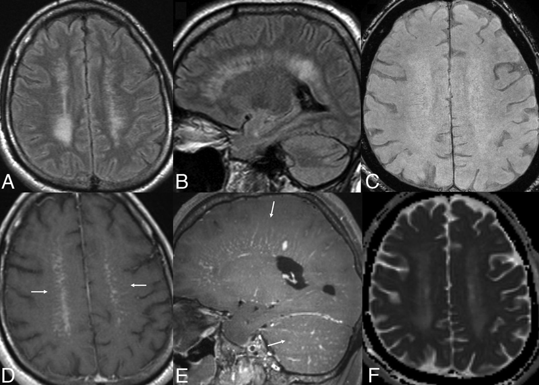
- FIG 1.
Characteristic MR imaging findings of GFAP astrocytopathy in a 68-year-old man with history of prostatic adenocarcinoma. Multiple foci of linear T2/FLAIR hyperintensity (A and B) are noted in the periventricular white matter with corresponding perivascular radial pattern enhancement (D and E, arrows). Similar pattern of enhancement is also noted in the cerebellum (D, arrow). SWI (C) is normal and ADC map (F) reveals T2 shine through artifacts in the periventricular white matter with no restricted diffusion. Patient had elevated WBC count of 200/mm3 in his CSF (lymphocyte predominant). Positive titer for GFAP-IgG was noted on CSF analysis by using cell-binding assay. Patient received methylprednisolone 1g daily for 5 days, with remarkable clinical recovery just after the first dose and was subsequently placed on oral taper. Near-complete radiographic response was noted on the follow-up MR imaging (2 weeks) along with negative GFAP IgG titers (Online Supplemental Data).
- FIG 2.
Histopathologic findings in 2 different patients with GFAP astrocytopathy (A and B) and intravascular CNS lymphoma (C and D). Hematoxylin-eosin (A) and CD20 (B) staining of brain biopsy in a patient with subacute course of encephalitis and CSF positive for GFAP autoantibodies shows extensive infiltration of inflammatory cells (A and B, arrows) mainly around the vessels with no evidence of transmural vessel wall inflammation (vasculitis). Similar stains in a different patient (C and D) show plugging of the lumen of small vessels by large atypical cells (C, arrow) that are positive for CD20 (D, arrow) and negative for CD3, consistent with intravascular large B cell lymphoma.
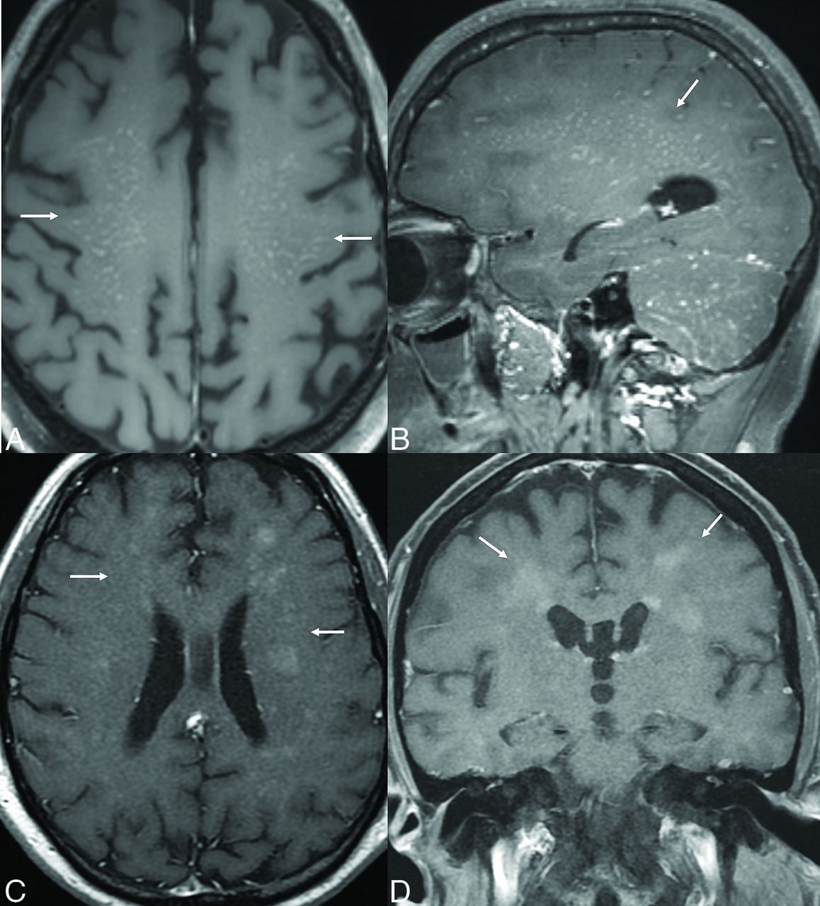
- FIG 3.
Common patterns of enhancement on MR imaging in 2 different patients with GFAP astrocytopathy. Contrast-enhanced MR imaging in 2 different patients with no significant prior history. Multiple foci of punctate enhancement noted in supratentorial and cerebellar white matter in a 42-year-old man (A and B, arrows). Multiple solid nodular foci of enhancement in the supratentorial white matter involving the centrum semiovale and corona radiata noted in a 35-year-old woman (C and D, arrows). Radiographic differentials in both cases included vasculitis, lymphoma, and neurosarcoidosis. The final diagnosis of GFAP encephalitis was made based on positive CSF and serologic titers of GFAP autoantibodies.
- FIG 4.
GFAP astrocytopathy in a 65-year-old man with predominant cerebellar, brainstem and spinal cord involvement. Multiple contrast-enhanced MR images reveal patchy striated enhancement in the brainstem, cervical cord (A, arrows), and the cerebellum (B, arrows). Enhancement involves primarily the central cord (A) with radial linear pattern in the cerebellar white matter and brainstem (B and C). Mild periventricular linear enhancement is also noted in the supratentorial white matter (C, black arrows), though less pronounced than the cerebellar enhancement. The final diagnosis of GFAP encephalomyelitis was made based on positive CSF and serologic titers of GFAP autoantibodies. Patient had a long-standing history of Crohn disease and ankylosing spondylitis and showed marked improvement in symptoms (ataxia and cognitive changes) after rituximab therapy.
- FIG 5.
GFAP astrocytopathy in a 34-year-old woman with longitudinally extensive transverse myelitis and optic neuritis mimicking NMOSD. Patient was admitted in neurology with acute onset vision changes and sensorimotor symptoms in the lower limbs. Long segment T2 hyperintensity is noted involving the central cord (A, arrow) with patchy enhancement (B, arrow) and sparing of the cord periphery (C, arrow). Contrast-enhanced image of the orbits (D) reveals intense patchy enhancement involving the entire intraorbital segments of bilateral optic nerves (D, arrows) and the right optic disc head (D, arrowhead) suggesting acute optic neuritis with papillitis. OCT examination reveals increased peripapillary retinal nerve fiber layer thickness (left > right) with subretinal fluid (E and F, arrows). GFAP astrocytopathy was confirmed based on CSF antibody positivity and brain biopsy. CSF titers were negative for AQP4 with absence of oligoclonal bands. She underwent plasmapheresis and IV solumedrol treatment and saw rapid improvement in her symptoms.
- FIG 6.
Intravascular large B cell lymphoma in a 57-year-old woman presenting with headaches, vision changes, and confusion. Multifocal discrete areas of T2/FLAIR hyperintensity (A) with corresponding restricted diffusion (B) with foci of low ADC values (C, arrows) noted in the supratentorial white matter with no significant mass effect. Contrast-enhanced axial (D) and sagittal (E) images reveal solid enhancement (E, arrows) corresponding to the areas of T2/FLAIR hyperintensities. No hemorrhagic changes are noted on SWI (F). Right frontal lobe biopsy revealed plugging of the lumen of multiple small vessels by large, atypical cells, which were positive for CD20 and PAX5 and negative for CD3 with BCL6 rearrangement in approximately 100% of nuclei consistent with intravascular lymphoma. No other organ involvement was seen on further work-up and the patient remains disease free, 4 years after the initial diagnosis, status post completion of 6 cycles of chemotherapy (MR-CHOP regimen).


Jump to section
Related Articles
Cited By...
- No citing articles found.