Advertisement

Main menu

  • Home
  • Content
    • Current Issue
    • Accepted Manuscripts
    • Article Preview
    • Past Issue Archive
    • Video Articles
    • AJNR Case Collection
    • Case of the Week Archive
    • Case of the Month Archive
    • Classic Case Archive
  • Special Collections
    • AJNR Awards
    • Low-Field MRI
    • Alzheimer Disease
    • ASNR Foundation Special Collection
    • Photon-Counting CT
    • View All
  • Multimedia
    • AJNR Podcasts
    • AJNR SCANtastic
    • Trainee Corner
    • MRI Safety Corner
    • Imaging Protocols
  • For Authors
    • Submit a Manuscript
    • Submit a Video Article
    • Submit an eLetter to the Editor/Response
    • Manuscript Submission Guidelines
    • Statistical Tips
    • Fast Publishing of Accepted Manuscripts
    • Graphical Abstract Preparation
    • Imaging Protocol Submission
    • Author Policies
  • About Us
    • About AJNR
    • Editorial Board
    • Editorial Board Alumni
  • More
    • Become a Reviewer/Academy of Reviewers
    • Subscribers
    • Permissions
    • Alerts
    • Feedback
    • Advertisers
    • ASNR Home

User menu

  • Alerts
  • Log in

Search

  • Advanced search
American Journal of Neuroradiology

American Journal of Neuroradiology

ASHNR American Society of Functional Neuroradiology ASHNR American Society of Pediatric Neuroradiology ASSR
  • Alerts
  • Log in

Advanced Search

  • Home
  • Content
    • Current Issue
    • Accepted Manuscripts
    • Article Preview
    • Past Issue Archive
    • Video Articles
    • AJNR Case Collection
    • Case of the Week Archive
    • Case of the Month Archive
    • Classic Case Archive
  • Special Collections
    • AJNR Awards
    • Low-Field MRI
    • Alzheimer Disease
    • ASNR Foundation Special Collection
    • Photon-Counting CT
    • View All
  • Multimedia
    • AJNR Podcasts
    • AJNR SCANtastic
    • Trainee Corner
    • MRI Safety Corner
    • Imaging Protocols
  • For Authors
    • Submit a Manuscript
    • Submit a Video Article
    • Submit an eLetter to the Editor/Response
    • Manuscript Submission Guidelines
    • Statistical Tips
    • Fast Publishing of Accepted Manuscripts
    • Graphical Abstract Preparation
    • Imaging Protocol Submission
    • Author Policies
  • About Us
    • About AJNR
    • Editorial Board
    • Editorial Board Alumni
  • More
    • Become a Reviewer/Academy of Reviewers
    • Subscribers
    • Permissions
    • Alerts
    • Feedback
    • Advertisers
    • ASNR Home
  • Follow AJNR on Twitter
  • Visit AJNR on Facebook
  • Follow AJNR on Instagram
  • Join AJNR on LinkedIn
  • RSS Feeds

AJNR Awards, New Junior Editors, and more. Read the latest AJNR updates

Research ArticleSpine Imaging and Spine Image-Guided Interventions

The Development and Application of a Cost-Effective Cervical Spine Phantom for Use in Fluoroscopically Guided Lateral C1–C2 Spinal Puncture Training

V. Risner, B. Huang, K. McCullagh, T. Benefield and Y.Z. Lee
American Journal of Neuroradiology July 2023, 44 (7) 873-878; DOI: https://doi.org/10.3174/ajnr.A7908
V. Risner
aFrom the Department of Radiology, University of North Carolina at Chapel Hill School of Medicine, Chapel Hill, North Carolina
  • Find this author on Google Scholar
  • Find this author on PubMed
  • Search for this author on this site
  • ORCID record for V. Risner
B. Huang
aFrom the Department of Radiology, University of North Carolina at Chapel Hill School of Medicine, Chapel Hill, North Carolina
  • Find this author on Google Scholar
  • Find this author on PubMed
  • Search for this author on this site
  • ORCID record for B. Huang
K. McCullagh
aFrom the Department of Radiology, University of North Carolina at Chapel Hill School of Medicine, Chapel Hill, North Carolina
  • Find this author on Google Scholar
  • Find this author on PubMed
  • Search for this author on this site
T. Benefield
aFrom the Department of Radiology, University of North Carolina at Chapel Hill School of Medicine, Chapel Hill, North Carolina
  • Find this author on Google Scholar
  • Find this author on PubMed
  • Search for this author on this site
  • ORCID record for T. Benefield
Y.Z. Lee
aFrom the Department of Radiology, University of North Carolina at Chapel Hill School of Medicine, Chapel Hill, North Carolina
  • Find this author on Google Scholar
  • Find this author on PubMed
  • Search for this author on this site
  • ORCID record for Y.Z. Lee
  • Article
  • Figures & Data
  • Info & Metrics
  • Responses
  • References
  • PDF
Loading

Article Figures & Data

Figures

  • Tables
  • FIG 1.
    • Download figure
    • Open in new tab
    • Download powerpoint
    FIG 1.

    Puncturing the spinal cord. The photograph demonstrates the result of puncturing the inner latex balloon, which represents the spinal cord. Red dye returns from the spinal needle.

  • FIG 2.
    • Download figure
    • Open in new tab
    • Download powerpoint
    FIG 2.

    Constructed cervical phantom model. The photograph shows the fully-constructed phantom model. The cervical spine model is covered with polyalginate. There is a latex tube running through the spinal canal of the cervical model, with an inner latex balloon within the tube. The inner latex balloon is filled with dyed water, and the outer tube is filled with clear water. Two hemostats on either end of the phantom are occluding the posterior third of the latex tube to ensure that the balloon is in a more accurate anatomic position relative to the spinal cord. The hemostat at the distal end of the latex tube is used to maintain water pressure within the latex tube.
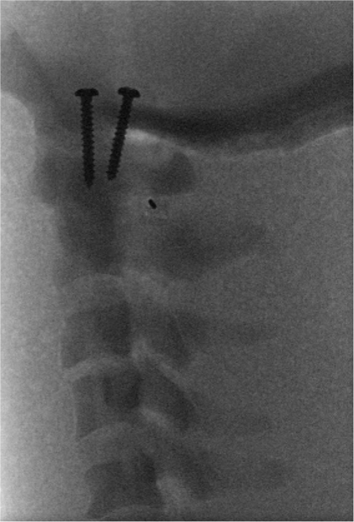
  • FIG 3.
    • Download figure
    • Open in new tab
    • Download powerpoint
    FIG 3.

    Fluoroscopic visualization of the phantom model shows the appearance of the model under fluoroscopy during a training session. The spinal needle can be seen within the C1–C2 space. The 2 screws seen in this image connect the skull base to the cervical spine and could not be removed in order to preserve integrity of the phantom.
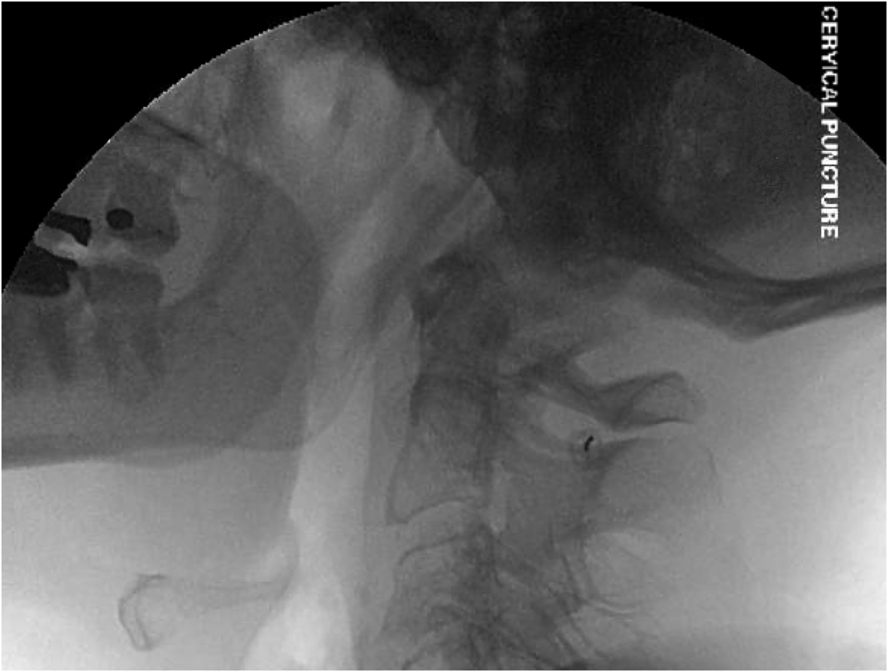
  • FIG 4.
    • Download figure
    • Open in new tab
    • Download powerpoint
    FIG 4.

    Real example of a fluoroscopic image of a lateral C1–C2 spinal puncture that shows a lateral view of a C1–C2 spinal puncture performed on a patient for comparing the performance of the phantom in replicating fluoroscopic views during the procedure.
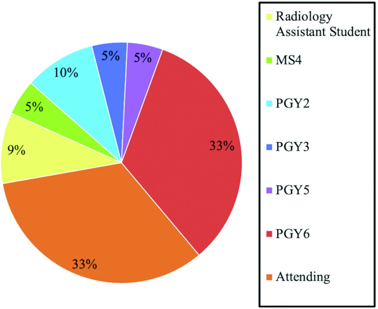
  • FIG 5.
    • Download figure
    • Open in new tab
    • Download powerpoint
    FIG 5.

    Stages of training of workshop participants. The graph shows the distribution of participant training levels during lateral C1–C2 spinal puncture training. A total of 21 individuals participated in training workshops, with 1 resident participating in 2 training workshops. PGY indicates postgraduate year; MS4, fourth-year medical student.
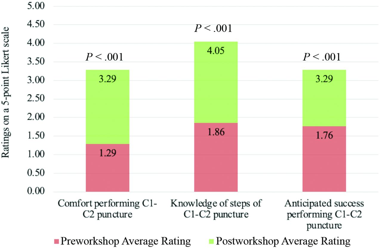
  • FIG 6.
    • Download figure
    • Open in new tab
    • Download powerpoint
    FIG 6.

    Pre- and postworkshop ratings of comfort, knowledge, and confidence of C1–C2 lateral cervical puncture. The image shows pre- and postsurvey results of participants’ ratings of their comfort performing, knowledge of steps, and anticipated success of lateral C1–C2 spinal puncture before and after taking part in an educational workshop using the cervical model developed in this article. Ratings were on a 5-point Likert scale.

Tables

  • Figures
  • Comparison of phantom performance in replicating actual lateral C1–C2 spinal punctures

    Component of Phantom/ProcedureComparison with Real-Life Experience of Lateral C1–C2 Spinal Punctures
    Feel of alginate/soft tissueSimilar
    Positioning of phantom/patientDifferent: The patient is usually positioned prone for this procedure, while the training simulated the patient being in the lateral decubitus position due to constraints of the fluoroscopy room available for training (not related to the phantom itself)
    Force required to penetrate outer latex tubing/duraDifferent: The amount of pressure required to pop through the outer latex tube dura was greater than in real life; this difference was emphasized to the trainees who participated
    Landmarks visualized under fluoroscopySimilar
    Speed of CSF egressVariable: depending on how much pressure we applied to outer latex tubing via a syringe and the PVC on/off valve; future directions include construction of a model with a pressure gauge so that we could fill the tubing to match normal CSF pressure (∼15 cm H20)
    • Note:—PVC indicates polyvinyl chloride.

PreviousNext
Back to top

In this issue

American Journal of Neuroradiology: 44 (7)
American Journal of Neuroradiology
Vol. 44, Issue 7
1 Jul 2023
  • Table of Contents
  • Index by author
  • Complete Issue (PDF)
Advertisement
Print
Download PDF
Email Article

Thank you for your interest in spreading the word on American Journal of Neuroradiology.

NOTE: We only request your email address so that the person you are recommending the page to knows that you wanted them to see it, and that it is not junk mail. We do not capture any email address.

Enter multiple addresses on separate lines or separate them with commas.
The Development and Application of a Cost-Effective Cervical Spine Phantom for Use in Fluoroscopically Guided Lateral C1–C2 Spinal Puncture Training
(Your Name) has sent you a message from American Journal of Neuroradiology
(Your Name) thought you would like to see the American Journal of Neuroradiology web site.
CAPTCHA
This question is for testing whether or not you are a human visitor and to prevent automated spam submissions.
Cite this article
V. Risner, B. Huang, K. McCullagh, T. Benefield, Y.Z. Lee
The Development and Application of a Cost-Effective Cervical Spine Phantom for Use in Fluoroscopically Guided Lateral C1–C2 Spinal Puncture Training
American Journal of Neuroradiology Jul 2023, 44 (7) 873-878; DOI: 10.3174/ajnr.A7908

Citation Manager Formats

  • BibTeX
  • Bookends
  • EasyBib
  • EndNote (tagged)
  • EndNote 8 (xml)
  • Medlars
  • Mendeley
  • Papers
  • RefWorks Tagged
  • Ref Manager
  • RIS
  • Zotero
0 Responses
Respond to this article
Share
Bookmark this article
Cervical Spine Phantom for Puncture Training
V. Risner, B. Huang, K. McCullagh, T. Benefield, Y.Z. Lee
American Journal of Neuroradiology Jul 2023, 44 (7) 873-878; DOI: 10.3174/ajnr.A7908
del.icio.us logo Twitter logo Facebook logo Mendeley logo
  • Tweet Widget
  • Facebook Like
  • Google Plus One
Purchase

Jump to section

  • Article
    • Abstract
    • MATERIALS AND METHODS
    • RESULTS
    • DISCUSSION
    • CONCLUSIONS
    • Footnotes
    • References
  • Figures & Data
  • Info & Metrics
  • Responses
  • References
  • PDF

Related Articles

  • No related articles found.
  • PubMed
  • Google Scholar

Cited By...

  • No citing articles found.
  • Crossref
  • Google Scholar

This article has not yet been cited by articles in journals that are participating in Crossref Cited-by Linking.

More in this TOC Section

  • Optimization of Photon Counting CT Myelography
  • Characteristics of SIH Type I Culprit Lesions
  • Management Outcomes For VO Spine Biopsy
Show more Spine Imaging and Spine Image-Guided Interventions

Similar Articles

Advertisement

Indexed Content

  • Current Issue
  • Accepted Manuscripts
  • Article Preview
  • Past Issues
  • Editorials
  • Editor's Choice
  • Fellows' Journal Club
  • Letters to the Editor
  • Video Articles

Cases

  • Case Collection
  • Archive - Case of the Week
  • Archive - Case of the Month
  • Archive - Classic Case

More from AJNR

  • Trainee Corner
  • Imaging Protocols
  • MRI Safety Corner
  • Book Reviews

Multimedia

  • AJNR Podcasts
  • AJNR Scantastics

Resources

  • Turnaround Time
  • Submit a Manuscript
  • Submit a Video Article
  • Submit an eLetter to the Editor/Response
  • Manuscript Submission Guidelines
  • Statistical Tips
  • Fast Publishing of Accepted Manuscripts
  • Graphical Abstract Preparation
  • Imaging Protocol Submission
  • Evidence-Based Medicine Level Guide
  • Publishing Checklists
  • Author Policies
  • Become a Reviewer/Academy of Reviewers
  • News and Updates

About Us

  • About AJNR
  • Editorial Board
  • Editorial Board Alumni
  • Alerts
  • Permissions
  • Not an AJNR Subscriber? Join Now
  • Advertise with Us
  • Librarian Resources
  • Feedback
  • Terms and Conditions
  • AJNR Editorial Board Alumni

American Society of Neuroradiology

  • Not an ASNR Member? Join Now

© 2025 by the American Society of Neuroradiology All rights, including for text and data mining, AI training, and similar technologies, are reserved.
Print ISSN: 0195-6108 Online ISSN: 1936-959X

Powered by HighWire