
Article Figures & Data
Figures
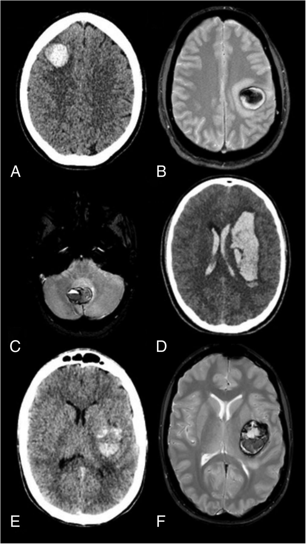
- FIG 1.
Assessment of hematoma shape. A, Spherical, acute lobar hematoma on plain CT of a patient with a pathologically proved hemorrhagic cerebral cavernoma. B, Spherical lobar hematoma on T2*-weighted imaging in a patient with pathologically proved hemorrhagic cerebral cavernoma. C, Ovoid cerebellar hematoma on T2*-weighted imaging of a patient with pathologically proved hemorrhagic cavernoma. D, Plain CT obtained at admission in a patient with an angiographically proved cerebral AVM. The elongated shape of the hematoma did not fulfill the criteria spherical or ovoid. Its margins were considered irregular because angular aspects were present. It was associated with intraventricular hemorrhage. E and F, Hemorrhagic cerebral cavernoma. Both plain CT (D) and T2* MR images (E) were available to assess the hematoma shape. The hematoma shape was classified as spherical/ovoid on the basis of MR imaging.
- FIG 2.
Assessment of hematoma margins. A and B, T1- and T2*-weighted MR imaging shows a hematoma with regular margins in a patient with pathologically proved hemorrhagic cavernoma in the setting of familial cavernomatosis. C and D, T1 and T2*-weighted MR imaging shows a hematoma in a patient with a pathologically proved cerebral cavernoma. The lesion was multiloculated with bumpy borders. The margins were classified as regular because no angular aspect was present in any part of the lesion. Hematoma with irregular margins presenting with angular aspects on plain CT (E) and T2*-weighted MR imaging (F) in 2 patients with arteriovenous shunts proved by cerebral angiography.
- FIG 3.
Flowchart of study population.
- FIG 4.
Decision tree model applied to the validation sample.


Tables
Univariate and multivariate logistic regression for the diagnosis of hemorrhagic cavernomas
Crude OR (95% CI) P Value Adjusted OR (95% CI) P Value Spherical/ovoid 36.60 (15.89–110.20) <.001 6.11 (2.64–15.08) <.001 Regular margins 34.81 (14.99–103.90) <.001 3.38 (1.44–8.61) .009 Extralesional hemorrhage 0.06 (0.02–0.15) <.001 0.39 (0.18–0.76) .01 Peripheral rim enhancement 0.18 (0.03–0.53) .009 0.29 (0.13–0.59) .002


Jump to section
Related Articles
Cited By...
- No citing articles found.