Advertisement

Main menu

  • Home
  • Content
    • Current Issue
    • Accepted Manuscripts
    • Article Preview
    • Past Issue Archive
    • Video Articles
    • AJNR Case Collection
    • Case of the Week Archive
    • Case of the Month Archive
    • Classic Case Archive
  • Special Collections
    • AJNR Awards
    • Low-Field MRI
    • Alzheimer Disease
    • ASNR Foundation Special Collection
    • Photon-Counting CT
    • View All
  • Multimedia
    • AJNR Podcasts
    • AJNR SCANtastic
    • Trainee Corner
    • MRI Safety Corner
    • Imaging Protocols
  • For Authors
    • Submit a Manuscript
    • Submit a Video Article
    • Submit an eLetter to the Editor/Response
    • Manuscript Submission Guidelines
    • Statistical Tips
    • Fast Publishing of Accepted Manuscripts
    • Graphical Abstract Preparation
    • Imaging Protocol Submission
    • Author Policies
  • About Us
    • About AJNR
    • Editorial Board
    • Editorial Board Alumni
  • More
    • Become a Reviewer/Academy of Reviewers
    • Subscribers
    • Permissions
    • Alerts
    • Feedback
    • Advertisers
    • ASNR Home

User menu

  • Alerts
  • Log in

Search

  • Advanced search
American Journal of Neuroradiology

American Journal of Neuroradiology

ASHNR American Society of Functional Neuroradiology ASHNR American Society of Pediatric Neuroradiology ASSR
  • Alerts
  • Log in

Advanced Search

  • Home
  • Content
    • Current Issue
    • Accepted Manuscripts
    • Article Preview
    • Past Issue Archive
    • Video Articles
    • AJNR Case Collection
    • Case of the Week Archive
    • Case of the Month Archive
    • Classic Case Archive
  • Special Collections
    • AJNR Awards
    • Low-Field MRI
    • Alzheimer Disease
    • ASNR Foundation Special Collection
    • Photon-Counting CT
    • View All
  • Multimedia
    • AJNR Podcasts
    • AJNR SCANtastic
    • Trainee Corner
    • MRI Safety Corner
    • Imaging Protocols
  • For Authors
    • Submit a Manuscript
    • Submit a Video Article
    • Submit an eLetter to the Editor/Response
    • Manuscript Submission Guidelines
    • Statistical Tips
    • Fast Publishing of Accepted Manuscripts
    • Graphical Abstract Preparation
    • Imaging Protocol Submission
    • Author Policies
  • About Us
    • About AJNR
    • Editorial Board
    • Editorial Board Alumni
  • More
    • Become a Reviewer/Academy of Reviewers
    • Subscribers
    • Permissions
    • Alerts
    • Feedback
    • Advertisers
    • ASNR Home
  • Follow AJNR on Twitter
  • Visit AJNR on Facebook
  • Follow AJNR on Instagram
  • Join AJNR on LinkedIn
  • RSS Feeds

AJNR Awards, New Junior Editors, and more. Read the latest AJNR updates

Research ArticlePediatric Neuroimaging

Subarachnoid Space Measurements in Apparently Healthy Fetuses Using MR Imaging

A. Wandel, T. Weissbach, E. Katorza and T. Ziv-Baran
American Journal of Neuroradiology June 2023, 44 (6) 716-721; DOI: https://doi.org/10.3174/ajnr.A7864
A. Wandel
aFrom the Sackler School of Medicine (A.W., T.W., E.K.)
cDepartment of Diagnostic Radiology (A.W.), Shamir Medical Center, Zerifin, Israel
  • Find this author on Google Scholar
  • Find this author on PubMed
  • Search for this author on this site
  • ORCID record for A. Wandel
T. Weissbach
aFrom the Sackler School of Medicine (A.W., T.W., E.K.)
dAntenatal Diagnostic Unit (T.W., E.K.)
  • Find this author on Google Scholar
  • Find this author on PubMed
  • Search for this author on this site
  • ORCID record for T. Weissbach
E. Katorza
aFrom the Sackler School of Medicine (A.W., T.W., E.K.)
dAntenatal Diagnostic Unit (T.W., E.K.)
eDepartment of Obstetrics and Gynecology, and Gertner Institute for Epidemiology and Health Policy Research (E.K.), Sheba Medical Center, Tel HaShomer, Israel
  • Find this author on Google Scholar
  • Find this author on PubMed
  • Search for this author on this site
  • ORCID record for E. Katorza
T. Ziv-Baran
bSchool of Public Health (T.Z.-B.), Sackler Faculty of Medicine, Tel Aviv University, Tel Aviv, Israel
  • Find this author on Google Scholar
  • Find this author on PubMed
  • Search for this author on this site
  • ORCID record for T. Ziv-Baran
  • Article
  • Figures & Data
  • Info & Metrics
  • Responses
  • References
  • PDF
Loading

Article Figures & Data

Figures

  • Tables
  • FIG 1.
    • Download figure
    • Open in new tab
    • Download powerpoint
    FIG 1.

    Axial T2-weighted fetal brain image demonstrating the measurements of the subarachnoid space: 1) Right frontal gyrus. 2) Left frontal gyrus. 3) Right insula gyrus. 4) Left insula gyrus.

  • FIG 2.
    • Download figure
    • Open in new tab
    • Download powerpoint
    FIG 2.

    Coronal T2-weighted fetal brain image demonstrating the measurements of the subarachnoid space: 1) Right frontal gyrus. 2) Left frontal gyrus. 3) Right insula gyrus. 4) Left insula gyrus. 5) Right inferior temporal gyrus. 6) Left inferior temporal gyrus.
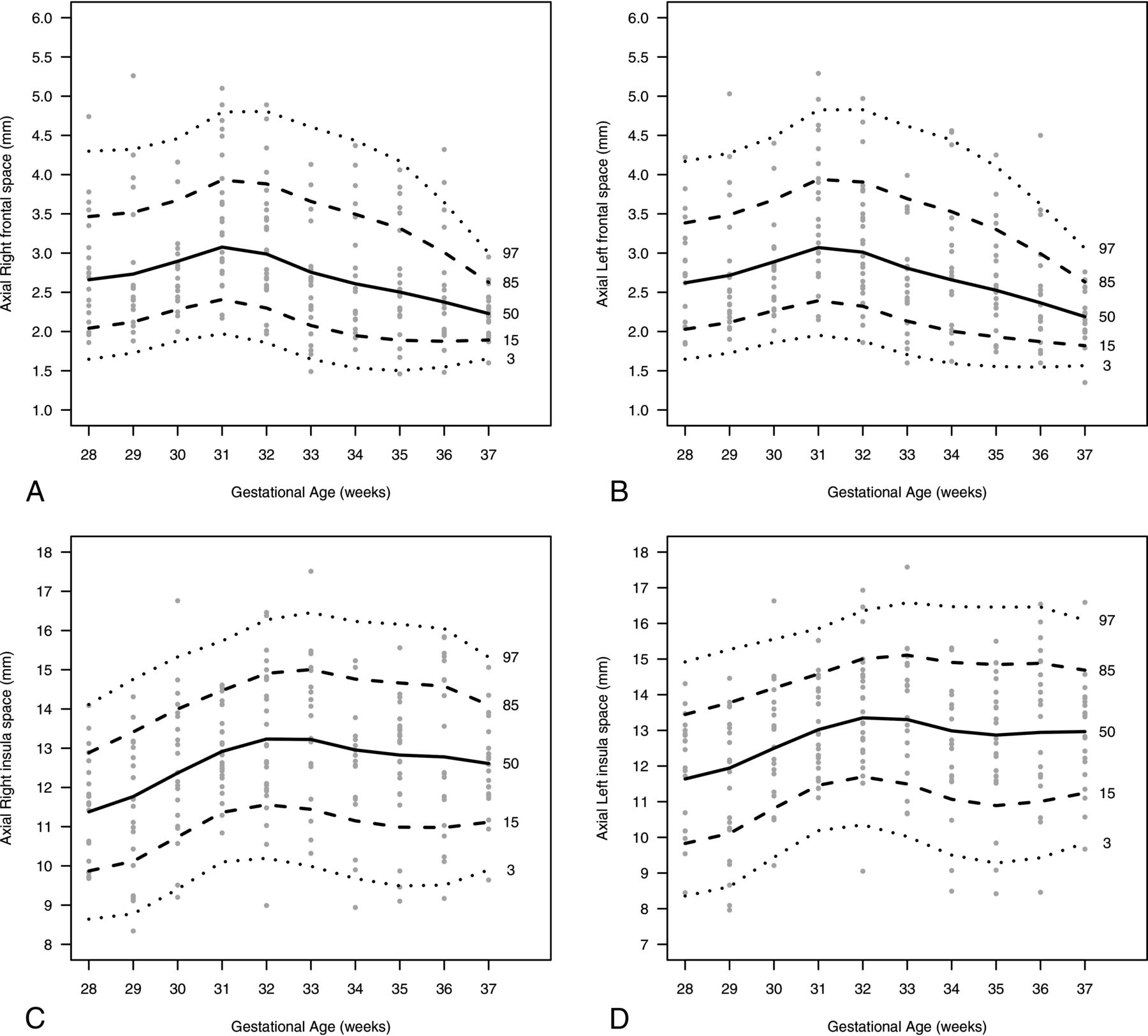
  • FIG 3.
    • Download figure
    • Open in new tab
    • Download powerpoint
    FIG 3.

    A nomogram presenting the SAS size (millimeters) in the axial plane according to the 3rd, 15th, 50th, 85th, 97th percentiles and GA (weeks). A, Right frontal. B, Left frontal. C, Right insula; D, Left insula.
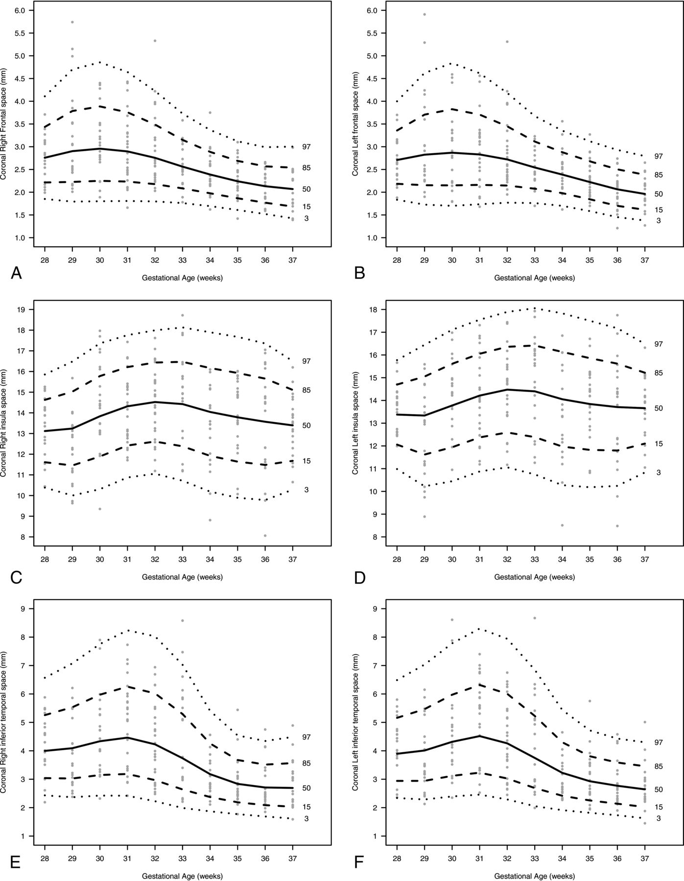
  • FIG 4.
    • Download figure
    • Open in new tab
    • Download powerpoint
    FIG 4.

    A nomogram presenting the SAS size (millimeters) in the coronal plane according to 3rd, 15th, 50th, 85th, 97th percentiles, and GA (weeks): A, Right frontal. B, Left frontal. C, Right insula, D, Left insula. E, Right inferior temporal. F, Left inferior temporal.

Tables

  • Figures
  • Intraobserver and interobserver agreement

    Type of Agreement and SAS LocationICC (95% CI)
    Intraobsever
     Ax. Rt. Fr.0.761 (0.573–0.870)
     Ax. Lt. Fr.0.670 (0.431–0.816)
     Ax. Rt. In.0.865 (0.760–0.926)
     Ax. Lt. In.0.869 (0.762–0.929)
     Cr. Rt. Fr.0.942 (0.889–0.969)
     Cr. Lt. Fr.0.950 (0.908–0.973)
     Cr. Rt. In.0.911 (0.737–0.962)
     Cr. Lt. In.0.933 (0.767–0.973)
     Cr. Rt. It.0.890 (0.803–0.940)
     Cr. Lt. It.0.877 (0.780–0.933)
    Interobserver
     Ax. Rt. Fr.0.742 (0.562–0.854)
     Ax. Lt. Fr.0.700 (0.499–0.830)
     Ax. Rt. In.0.858 (0.740–0.923)
     Ax. Lt. In.0.877 (0.718–0.941)
     Cr. Rt. Fr.0.913 (0.842–0.953)
     Cr. Lt. Fr.0.893 (0.807–0.942)
     Cr. Rt. In.0.884 (0.792–0.937)
     Cr. Lt. In.0.919 (0.852–0.956)
     Cr. Rt. It.0.893 (0.808–0.942)
     Cr. Lt. It.0.890 (0.798–0.941)
    • Note:–Ax. indicates axial; Cr., coronal; Fr., frontal; It., inferior temporal; In., Insula; Lt., left; Rt., right.

PreviousNext
Back to top

In this issue

American Journal of Neuroradiology: 44 (6)
American Journal of Neuroradiology
Vol. 44, Issue 6
1 Jun 2023
  • Table of Contents
  • Index by author
  • Complete Issue (PDF)
Advertisement
Print
Download PDF
Email Article

Thank you for your interest in spreading the word on American Journal of Neuroradiology.

NOTE: We only request your email address so that the person you are recommending the page to knows that you wanted them to see it, and that it is not junk mail. We do not capture any email address.

Enter multiple addresses on separate lines or separate them with commas.
Subarachnoid Space Measurements in Apparently Healthy Fetuses Using MR Imaging
(Your Name) has sent you a message from American Journal of Neuroradiology
(Your Name) thought you would like to see the American Journal of Neuroradiology web site.
CAPTCHA
This question is for testing whether or not you are a human visitor and to prevent automated spam submissions.
Cite this article
A. Wandel, T. Weissbach, E. Katorza, T. Ziv-Baran
Subarachnoid Space Measurements in Apparently Healthy Fetuses Using MR Imaging
American Journal of Neuroradiology Jun 2023, 44 (6) 716-721; DOI: 10.3174/ajnr.A7864

Citation Manager Formats

  • BibTeX
  • Bookends
  • EasyBib
  • EndNote (tagged)
  • EndNote 8 (xml)
  • Medlars
  • Mendeley
  • Papers
  • RefWorks Tagged
  • Ref Manager
  • RIS
  • Zotero
0 Responses
Respond to this article
Share
Bookmark this article
Subarachnoid Space Measurements in Healthy Fetuses
A. Wandel, T. Weissbach, E. Katorza, T. Ziv-Baran
American Journal of Neuroradiology Jun 2023, 44 (6) 716-721; DOI: 10.3174/ajnr.A7864
del.icio.us logo Twitter logo Facebook logo Mendeley logo
  • Tweet Widget
  • Facebook Like
  • Google Plus One
Purchase

Jump to section

  • Article
    • Abstract
    • ABBREVIATIONS:
    • MATERIALS AND METHODS
    • RESULTS
    • DISCUSSION
    • CONCLUSIONS
    • Footnotes
    • References
  • Figures & Data
  • Info & Metrics
  • Responses
  • References
  • PDF

Related Articles

  • No related articles found.
  • PubMed
  • Google Scholar

Cited By...

  • No citing articles found.
  • Crossref
  • Google Scholar

This article has not yet been cited by articles in journals that are participating in Crossref Cited-by Linking.

More in this TOC Section

  • Frontal Paraventricular Cysts
  • Sodium MRI in Pediatric Brain Tumors
  • FRACTURE MR in Congenital Vertebral Anomalies
Show more Pediatric Neuroimaging

Similar Articles

Advertisement

Indexed Content

  • Current Issue
  • Accepted Manuscripts
  • Article Preview
  • Past Issues
  • Editorials
  • Editor's Choice
  • Fellows' Journal Club
  • Letters to the Editor
  • Video Articles

Cases

  • Case Collection
  • Archive - Case of the Week
  • Archive - Case of the Month
  • Archive - Classic Case

More from AJNR

  • Trainee Corner
  • Imaging Protocols
  • MRI Safety Corner
  • Book Reviews

Multimedia

  • AJNR Podcasts
  • AJNR Scantastics

Resources

  • Turnaround Time
  • Submit a Manuscript
  • Submit a Video Article
  • Submit an eLetter to the Editor/Response
  • Manuscript Submission Guidelines
  • Statistical Tips
  • Fast Publishing of Accepted Manuscripts
  • Graphical Abstract Preparation
  • Imaging Protocol Submission
  • Evidence-Based Medicine Level Guide
  • Publishing Checklists
  • Author Policies
  • Become a Reviewer/Academy of Reviewers
  • News and Updates

About Us

  • About AJNR
  • Editorial Board
  • Editorial Board Alumni
  • Alerts
  • Permissions
  • Not an AJNR Subscriber? Join Now
  • Advertise with Us
  • Librarian Resources
  • Feedback
  • Terms and Conditions
  • AJNR Editorial Board Alumni

American Society of Neuroradiology

  • Not an ASNR Member? Join Now

© 2025 by the American Society of Neuroradiology All rights, including for text and data mining, AI training, and similar technologies, are reserved.
Print ISSN: 0195-6108 Online ISSN: 1936-959X

Powered by HighWire