
Article Figures & Data
Figures
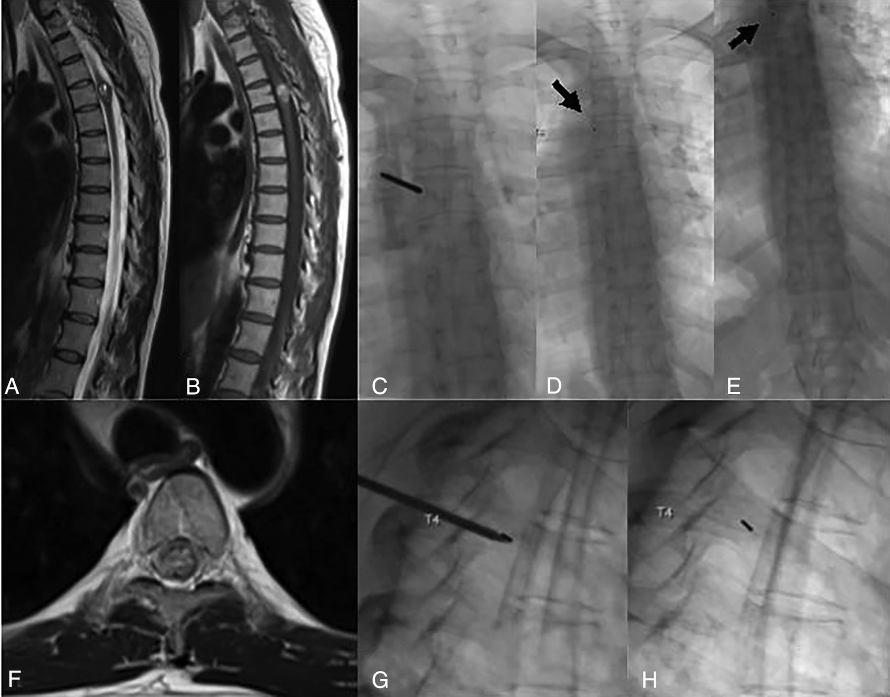
- FIG 1.
Biplane FL was used to place a gold fiducial marker at T4. Sagittal T2WI (A), sagittal T1WI pregadolinium (B), and axial T2WI (F) demonstrate a multilobulated T1-hyperintense, intramedullary mass with fluid-fluid levels and peripheral T2 hypointensity, which was resected and pathologically proved to be a cavernous malformation. C, Anterior-posterior FL image demonstrates the bone access needle traversing the left pedicle of T4. D and E, Postprocedural images for counting purposes show the first and last ribs, respectively, with the fiducial marker in place (arrow). G, Intraprocedural image demonstrates the coaxial bone access needle with the stylet in place and the gold fiducial at the tip of the needle. H, Subsequently, the needle has been removed, and the gold fiducial remains in place at the junction of the pedicle and vertebral body.
- FIG 2.
CT-guided gold fiducial marker placement. A and B, Sagittal T2WI and fat-saturated postcontrast T1WI demonstrate an intradural extramedullary, lobulated, homogeneously intensely enhancing mass that was resected and found to be a schwannoma on pathology. D, Intraprocedural axial CT bone windows at T12 demonstrate a coaxial bone access needle located within the left pedicle of T12. C and E, Postprocedural CT MIP and sagittal reconstructions confirm the gold fiducial marker placement in the left T12 pedicle. Arrow points toward gold fiducial marker.


Tables
Demographics Mean age (range) 57 (12–96) Sex Male = 75 Female = 104 Level of gold fiducial marker placement C7 = 1 T1 = 5 T2 = 5 T3 = 14 T4 = 14 T5 = 18 T6 = 21 T7 = 26 T8 = 21 T9 = 15 T10 = 19 T11 = 15 T12 = 7 L3 = 1 L4 = 1 Method CT = 36 FL = 143 Mean radiation dose (range) (mGy) 183.43 (13–805) Mean FL time (range) (min) 3.92 (0.4–15.2) No. of wrong-level surgeries 0 No. of different radiologists who performed a gold fiducial marker placement 13 Mean BMI (range) 28.9 (16.1–55.8) Osteoporosis diagnosis No = 159 Yes = 20 Trainee present? No = 107 Yes = 72 Mean procedure duration (range) (min)a 51.9 (20–114) Mean No. of days when operation was performed after placement (range) 3.6 days (0–92) Note:—BMI indicates body mass index.
↵a Entry of the room by physician to room exit time.
CT FL Total cases 36 143 Level of gold fiducial marker placement C7 = 1 C7 = 0 T1 = 4 T1 = 1 T2 = 3 T2 = 2 T3 = 6 T3 = 8 T4 = 6 T4 = 8 T5 = 2 T5 = 16 T6 = 2 T6 = 19 T7 = 2 T7 = 24 T8 = 2 T8 = 19 T9 = 1 T9 = 14 T10 = 4 T10 = 15 T11 = 1 T11 = 14 T12 = 2 T12 = 5 L3 = 0 L3 = 1 L4 = 0 L4 = 1 Average radiation dose (range) (mGy) 258.92 (80.69–563.56) 164.43 (13–805) Average procedural time (range) (min) 51.19 (31–114) 52.09 (20–89)


