Advertisement

Main menu

  • Home
  • Content
    • Current Issue
    • Accepted Manuscripts
    • Article Preview
    • Past Issue Archive
    • Video Articles
    • AJNR Case Collection
    • Case of the Week Archive
    • Case of the Month Archive
    • Classic Case Archive
  • Special Collections
    • AJNR Awards
    • Low-Field MRI
    • Alzheimer Disease
    • ASNR Foundation Special Collection
    • Photon-Counting CT
    • View All
  • Multimedia
    • AJNR Podcasts
    • AJNR SCANtastic
    • Trainee Corner
    • MRI Safety Corner
    • Imaging Protocols
  • For Authors
    • Submit a Manuscript
    • Submit a Video Article
    • Submit an eLetter to the Editor/Response
    • Manuscript Submission Guidelines
    • Statistical Tips
    • Fast Publishing of Accepted Manuscripts
    • Graphical Abstract Preparation
    • Imaging Protocol Submission
    • Author Policies
  • About Us
    • About AJNR
    • Editorial Board
    • Editorial Board Alumni
  • More
    • Become a Reviewer/Academy of Reviewers
    • Subscribers
    • Permissions
    • Alerts
    • Feedback
    • Advertisers
    • ASNR Home

User menu

  • Alerts
  • Log in

Search

  • Advanced search
American Journal of Neuroradiology

American Journal of Neuroradiology

ASHNR American Society of Functional Neuroradiology ASHNR American Society of Pediatric Neuroradiology ASSR
  • Alerts
  • Log in

Advanced Search

  • Home
  • Content
    • Current Issue
    • Accepted Manuscripts
    • Article Preview
    • Past Issue Archive
    • Video Articles
    • AJNR Case Collection
    • Case of the Week Archive
    • Case of the Month Archive
    • Classic Case Archive
  • Special Collections
    • AJNR Awards
    • Low-Field MRI
    • Alzheimer Disease
    • ASNR Foundation Special Collection
    • Photon-Counting CT
    • View All
  • Multimedia
    • AJNR Podcasts
    • AJNR SCANtastic
    • Trainee Corner
    • MRI Safety Corner
    • Imaging Protocols
  • For Authors
    • Submit a Manuscript
    • Submit a Video Article
    • Submit an eLetter to the Editor/Response
    • Manuscript Submission Guidelines
    • Statistical Tips
    • Fast Publishing of Accepted Manuscripts
    • Graphical Abstract Preparation
    • Imaging Protocol Submission
    • Author Policies
  • About Us
    • About AJNR
    • Editorial Board
    • Editorial Board Alumni
  • More
    • Become a Reviewer/Academy of Reviewers
    • Subscribers
    • Permissions
    • Alerts
    • Feedback
    • Advertisers
    • ASNR Home
  • Follow AJNR on Twitter
  • Visit AJNR on Facebook
  • Follow AJNR on Instagram
  • Join AJNR on LinkedIn
  • RSS Feeds

AJNR Awards, New Junior Editors, and more. Read the latest AJNR updates

Research ArticlePediatric Neuroimaging
Open Access

Longitudinal MRI Evaluation of Brain Development in Fetuses with Congenital Diaphragmatic Hernia around the Time of Fetal Endotracheal Occlusion

D. Emam, M. Aertsen, L. Van der Veeken, L. Fidon, P. Patkee, V. Kyriakopoulou, L. De Catte, F. Russo, P. Demaerel, T. Vercauteren, M. Rutherford and J. Deprest
American Journal of Neuroradiology February 2023, 44 (2) 205-211; DOI: https://doi.org/10.3174/ajnr.A7760
D. Emam
aFrom the Department of Development and Regeneration (D.E., L.V.d.V., L.D.C., F.R., J.D.), Cluster Woman and Child, Group Biomedical Sciences, KU Leuven University of Leuven, Leuven, Belgium
bDepartment Obstetrics and Gynaecology (D.E., L.F.), Faculty of Medicine, Tanta University, Tanta, Egypt
  • Find this author on Google Scholar
  • Find this author on PubMed
  • Search for this author on this site
  • ORCID record for D. Emam
M. Aertsen
cDepartment of Imaging and Pathology (M.A., P.D.), Clinical Department of Radiology, University Hospitals, KU Leuven, Leuven, Belgium
  • Find this author on Google Scholar
  • Find this author on PubMed
  • Search for this author on this site
  • ORCID record for M. Aertsen
L. Van der Veeken
aFrom the Department of Development and Regeneration (D.E., L.V.d.V., L.D.C., F.R., J.D.), Cluster Woman and Child, Group Biomedical Sciences, KU Leuven University of Leuven, Leuven, Belgium
dClinical Department Obstetrics and Gynaecology (L.V.d.V., L.D.C., F.R., J.D.), University Hospitals Leuven, Leuven, Belgium
  • Find this author on Google Scholar
  • Find this author on PubMed
  • Search for this author on this site
  • ORCID record for L. Van der Veeken
L. Fidon
bDepartment Obstetrics and Gynaecology (D.E., L.F.), Faculty of Medicine, Tanta University, Tanta, Egypt
fDivision of Imaging Sciences and Biomedical Engineering, Perinatal Imaging and Health and School of Biomedical Engineering and Imaging Sciences (L.F., T.V., J.D.), King’s College London, King’s Health Partners, St. Thomas’ Hospital, London, UK
  • Find this author on Google Scholar
  • Find this author on PubMed
  • Search for this author on this site
  • ORCID record for L. Fidon
P. Patkee
eCentre for the Developing Brain (P.P., V.K., M.R., J.D.)
  • Find this author on Google Scholar
  • Find this author on PubMed
  • Search for this author on this site
  • ORCID record for P. Patkee
V. Kyriakopoulou
eCentre for the Developing Brain (P.P., V.K., M.R., J.D.)
  • Find this author on Google Scholar
  • Find this author on PubMed
  • Search for this author on this site
  • ORCID record for V. Kyriakopoulou
L. De Catte
aFrom the Department of Development and Regeneration (D.E., L.V.d.V., L.D.C., F.R., J.D.), Cluster Woman and Child, Group Biomedical Sciences, KU Leuven University of Leuven, Leuven, Belgium
dClinical Department Obstetrics and Gynaecology (L.V.d.V., L.D.C., F.R., J.D.), University Hospitals Leuven, Leuven, Belgium
  • Find this author on Google Scholar
  • Find this author on PubMed
  • Search for this author on this site
  • ORCID record for L. De Catte
F. Russo
aFrom the Department of Development and Regeneration (D.E., L.V.d.V., L.D.C., F.R., J.D.), Cluster Woman and Child, Group Biomedical Sciences, KU Leuven University of Leuven, Leuven, Belgium
dClinical Department Obstetrics and Gynaecology (L.V.d.V., L.D.C., F.R., J.D.), University Hospitals Leuven, Leuven, Belgium
  • Find this author on Google Scholar
  • Find this author on PubMed
  • Search for this author on this site
P. Demaerel
cDepartment of Imaging and Pathology (M.A., P.D.), Clinical Department of Radiology, University Hospitals, KU Leuven, Leuven, Belgium
  • Find this author on Google Scholar
  • Find this author on PubMed
  • Search for this author on this site
  • ORCID record for P. Demaerel
T. Vercauteren
fDivision of Imaging Sciences and Biomedical Engineering, Perinatal Imaging and Health and School of Biomedical Engineering and Imaging Sciences (L.F., T.V., J.D.), King’s College London, King’s Health Partners, St. Thomas’ Hospital, London, UK
  • Find this author on Google Scholar
  • Find this author on PubMed
  • Search for this author on this site
  • ORCID record for T. Vercauteren
M. Rutherford
eCentre for the Developing Brain (P.P., V.K., M.R., J.D.)
  • Find this author on Google Scholar
  • Find this author on PubMed
  • Search for this author on this site
  • ORCID record for M. Rutherford
J. Deprest
aFrom the Department of Development and Regeneration (D.E., L.V.d.V., L.D.C., F.R., J.D.), Cluster Woman and Child, Group Biomedical Sciences, KU Leuven University of Leuven, Leuven, Belgium
dClinical Department Obstetrics and Gynaecology (L.V.d.V., L.D.C., F.R., J.D.), University Hospitals Leuven, Leuven, Belgium
eCentre for the Developing Brain (P.P., V.K., M.R., J.D.)
fDivision of Imaging Sciences and Biomedical Engineering, Perinatal Imaging and Health and School of Biomedical Engineering and Imaging Sciences (L.F., T.V., J.D.), King’s College London, King’s Health Partners, St. Thomas’ Hospital, London, UK
gInstitute for Women’s Health (J.D.), University College London, London, UK
  • Find this author on Google Scholar
  • Find this author on PubMed
  • Search for this author on this site
  • ORCID record for J. Deprest
  • Article
  • Figures & Data
  • Info & Metrics
  • Responses
  • References
  • PDF
Loading

Article Figures & Data

Figures

  • Tables
  • FIG 1.
    • Download figure
    • Open in new tab
    • Download powerpoint
    FIG 1.

    T2-weighted images in the axial (A), coronal (B), and sagittal (C) planes illustrating automated segmentations of the extra-axial space (yellow), white matter (red), ventricular system (green), and cerebellum (blue).
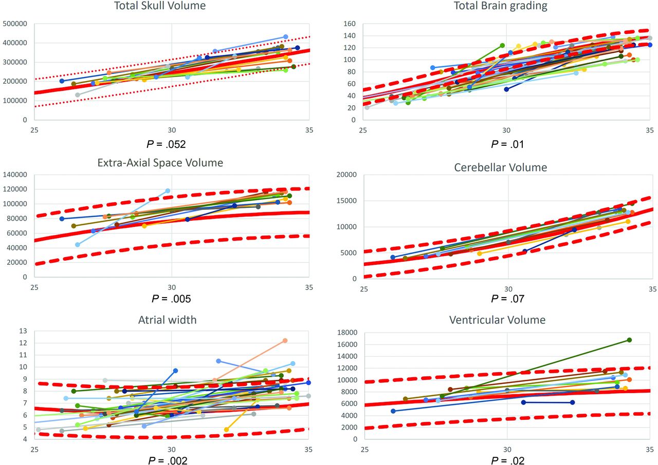
  • FIG 2.
    • Download figure
    • Open in new tab
    • Download powerpoint
    FIG 2.

    Graphs demonstrating the paired observations of the total skull volume, total brain grading, extra-axial space volume, cerebellar volume, atrial width, and ventricular volume. The provided P value is the significance level of the Wilcoxon-Mann-Whitney test comparing the observations at the second time point in fetuses with congenital diaphragmatic hernia with that in healthy controls. The mean values of the control populations (full red line) with the 95% confidence intervals (dashed red line) are also shown.

  • FIG 3.
    • Download figure
    • Open in new tab
    • Download powerpoint
    FIG 3.

    Three orthogonal T2-weighted spin-echo images in a healthy fetus (A–C) and a fetus with a left-sided diaphragmatic hernia (D–F), both at a GA of 30 weeks 4 days. In the axial plane (A and D), the enlarged pericerebral space is evident (arrowheads). The difference in gyrification is most evident in the parietal area and best seen in the coronal plane (circle in B and E) and to a lesser extent in the sagittal plane (arrows in C and F). The enlarged pericerebral space in the coronal and sagittal planes is marked with arrowheads.

Tables

  • Figures
  • General characteristics of the congenital diaphragmatic hernia group

    General Characteristics
    Left/right/bilateral28 (66.7%)/12 (28.6%)/2 (4.8%)
    o/e TFLV at first MR imaging
     <15%6 (14.2%)
     16%–25%13 (31%)
     26%–45%22 (52.4%)
     >45%1 (2.4%)
    Liver herniation40 (95.2%)
    Liver/thorax ratio30.13 (9.67)
    Fetal position at MR imaging 1 (cephalic/breech/transverse)28 (67%)/13 (31%)/1 (2%)
    Fetal position at MR imaging 2 (cephalic/breech/transverse)37 (88%)/5 (12%) / 0
    GA at MR imaging 1 (mean)28.0 (SD, 2.1)
    GA at MR imaging 2 (mean)33.2 (SD, 1.3)
    Individual interval4.83 (1.85)
    Fetal endoluminal tracheal occlusion40 (95%)
    • Note:—o/e indicates observed over expected ratio.

PreviousNext
Back to top

In this issue

American Journal of Neuroradiology: 44 (2)
American Journal of Neuroradiology
Vol. 44, Issue 2
1 Feb 2023
  • Table of Contents
  • Index by author
  • Complete Issue (PDF)
Advertisement
Print
Download PDF
Email Article

Thank you for your interest in spreading the word on American Journal of Neuroradiology.

NOTE: We only request your email address so that the person you are recommending the page to knows that you wanted them to see it, and that it is not junk mail. We do not capture any email address.

Enter multiple addresses on separate lines or separate them with commas.
Longitudinal MRI Evaluation of Brain Development in Fetuses with Congenital Diaphragmatic Hernia around the Time of Fetal Endotracheal Occlusion
(Your Name) has sent you a message from American Journal of Neuroradiology
(Your Name) thought you would like to see the American Journal of Neuroradiology web site.
CAPTCHA
This question is for testing whether or not you are a human visitor and to prevent automated spam submissions.
Cite this article
D. Emam, M. Aertsen, L. Van der Veeken, L. Fidon, P. Patkee, V. Kyriakopoulou, L. De Catte, F. Russo, P. Demaerel, T. Vercauteren, M. Rutherford, J. Deprest
Longitudinal MRI Evaluation of Brain Development in Fetuses with Congenital Diaphragmatic Hernia around the Time of Fetal Endotracheal Occlusion
American Journal of Neuroradiology Feb 2023, 44 (2) 205-211; DOI: 10.3174/ajnr.A7760

Citation Manager Formats

  • BibTeX
  • Bookends
  • EasyBib
  • EndNote (tagged)
  • EndNote 8 (xml)
  • Medlars
  • Mendeley
  • Papers
  • RefWorks Tagged
  • Ref Manager
  • RIS
  • Zotero
0 Responses
Respond to this article
Share
Bookmark this article
Brain in Fetuses with Diaphragmatic Hernia
D. Emam, M. Aertsen, L. Van der Veeken, L. Fidon, P. Patkee, V. Kyriakopoulou, L. De Catte, F. Russo, P. Demaerel, T. Vercauteren, M. Rutherford, J. Deprest
American Journal of Neuroradiology Feb 2023, 44 (2) 205-211; DOI: 10.3174/ajnr.A7760
del.icio.us logo Twitter logo Facebook logo Mendeley logo
  • Tweet Widget
  • Facebook Like
  • Google Plus One
Purchase

Jump to section

  • Article
    • Abstract
    • ABBREVIATIONS:
    • MATERIALS AND METHODS
    • RESULTS
    • DISCUSSION
    • CONCLUSIONS
    • Footnotes
    • References
  • Figures & Data
  • Info & Metrics
  • Responses
  • References
  • PDF

Related Articles

  • No related articles found.
  • PubMed
  • Google Scholar

Cited By...

  • Normative range of MRI-derived fetal brain volume throughout gestation: a prospective study
  • Unveiling fetal brain changes in congenital diaphragmatic hernia: hypoxic injury with loss of progenitor cells, neurons, and oligodendrocytes
  • Crossref
  • Google Scholar

This article has not yet been cited by articles in journals that are participating in Crossref Cited-by Linking.

More in this TOC Section

  • Frontal Paraventricular Cysts
  • Sodium MRI in Pediatric Brain Tumors
  • FRACTURE MR in Congenital Vertebral Anomalies
Show more Pediatric Neuroimaging

Similar Articles

Advertisement

Indexed Content

  • Current Issue
  • Accepted Manuscripts
  • Article Preview
  • Past Issues
  • Editorials
  • Editor's Choice
  • Fellows' Journal Club
  • Letters to the Editor
  • Video Articles

Cases

  • Case Collection
  • Archive - Case of the Week
  • Archive - Case of the Month
  • Archive - Classic Case

More from AJNR

  • Trainee Corner
  • Imaging Protocols
  • MRI Safety Corner
  • Book Reviews

Multimedia

  • AJNR Podcasts
  • AJNR Scantastics

Resources

  • Turnaround Time
  • Submit a Manuscript
  • Submit a Video Article
  • Submit an eLetter to the Editor/Response
  • Manuscript Submission Guidelines
  • Statistical Tips
  • Fast Publishing of Accepted Manuscripts
  • Graphical Abstract Preparation
  • Imaging Protocol Submission
  • Evidence-Based Medicine Level Guide
  • Publishing Checklists
  • Author Policies
  • Become a Reviewer/Academy of Reviewers
  • News and Updates

About Us

  • About AJNR
  • Editorial Board
  • Editorial Board Alumni
  • Alerts
  • Permissions
  • Not an AJNR Subscriber? Join Now
  • Advertise with Us
  • Librarian Resources
  • Feedback
  • Terms and Conditions
  • AJNR Editorial Board Alumni

American Society of Neuroradiology

  • Not an ASNR Member? Join Now

© 2025 by the American Society of Neuroradiology All rights, including for text and data mining, AI training, and similar technologies, are reserved.
Print ISSN: 0195-6108 Online ISSN: 1936-959X

Powered by HighWire