
Article Figures & Data
Figures
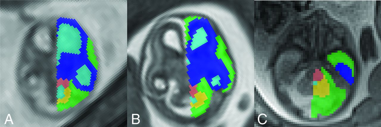
- FIG 1.
Representative example of ROIs in the brain of 12-week (A), 15-week (B), and 19-week-old fetuses (C). The supratentorial brain is labeled in dark blue; the ventricular CSF, in light blue; the extra-axial CSF, in green; the brainstem, in salmon; and the cerebellum, in yellow.
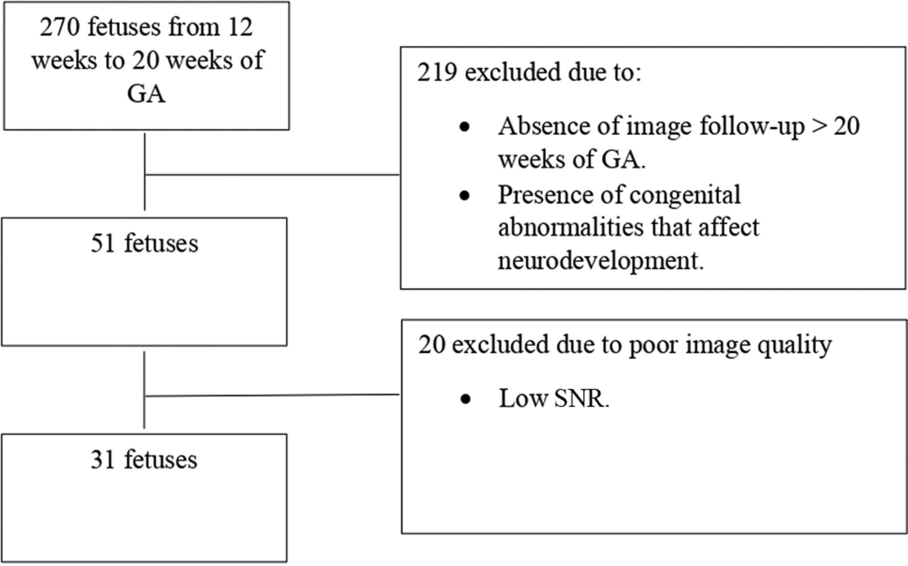
- FIG 2.
Application of inclusion and exclusion criteria in a flow diagram.
- FIG 3.
Absolute and relative volume growth per week. For each structure, absolute volume growth per week of GA (A) and relative volume growth per week (B) demonstrate relatively larger supratentorial growth during these early GAs.


Tables
Population Characteristics Median (Range) Maternal age (yr) 32 (18–41) Median GA (weeks) 18 (12–19) Mean fetal growth percentile 44.62 (10.6–96.3) Sexa Female 12 (38.70%) Male 19 (61.29%) Fetal diagnosis Congenital diaphragmatic hernia 7 (21.87%) Normal 7 (21.87%) Omphalocele 4 (12.5%) Congenital pulmonary airway malformation 3 (9.37%) Cleft lip 2 (6.25%) Congenital heart disease 2 (6.25%) Patent urachus 2 (6.25%) Lymphatic malformation 1 (3.12%) Occipital subcutaneous mass 1 (3.12%) Hepatic hemangioma 1 (3.12%) Hepatic pseudocyst 1 (3.12%) Cardiac rhabdomyoma 1 (3.12%) ↵a Data from this point down are No. of subjects (%).
Structure Initial Relative Volume Final Relative Volume Change in Relative Volume per Week (95% CI) P Value Intraventricular CSF 23.52% (20.912%–26.12%) 8.64% (7.45%–9.84%) −2.12 (−2.58 to −1.66) .001 Supratentorial parenchyma 38.06% (34.25%–41.87%) 48.15% (46.39%–49.90%) 1.44 (0.77–2.11) .001 Extra-axial CSF 32.26% (27.65%–36.86%) 39.62% (37.50%–41.74%) 1.05 (0.24–1.87) .02 Brainstem 3.57% (2.97%–4.18%) 1.93% (1.66%–2.21%) −0.23 (−0.34 to −0.13) .001 Cerebellum 2.57% (2.16%–2.98%) 1.63% (1.44%–1.8%2) −0.13 (−0.21 to −0.06) .001 ↵a Relative volume is expressed in terms of percentage. Data in parentheses are 95% CIs. ROIs are sorted in descending order based on the magnitude of the change of relative volume. The initial relative volume represents an estimate of 12 weeks' GA, and the final relative volume represents an estimate of 19 weeks' GA.


