Advertisement

Main menu

  • Home
  • Content
    • Current Issue
    • Accepted Manuscripts
    • Article Preview
    • Past Issue Archive
    • Video Articles
    • AJNR Case Collection
    • Case of the Week Archive
    • Case of the Month Archive
    • Classic Case Archive
  • Special Collections
    • AJNR Awards
    • Low-Field MRI
    • Alzheimer Disease
    • ASNR Foundation Special Collection
    • Photon-Counting CT
    • View All
  • Multimedia
    • AJNR Podcasts
    • AJNR SCANtastic
    • Trainee Corner
    • MRI Safety Corner
    • Imaging Protocols
  • For Authors
    • Submit a Manuscript
    • Submit a Video Article
    • Submit an eLetter to the Editor/Response
    • Manuscript Submission Guidelines
    • Statistical Tips
    • Fast Publishing of Accepted Manuscripts
    • Graphical Abstract Preparation
    • Imaging Protocol Submission
    • Author Policies
  • About Us
    • About AJNR
    • Editorial Board
    • Editorial Board Alumni
  • More
    • Become a Reviewer/Academy of Reviewers
    • Subscribers
    • Permissions
    • Alerts
    • Feedback
    • Advertisers
    • ASNR Home

User menu

  • Alerts
  • Log in

Search

  • Advanced search
American Journal of Neuroradiology

American Journal of Neuroradiology

ASHNR American Society of Functional Neuroradiology ASHNR American Society of Pediatric Neuroradiology ASSR
  • Alerts
  • Log in

Advanced Search

  • Home
  • Content
    • Current Issue
    • Accepted Manuscripts
    • Article Preview
    • Past Issue Archive
    • Video Articles
    • AJNR Case Collection
    • Case of the Week Archive
    • Case of the Month Archive
    • Classic Case Archive
  • Special Collections
    • AJNR Awards
    • Low-Field MRI
    • Alzheimer Disease
    • ASNR Foundation Special Collection
    • Photon-Counting CT
    • View All
  • Multimedia
    • AJNR Podcasts
    • AJNR SCANtastic
    • Trainee Corner
    • MRI Safety Corner
    • Imaging Protocols
  • For Authors
    • Submit a Manuscript
    • Submit a Video Article
    • Submit an eLetter to the Editor/Response
    • Manuscript Submission Guidelines
    • Statistical Tips
    • Fast Publishing of Accepted Manuscripts
    • Graphical Abstract Preparation
    • Imaging Protocol Submission
    • Author Policies
  • About Us
    • About AJNR
    • Editorial Board
    • Editorial Board Alumni
  • More
    • Become a Reviewer/Academy of Reviewers
    • Subscribers
    • Permissions
    • Alerts
    • Feedback
    • Advertisers
    • ASNR Home
  • Follow AJNR on Twitter
  • Visit AJNR on Facebook
  • Follow AJNR on Instagram
  • Join AJNR on LinkedIn
  • RSS Feeds

AJNR Awards, New Junior Editors, and more. Read the latest AJNR updates

Research ArticleSpine Imaging and Spine Image-Guided Interventions

Likelihood of Discovering a CSF Leak Based on Intracranial MRI Findings in Patients without a Spinal Longitudinal Extradural Collection: A New Probabilistic Scoring System

John C. Benson, Ajay A. Madhavan, Ian T. Mark, Jeremy K. Cutsforth-Gregory, Waleed Brinjikji and Jared T. Verdoorn
American Journal of Neuroradiology November 2023, 44 (11) 1339-1344; DOI: https://doi.org/10.3174/ajnr.A8030
John C. Benson
aFrom the Department of Radiology (J.C.B., A.A.M., I.T.M., W.B., J.T.V.), Mayo Clinic, Rochester, Minnesota
  • Find this author on Google Scholar
  • Find this author on PubMed
  • Search for this author on this site
  • ORCID record for John C. Benson
Ajay A. Madhavan
aFrom the Department of Radiology (J.C.B., A.A.M., I.T.M., W.B., J.T.V.), Mayo Clinic, Rochester, Minnesota
  • Find this author on Google Scholar
  • Find this author on PubMed
  • Search for this author on this site
  • ORCID record for Ajay A. Madhavan
Ian T. Mark
aFrom the Department of Radiology (J.C.B., A.A.M., I.T.M., W.B., J.T.V.), Mayo Clinic, Rochester, Minnesota
  • Find this author on Google Scholar
  • Find this author on PubMed
  • Search for this author on this site
  • ORCID record for Ian T. Mark
Jeremy K. Cutsforth-Gregory
bDepartment of Neurology (J.K.C.-G.), Mayo Clinic, Rochester, Minnesota
  • Find this author on Google Scholar
  • Find this author on PubMed
  • Search for this author on this site
  • ORCID record for Jeremy K. Cutsforth-Gregory
Waleed Brinjikji
aFrom the Department of Radiology (J.C.B., A.A.M., I.T.M., W.B., J.T.V.), Mayo Clinic, Rochester, Minnesota
  • Find this author on Google Scholar
  • Find this author on PubMed
  • Search for this author on this site
  • ORCID record for Waleed Brinjikji
Jared T. Verdoorn
aFrom the Department of Radiology (J.C.B., A.A.M., I.T.M., W.B., J.T.V.), Mayo Clinic, Rochester, Minnesota
  • Find this author on Google Scholar
  • Find this author on PubMed
  • Search for this author on this site
  • ORCID record for Jared T. Verdoorn
  • Article
  • Figures & Data
  • Info & Metrics
  • Responses
  • References
  • PDF
Loading

Article Figures & Data

Figures

  • Tables
  • FIG 1.
    • Download figure
    • Open in new tab
    • Download powerpoint
    FIG 1.

    An example of a distal nerve root sleeve tear (type 4 leak) diagnosed using a combination of DSM and delayed CTM findings. Left lateral decubitus unsubtracted image from a DSM (A) demonstrates contained contrast within a left T12 meningeal diverticulum (A, arrow). On a 30-minute delayed left decubitus CTM (B), there is a subtle contrast leak posterior to the diverticulum (B, arrow). The combined findings from a DSM and delayed CTM are often necessary to confidently diagnose type 4 leaks.
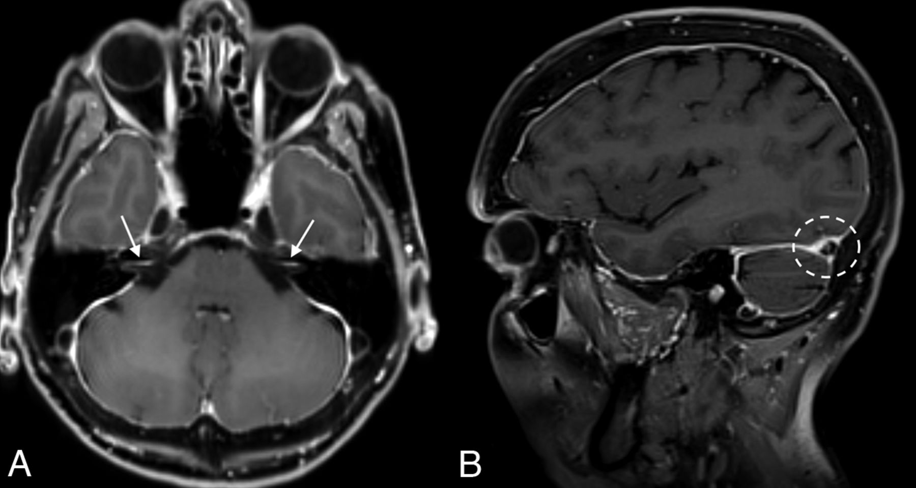
  • FIG 2.
    • Download figure
    • Open in new tab
    • Download powerpoint
    FIG 2.

    An example of multiple intracranial sequelae of SIH in a 69-year-old woman. Axial (A) and sagittal (B) postcontrast images demonstrate diffuse smooth dural enhancement, with involvement of the IACs (arrows, A). The right transverse sinus is engorged (dashed circle, B).

  • FIG 3.
    • Download figure
    • Open in new tab
    • Download powerpoint
    FIG 3.

    Schematics of intracranial findings used for the probabilistic scoring system developed in this study. Pertinent findings included diffuse dural enhancement and dural enhancement involving the walls of the IACs (pink lines, A), engorgement of the transverse sinus and non-Chiari cerebellar descent (B), and pituitary engorgement, effacement of the suprasellar cistern, and descent of the aqueduct iter below the incisural line (dotted line, C). Used with permission of Mayo Foundation for Medical Education and Research, all rights reserved.

Tables

  • Figures
    • View popup
    Table 1:

    Frequency and measurements of intracranial findingsa

    CSF Leak (n = 76)No CSF Leak (n = 98)P Value
    Smooth dural enhancement54 (57.4%)9 (12.5%)P < .001b
    Dural enhancement in IACs34 (36.2%)4 (5.6%)P < .001b
    Subdural fluid collections9 (9.2%)4 (5.3%)P = .32
    Superficial siderosis3 (3.1%)1 (1.3%)P = .44
    Pituitary engorgement56 (57.1%)17 (22.4%)P < .001b
    Non-Chiari cerebellar tonsillar descent of >5 mm22 (22.4%)8 (10.5%)P = .04b
    Dural venous sinus engorgementYes = 25 (25.8%) Indeterminate = 27 (27.8%)Yes = 5 (6.6%) Indeterminate = 4 (5.3%)P < .001b
    Layered hyperostosis8 (8.2%)3 (3.9%)P = .25
    Cerebral aqueduct iter below incisural line33 (33.7%)15 (19.7%)P = .04b
    Average mamillopontine distance (mm)5.5 (SD, 2.1) mm5.9 (SD, 1.7) mmP = .13
    Average prepontine cistern size (mm)4 (SD, 1.5) mm4.2 (SD, 1.6) mmP = .35
    Average suprasellar cistern size (mm)3.2 (SD, 2.1) mm4.8 (SD, 2.7) mmP < .001b
    • ↵a Indeterminate dural sinus engorgement is counted as positive in this analysis.

    • ↵b Statistically significant P values.

    • View popup
    Table 2:

    Scoring system based on 7 intracranial imaging findings

    FindingPoints
    Smooth dural enhancement1
    Dural enhancement in the IACs1
    Pituitary engorgement1
    Non-Chiari cerebellar descent of >5 mm1
    Dural venous sinus engorgement1
    Cerebral aqueduct iter below incisural line1
    Suprasellar cistern ≤2.5 mm1
    • View popup
    Table 3:

    Proposed probabilistic scoring system for determining whether a spinal CSF leak is present

    ScoreProbability of CSF Leak
    0–2Low
    ≥3Intermediate to high
PreviousNext
Back to top

In this issue

American Journal of Neuroradiology: 44 (11)
American Journal of Neuroradiology
Vol. 44, Issue 11
1 Nov 2023
  • Table of Contents
  • Index by author
  • Complete Issue (PDF)
Advertisement
Print
Download PDF
Email Article

Thank you for your interest in spreading the word on American Journal of Neuroradiology.

NOTE: We only request your email address so that the person you are recommending the page to knows that you wanted them to see it, and that it is not junk mail. We do not capture any email address.

Enter multiple addresses on separate lines or separate them with commas.
Likelihood of Discovering a CSF Leak Based on Intracranial MRI Findings in Patients without a Spinal Longitudinal Extradural Collection: A New Probabilistic Scoring System
(Your Name) has sent you a message from American Journal of Neuroradiology
(Your Name) thought you would like to see the American Journal of Neuroradiology web site.
CAPTCHA
This question is for testing whether or not you are a human visitor and to prevent automated spam submissions.
Cite this article
John C. Benson, Ajay A. Madhavan, Ian T. Mark, Jeremy K. Cutsforth-Gregory, Waleed Brinjikji, Jared T. Verdoorn
Likelihood of Discovering a CSF Leak Based on Intracranial MRI Findings in Patients without a Spinal Longitudinal Extradural Collection: A New Probabilistic Scoring System
American Journal of Neuroradiology Nov 2023, 44 (11) 1339-1344; DOI: 10.3174/ajnr.A8030

Citation Manager Formats

  • BibTeX
  • Bookends
  • EasyBib
  • EndNote (tagged)
  • EndNote 8 (xml)
  • Medlars
  • Mendeley
  • Papers
  • RefWorks Tagged
  • Ref Manager
  • RIS
  • Zotero
0 Responses
Respond to this article
Share
Bookmark this article
MRI-Based Scoring for CSF Leak Likelihood
John C. Benson, Ajay A. Madhavan, Ian T. Mark, Jeremy K. Cutsforth-Gregory, Waleed Brinjikji, Jared T. Verdoorn
American Journal of Neuroradiology Nov 2023, 44 (11) 1339-1344; DOI: 10.3174/ajnr.A8030
del.icio.us logo Twitter logo Facebook logo Mendeley logo
  • Tweet Widget
  • Facebook Like
  • Google Plus One
Purchase

Jump to section

  • Article
    • Abstract
    • ABBREVIATIONS:
    • MATERIALS AND METHODS
    • RESULTS
    • DISCUSSION
    • CONCLUSIONS
    • Footnotes
    • References
  • Figures & Data
  • Info & Metrics
  • Responses
  • References
  • PDF

Related Articles

  • No related articles found.
  • PubMed
  • Google Scholar

Cited By...

  • Evaluation of Spontaneous Intracranial Hypotension Probabilistic Brain MRI Scoring Systems in Normal Patients
  • {beta}-Trace Protein as a Potential Biomarker for CSF-Venous Fistulas
  • Spinal CSF Leaks: The Neuroradiologist Transforming Care
  • Myelographic Techniques for the Localization of CSF-Venous Fistulas: Updates in 2024
  • Crossref
  • Google Scholar

This article has not yet been cited by articles in journals that are participating in Crossref Cited-by Linking.

More in this TOC Section

  • Optimization of Photon Counting CT Myelography
  • Characteristics of SIH Type I Culprit Lesions
  • Management Outcomes For VO Spine Biopsy
Show more Spine Imaging and Spine Image-Guided Interventions

Similar Articles

Advertisement

Indexed Content

  • Current Issue
  • Accepted Manuscripts
  • Article Preview
  • Past Issues
  • Editorials
  • Editor's Choice
  • Fellows' Journal Club
  • Letters to the Editor
  • Video Articles

Cases

  • Case Collection
  • Archive - Case of the Week
  • Archive - Case of the Month
  • Archive - Classic Case

More from AJNR

  • Trainee Corner
  • Imaging Protocols
  • MRI Safety Corner
  • Book Reviews

Multimedia

  • AJNR Podcasts
  • AJNR Scantastics

Resources

  • Turnaround Time
  • Submit a Manuscript
  • Submit a Video Article
  • Submit an eLetter to the Editor/Response
  • Manuscript Submission Guidelines
  • Statistical Tips
  • Fast Publishing of Accepted Manuscripts
  • Graphical Abstract Preparation
  • Imaging Protocol Submission
  • Evidence-Based Medicine Level Guide
  • Publishing Checklists
  • Author Policies
  • Become a Reviewer/Academy of Reviewers
  • News and Updates

About Us

  • About AJNR
  • Editorial Board
  • Editorial Board Alumni
  • Alerts
  • Permissions
  • Not an AJNR Subscriber? Join Now
  • Advertise with Us
  • Librarian Resources
  • Feedback
  • Terms and Conditions
  • AJNR Editorial Board Alumni

American Society of Neuroradiology

  • Not an ASNR Member? Join Now

© 2025 by the American Society of Neuroradiology All rights, including for text and data mining, AI training, and similar technologies, are reserved.
Print ISSN: 0195-6108 Online ISSN: 1936-959X

Powered by HighWire