Advertisement

Main menu

  • Home
  • Content
    • Current Issue
    • Accepted Manuscripts
    • Article Preview
    • Past Issue Archive
    • Video Articles
    • AJNR Case Collection
    • Case of the Week Archive
    • Case of the Month Archive
    • Classic Case Archive
  • Special Collections
    • AJNR Awards
    • Low-Field MRI
    • Alzheimer Disease
    • ASNR Foundation Special Collection
    • Photon-Counting CT
    • View All
  • Multimedia
    • AJNR Podcasts
    • AJNR SCANtastic
    • Trainee Corner
    • MRI Safety Corner
    • Imaging Protocols
  • For Authors
    • Submit a Manuscript
    • Submit a Video Article
    • Submit an eLetter to the Editor/Response
    • Manuscript Submission Guidelines
    • Statistical Tips
    • Fast Publishing of Accepted Manuscripts
    • Graphical Abstract Preparation
    • Imaging Protocol Submission
    • Author Policies
  • About Us
    • About AJNR
    • Editorial Board
    • Editorial Board Alumni
  • More
    • Become a Reviewer/Academy of Reviewers
    • Subscribers
    • Permissions
    • Alerts
    • Feedback
    • Advertisers
    • ASNR Home

User menu

  • Alerts
  • Log in

Search

  • Advanced search
American Journal of Neuroradiology

American Journal of Neuroradiology

ASHNR American Society of Functional Neuroradiology ASHNR American Society of Pediatric Neuroradiology ASSR
  • Alerts
  • Log in

Advanced Search

  • Home
  • Content
    • Current Issue
    • Accepted Manuscripts
    • Article Preview
    • Past Issue Archive
    • Video Articles
    • AJNR Case Collection
    • Case of the Week Archive
    • Case of the Month Archive
    • Classic Case Archive
  • Special Collections
    • AJNR Awards
    • Low-Field MRI
    • Alzheimer Disease
    • ASNR Foundation Special Collection
    • Photon-Counting CT
    • View All
  • Multimedia
    • AJNR Podcasts
    • AJNR SCANtastic
    • Trainee Corner
    • MRI Safety Corner
    • Imaging Protocols
  • For Authors
    • Submit a Manuscript
    • Submit a Video Article
    • Submit an eLetter to the Editor/Response
    • Manuscript Submission Guidelines
    • Statistical Tips
    • Fast Publishing of Accepted Manuscripts
    • Graphical Abstract Preparation
    • Imaging Protocol Submission
    • Author Policies
  • About Us
    • About AJNR
    • Editorial Board
    • Editorial Board Alumni
  • More
    • Become a Reviewer/Academy of Reviewers
    • Subscribers
    • Permissions
    • Alerts
    • Feedback
    • Advertisers
    • ASNR Home
  • Follow AJNR on Twitter
  • Visit AJNR on Facebook
  • Follow AJNR on Instagram
  • Join AJNR on LinkedIn
  • RSS Feeds

AJNR Awards, New Junior Editors, and more. Read the latest AJNR updates

Research ArticleAdult Brain
Open Access

Validation of a Denoising Method Using Deep Learning–Based Reconstruction to Quantify Multiple Sclerosis Lesion Load on Fast FLAIR Imaging

T. Yamamoto, C. Lacheret, H. Fukutomi, R.A. Kamraoui, L. Denat, B. Zhang, V. Prevost, L. Zhang, A. Ruet, B. Triaire, V. Dousset, P. Coupé and T. Tourdias
American Journal of Neuroradiology August 2022, 43 (8) 1099-1106; DOI: https://doi.org/10.3174/ajnr.A7589
T. Yamamoto
aFrom the Institut de Bio-imagerie (T.Y., H.F., L.D., V.D., T.T.), University Bordeaux, Bordeaux, France
  • Find this author on Google Scholar
  • Find this author on PubMed
  • Search for this author on this site
  • ORCID record for T. Yamamoto
C. Lacheret
bNeuroimagerie Diagnostique et Thérapeutique (C.L., V.D., T.T.)
  • Find this author on Google Scholar
  • Find this author on PubMed
  • Search for this author on this site
  • ORCID record for C. Lacheret
H. Fukutomi
aFrom the Institut de Bio-imagerie (T.Y., H.F., L.D., V.D., T.T.), University Bordeaux, Bordeaux, France
  • Find this author on Google Scholar
  • Find this author on PubMed
  • Search for this author on this site
  • ORCID record for H. Fukutomi
R.A. Kamraoui
dLaboratoire Bordelais de Recherche en Informatique (R.A.K., P.C.), University Bordeaux, Le Centre National de la Recherche Scientifique, Bordeaux Institut National Polytechnique, Talence, France
  • Find this author on Google Scholar
  • Find this author on PubMed
  • Search for this author on this site
  • ORCID record for R.A. Kamraoui
L. Denat
aFrom the Institut de Bio-imagerie (T.Y., H.F., L.D., V.D., T.T.), University Bordeaux, Bordeaux, France
  • Find this author on Google Scholar
  • Find this author on PubMed
  • Search for this author on this site
  • ORCID record for L. Denat
B. Zhang
eCanon Medical Systems Europe (B.Z.), Zoetermeer, the Netherlands
  • Find this author on Google Scholar
  • Find this author on PubMed
  • Search for this author on this site
  • ORCID record for B. Zhang
V. Prevost
fCanon Medical Systems (V.P., B.T.), Tochigi, Japan
  • Find this author on Google Scholar
  • Find this author on PubMed
  • Search for this author on this site
  • ORCID record for V. Prevost
L. Zhang
gCanon Medical Systems China (L.Z.), Beijing, China
  • Find this author on Google Scholar
  • Find this author on PubMed
  • Search for this author on this site
  • ORCID record for L. Zhang
A. Ruet
cService de Neurologie (A.R.), Centre Hospitalier Universitaire de Bordeaux, Bordeaux, France
  • Find this author on Google Scholar
  • Find this author on PubMed
  • Search for this author on this site
  • ORCID record for A. Ruet
B. Triaire
fCanon Medical Systems (V.P., B.T.), Tochigi, Japan
  • Find this author on Google Scholar
  • Find this author on PubMed
  • Search for this author on this site
  • ORCID record for B. Triaire
V. Dousset
aFrom the Institut de Bio-imagerie (T.Y., H.F., L.D., V.D., T.T.), University Bordeaux, Bordeaux, France
bNeuroimagerie Diagnostique et Thérapeutique (C.L., V.D., T.T.)
hNeurocentreMagendie (V.D., T.T.), University of Bordeaux, L’Institut National de la Santé et de la Recherche Médicale, Bordeaux, France
  • Find this author on Google Scholar
  • Find this author on PubMed
  • Search for this author on this site
  • ORCID record for V. Dousset
P. Coupé
dLaboratoire Bordelais de Recherche en Informatique (R.A.K., P.C.), University Bordeaux, Le Centre National de la Recherche Scientifique, Bordeaux Institut National Polytechnique, Talence, France
  • Find this author on Google Scholar
  • Find this author on PubMed
  • Search for this author on this site
  • ORCID record for P. Coupé
T. Tourdias
aFrom the Institut de Bio-imagerie (T.Y., H.F., L.D., V.D., T.T.), University Bordeaux, Bordeaux, France
bNeuroimagerie Diagnostique et Thérapeutique (C.L., V.D., T.T.)
hNeurocentreMagendie (V.D., T.T.), University of Bordeaux, L’Institut National de la Santé et de la Recherche Médicale, Bordeaux, France
  • Find this author on Google Scholar
  • Find this author on PubMed
  • Search for this author on this site
  • ORCID record for T. Tourdias
  • Article
  • Figures & Data
  • Supplemental
  • Info & Metrics
  • Responses
  • References
  • PDF
Loading

Article Figures & Data

Figures

  • Tables
  • FIG 1.
    • Download figure
    • Open in new tab
    • Download powerpoint
    FIG 1.

    A, Boxplots of image-quality evaluation. The median values are connected with dotted lines. dDLR generally improved the image quality. Even images of inferior quality were retrieved to moderate quality. B, Representative reconstructed axial slices of the different FLAIR acquisitions without (upper row) and with dDLR (lower row). The amount of noise and the progressive shadowing of lesions when the scan time decreased can be appreciated. One, 2, and 3 asterisks indicate P < .05, P < .01, and P < .001, respectively.
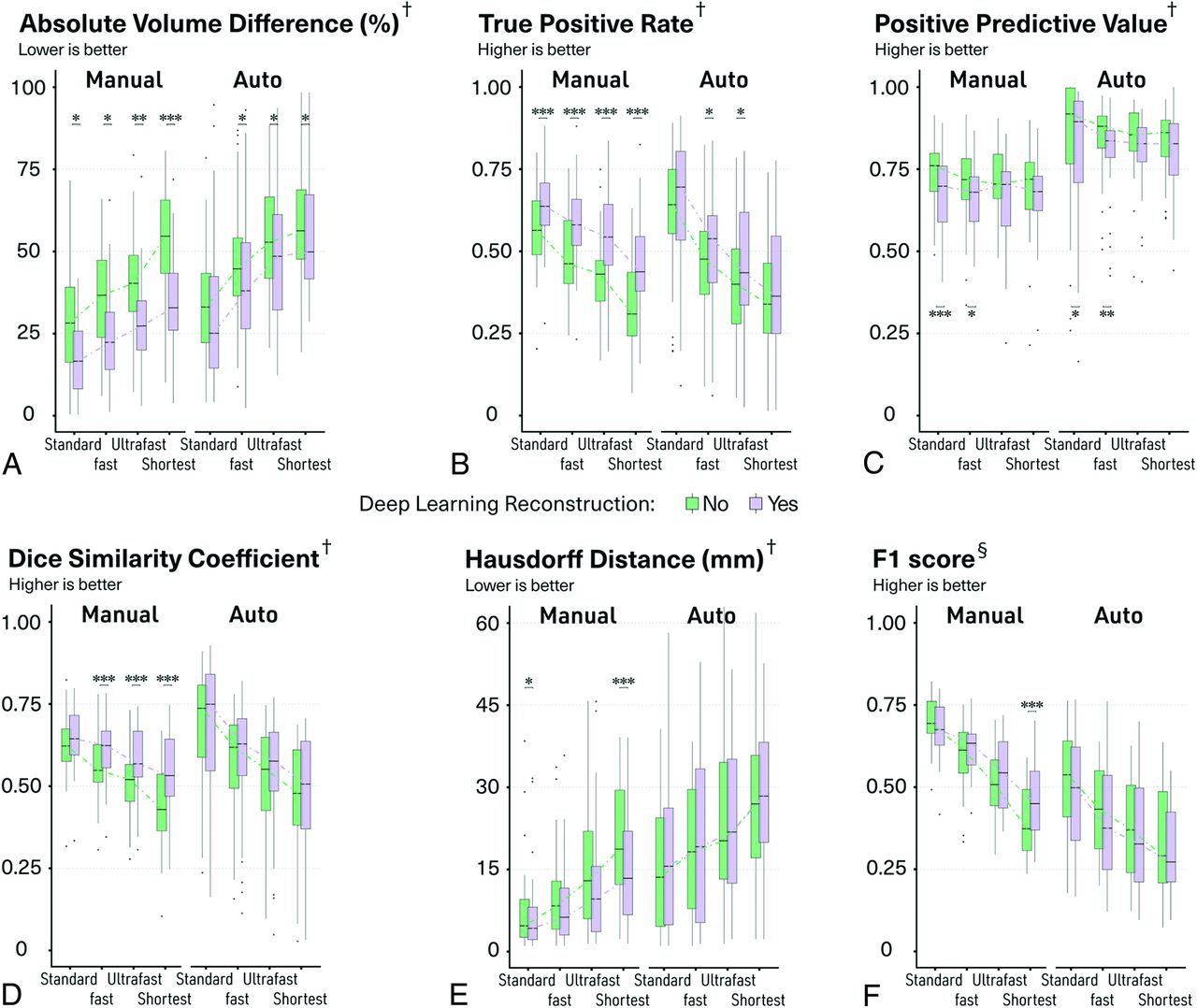
  • FIG 2.
    • Download figure
    • Open in new tab
    • Download powerpoint
    FIG 2.

    Boxplots of quantitative metrics to evaluate the manual and automatic segmentation. The median values are connected with dotted lines. One, 2, and 3 asterisks indicate P < .05, P < .01, and P < .001, respectively, for post hoc paired comparisons with Wilcoxon signed-rank test after 2-way ANOVA with the aligned rank transform procedure. † indicates voxelwise metric; §, lesion-wise metric.
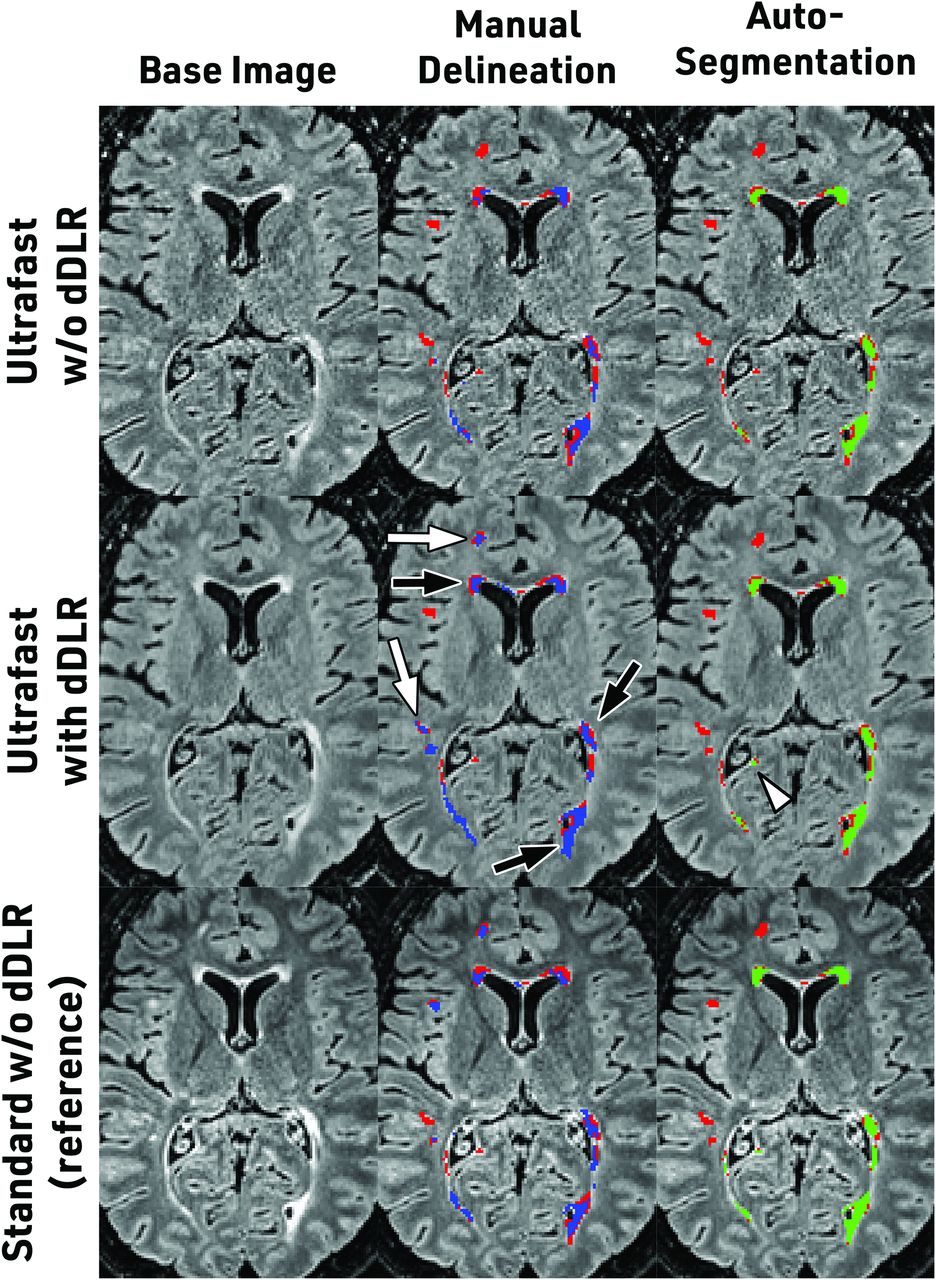
  • FIG 3.
    • Download figure
    • Open in new tab
    • Download powerpoint
    FIG 3.

    Illustrative axial slices of ultrafast FLAIR without and with dDLR. Standard FLAIR without dDLR is also shown for reference. The red mask represents the criterion standard that comes from the delineation of standard FLAIR by 2 expert readers; the blue mask is the manual delineation from a third reader; and the green mask is the automatic segmentation from volBrain software. After we applied dDLR, some lesions showed contours closer to the criterion standard (black arrows). dDLR also retrieved lesions that were missed on the original image (white arrows and arrowhead).

Tables

  • Figures
  • Parameters of MR imaging acquisitions

    3D FLAIRMPRAGE
    StandardFastUltrafastShortest
    TR (ms)70005000400030006.3
    TE (ms)/effective TE (ms)445.5/145.0445.5/145.0445.5/145.0445.5/145.02.8
    TI (ms)207015801270910950
    BW (Hz)558558558558279
    Echo space (ms)4.54.54.54.5
    FOV (mm)230 × 230230 × 230230 × 230230 × 230230 × 230
    Matrix224 × 224224 × 224224 × 224224 × 224224 × 224
    Thickness (mm)11111
    Parallel imaging2.0 × 3.02.0 × 3.02.0 × 3.02.0 × 3.01.8 × 1.5
    Frequency oversampling1.41.21.21.21.2
    Half Fourier85%70%70%
    Flip angle90°90°90°90°9°
    Refocus angleVFAVFAVFAVFA
    Shot interval (ms)2500
    Recovery time (ms)744
    Scan time (minute : second)4:542:351:401:156:18
    • Note:—VFA indicates variable flip angle; BW, Bandwidth.

PreviousNext
Back to top

In this issue

American Journal of Neuroradiology: 43 (8)
American Journal of Neuroradiology
Vol. 43, Issue 8
1 Aug 2022
  • Table of Contents
  • Index by author
  • Complete Issue (PDF)
Advertisement
Print
Download PDF
Email Article

Thank you for your interest in spreading the word on American Journal of Neuroradiology.

NOTE: We only request your email address so that the person you are recommending the page to knows that you wanted them to see it, and that it is not junk mail. We do not capture any email address.

Enter multiple addresses on separate lines or separate them with commas.
Validation of a Denoising Method Using Deep Learning–Based Reconstruction to Quantify Multiple Sclerosis Lesion Load on Fast FLAIR Imaging
(Your Name) has sent you a message from American Journal of Neuroradiology
(Your Name) thought you would like to see the American Journal of Neuroradiology web site.
CAPTCHA
This question is for testing whether or not you are a human visitor and to prevent automated spam submissions.
Cite this article
T. Yamamoto, C. Lacheret, H. Fukutomi, R.A. Kamraoui, L. Denat, B. Zhang, V. Prevost, L. Zhang, A. Ruet, B. Triaire, V. Dousset, P. Coupé, T. Tourdias
Validation of a Denoising Method Using Deep Learning–Based Reconstruction to Quantify Multiple Sclerosis Lesion Load on Fast FLAIR Imaging
American Journal of Neuroradiology Aug 2022, 43 (8) 1099-1106; DOI: 10.3174/ajnr.A7589

Citation Manager Formats

  • BibTeX
  • Bookends
  • EasyBib
  • EndNote (tagged)
  • EndNote 8 (xml)
  • Medlars
  • Mendeley
  • Papers
  • RefWorks Tagged
  • Ref Manager
  • RIS
  • Zotero
0 Responses
Respond to this article
Share
Bookmark this article
Deep Learning for MS Lesion Quantification
T. Yamamoto, C. Lacheret, H. Fukutomi, R.A. Kamraoui, L. Denat, B. Zhang, V. Prevost, L. Zhang, A. Ruet, B. Triaire, V. Dousset, P. Coupé, T. Tourdias
American Journal of Neuroradiology Aug 2022, 43 (8) 1099-1106; DOI: 10.3174/ajnr.A7589
del.icio.us logo Twitter logo Facebook logo Mendeley logo
  • Tweet Widget
  • Facebook Like
  • Google Plus One
Purchase

Jump to section

  • Article
    • Abstract
    • ABBREVIATIONS:
    • MATERIALS AND METHODS
    • RESULTS
    • DISCUSSION
    • CONCLUSIONS
    • Footnotes
    • References
  • Figures & Data
  • Supplemental
  • Info & Metrics
  • Responses
  • References
  • PDF

Related Articles

  • PubMed
  • Google Scholar

Cited By...

  • DANTE-CAIPI Accelerated Contrast-Enhanced 3D T1: Deep Learning-Based Image Quality Improvement for Vessel Wall MRI
  • Crossref
  • Google Scholar

This article has not yet been cited by articles in journals that are participating in Crossref Cited-by Linking.

More in this TOC Section

Adult Brain

  • Diagnostic Neuroradiology of Monoclonal Antibodies
  • Clinical Outcomes After Chiari I Decompression
  • Segmentation of Brain Metastases with BLAST
Show more Adult Brain

Functional

  • Kurtosis and Epileptogenic Tubers: A Pilot Study
  • Glutaric Aciduria Type 1: DK vs. Conventional MRI
  • WM Neuroaxonal Loss in Type 1 Diabetes
Show more Functional

Similar Articles

Advertisement

Indexed Content

  • Current Issue
  • Accepted Manuscripts
  • Article Preview
  • Past Issues
  • Editorials
  • Editor's Choice
  • Fellows' Journal Club
  • Letters to the Editor
  • Video Articles

Cases

  • Case Collection
  • Archive - Case of the Week
  • Archive - Case of the Month
  • Archive - Classic Case

More from AJNR

  • Trainee Corner
  • Imaging Protocols
  • MRI Safety Corner
  • Book Reviews

Multimedia

  • AJNR Podcasts
  • AJNR Scantastics

Resources

  • Turnaround Time
  • Submit a Manuscript
  • Submit a Video Article
  • Submit an eLetter to the Editor/Response
  • Manuscript Submission Guidelines
  • Statistical Tips
  • Fast Publishing of Accepted Manuscripts
  • Graphical Abstract Preparation
  • Imaging Protocol Submission
  • Evidence-Based Medicine Level Guide
  • Publishing Checklists
  • Author Policies
  • Become a Reviewer/Academy of Reviewers
  • News and Updates

About Us

  • About AJNR
  • Editorial Board
  • Editorial Board Alumni
  • Alerts
  • Permissions
  • Not an AJNR Subscriber? Join Now
  • Advertise with Us
  • Librarian Resources
  • Feedback
  • Terms and Conditions
  • AJNR Editorial Board Alumni

American Society of Neuroradiology

  • Not an ASNR Member? Join Now

© 2025 by the American Society of Neuroradiology All rights, including for text and data mining, AI training, and similar technologies, are reserved.
Print ISSN: 0195-6108 Online ISSN: 1936-959X

Powered by HighWire