
Article Figures & Data
Figures
- FIG 1.
Diagnostic approach for glioneuronal and low-grade glial tumors with cystic components (high-grade glial neoplasms were not considered for the creation of this flow chart). Predominantly cystic tumors (A), cystic with mural nodule (B), and mixed solid and cystic tumors (C) are presented. PLNTY indicates polymorphous low‐grade neuroepithelial tumor of the young; mI, myo-inositol.
- FIG 2.
An anaplastic ganglioglioma in a 15-year-old boy with treated cerebellar meduloblastoma 10 years ago. An infiltrative parieto-occipital mass lesion with heterogeneous enhancement and vasogenic edema, both extending to the contralateral hemisphere through the splenium of the corpus callosum. Also note blood by-products (C and D). Immunohistochemistry was positive for GFAP, p53, synaptophysin, and an MIB1 labeling index of 80% (not shown). Axial T2-weighted image (A). Axial T1-weighted fat-saturated contrast-enhanced image (B). Axial gradient-echo T2*-weighted image (C). Axial CT image (D).
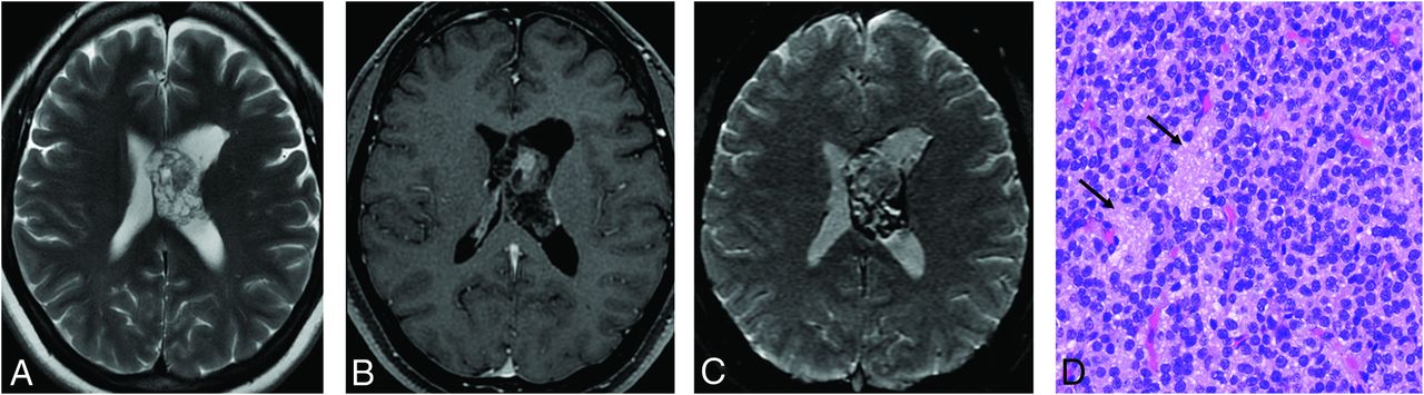
- FIG 3.
Papillary glioneuronal tumor in a 45-year-old man with migraine. A mixed solid and cystic periventricular tumor associated with vasogenic edema, blood products, and localized superficial siderosis. Microscopic findings are pseudopapillary structures composed of hyalinized vessels (long arrow) and surrounded by flattened/cuboidal glial cells. Round cells between pseudopapillae correspond to neurocytic cells (short arrow). Axial T2-weighted image (A). Axial T1-weighted fat-saturated contrast-enhanced image (B). Axial gradient-echo T2*-weighted image (C). Hematoxylin and eosin, original magnification ×100 (D).
- FIG 4.
Rosette-forming glioneuronal tumor. A lobulated multicystic tumor in the tectal plate extending to the mesencephalic aqueduct, with nodular contrast enhancement (short arrow) and displacement of a supracerebellar vein (long arrow) (A and B). Microscopic findings are small, round cells (neurocytic cells) (short arrow) surrounding mucinous extracellular matrix (DNET-like morphology) (long arrow). Sagittal T2-weighted CISS image (A). Sagittal T1-weighted fat-saturated contrast-enhanced image (B). Hematoxylin and eosin, original magnification ×100 (C).
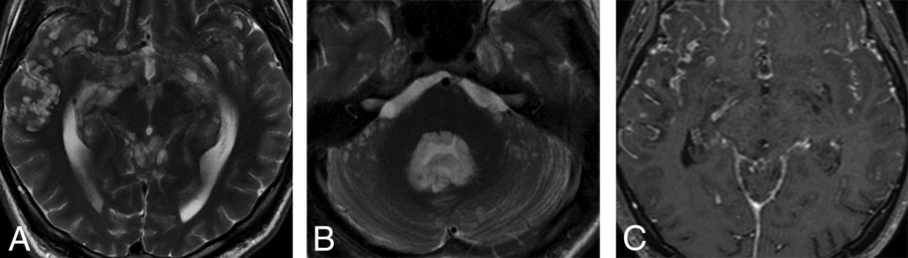
- FIG 5.
Diffuse leptomeningeal glioneuronal tumor in a 20-year-old man previously diagnosed 10 years earlier. Multiple superficial small cystlike lesions are seen in the brain sulci, basal cisterns, cerebellar folia, and filling the fourth ventricle (A and B). Also note extensive irregular leptomeningeal enhancement (C). Axial T2-weighted images (A and B). Axial T1-weighted fat-saturated contrast-enhanced image (C).
- FIG 6.
Gangliocytoma in a 40-year-old man with epilepsy. A lobulated tumor extending along the course of the left olfactory bulb and tract with foci of contrast enhancement (A and B). Microscopic findings are tumor composed of large (long arrow) and small (short arrow) ganglioneuronal cells, haphazardly distributed. Coronal T2-weighted image (A). Sagittal T1-weighted fat-saturated contrast-enhanced image (B). Hematoxylin and eosin, original magnification ×100 (C).
- FIG 7.
Dysplastic gangliocytoma of the cerebellum in a 19-year-old man with Cowden syndrome. Grossly thickened cerebellar folia with a tiger-striped pattern and rare patchy enhancement in the superior vermis and left cerebellar hemisphere. Axial T2-weighted image (A). Axial T1-weighted fat-saturated contrast-enhanced image (B).
- FIG 8.
Multinodular and vacuolating neuronal tumor of the cerebrum in a 33-year-old asymptomatic woman. Confluent, small, round lesions in a subcortical location with hypersignal in a T2-weighted FLAIR image (A). They clearly show different signal from the adjacent CSF in a T2-weighted FIESTA image, differentiating them from perivascular spaces (B).
- FIG 9.
Central neurocytoma. An intraventricular bubbly lesion adjacent to the septum showing honeycombing contrast enhancement and extensive signs of calcification and hemorrhage (A–C). Microscopic findings are sheets of round cells with scant cytoplasm. Neurocytic rosettes are present (arrows). Axial T2-weighted image (A). Axial T1-weighted fat-saturated contrast-enhanced image (B). Axial gradient-echo T2*-weighted image (C). Hematoxylin and eosin, original magnification ×200 (D).
- FIG 10.
Cerebellar liponeurocytoma in a 71-year-old woman with transient neurologic deficits. A heterogeneously enhancing nodule (long arrow) in the superior vermis associated with a linear component compatible with macroscopic fat (short arrow). Axial T1-weighted image (A). Axial T1-weighted fat-saturated contrast-enhanced image (B).


Jump to section
Related Articles
Cited By...
- No citing articles found.