
Article Figures & Data
Figures
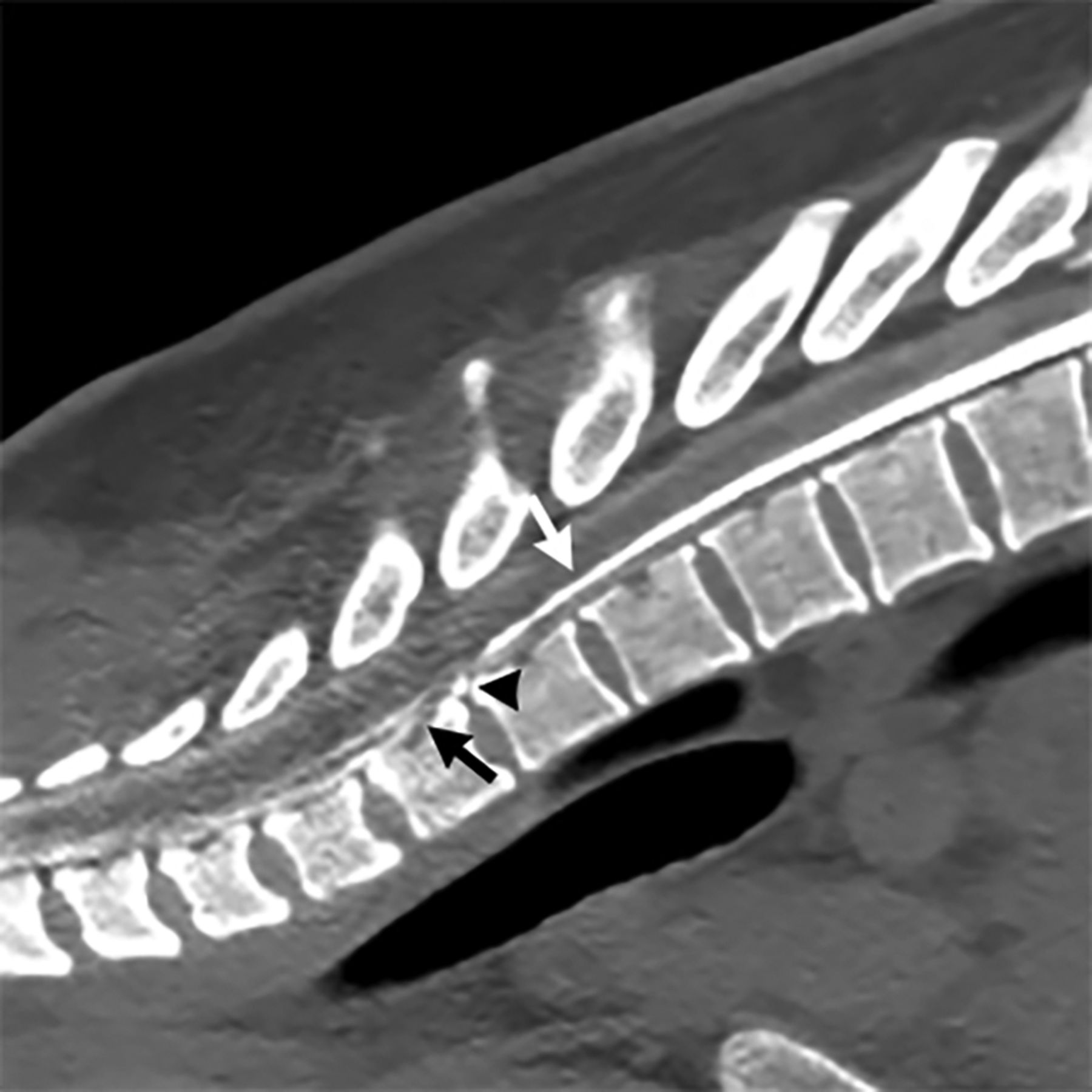
- FIG 1.
Type 1 CSF leak (dural tear): dynamic CT prone myelogram in the sagittal plane shows normal myelographic contrast in the subarachnoid space (white arrow) until there is a transition point at the T1–T2 level where there is a calcified disc (black arrowhead) that results in a split of contrast between the subarachnoid and ventral extradural spaces (black arrow).
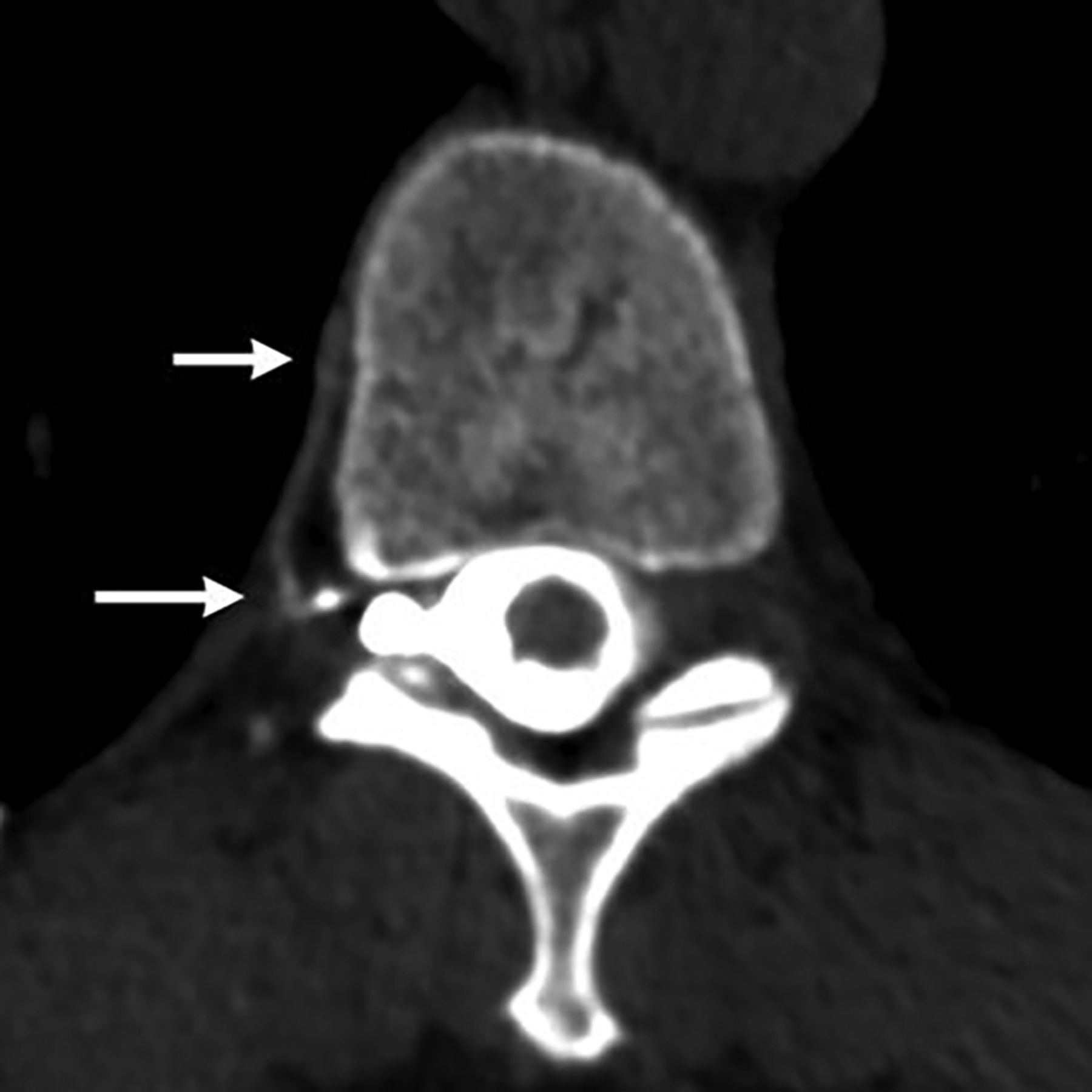
- FIG 2.
Type 2 CSF leak (ruptured meningeal diverticulum): left decubitus CT myelogram in the axial plane shows contrast leaking from the left T9–T10 meningeal diverticulum into the neural foramen (arrow).
- FIG 3.
Type 3 CSF leak (CVF): right decubitus CT myelogram in the axial plane shows a paraspinal vein (arrows) that was contiguous with the right T10–T11 meningeal diverticulum.


Tables
Patient demographics of spinal CSF leaks
Demographics All Patients Type 1 Type 2 Type 3 P Valuea No. of leaks 65 25 (39%) 4 (6%) 36 (55%) Age at symptoms onset (yr) <.00001 Mean (SD) 52.4 (13.1) 44.5 (10.3) 45.5 (8.1) 58.8 (11.9) 95% CI 49.3–55.6 40.4–48.5 37.6–53.4 54.9–62.6 Range 28–91 28–68 34–53 39–91 Sex (No.) .81 Female 37 14 4 19 Male 28 11 0 17 BMI .015 Mean (SD) 26.0 (4.5) 24.3 (3.1) 27.5 (7.9) 27.0 (4.7) 95% CI 24.9–27.1 23.1–25.5 19.7–35.3 25.4–28.5 Range 16–39 18–31 21–39 16–39 Leak location (No.) <.0002 Cervical 2 2 0 0 Upper thoracic (T1–T6) 31 18 0 13 Lower thoracic (T7–T12) 29 5 4 20 Lumbar 3 0 0 3 Bern SIH score .44 Mean (SD) 5.7 (2.4) 5.4 (2.0) 5.8 (3.3) 5.9 (2.6) Range 0–9 0–8 1–8 0–9 ↵a P values were compared between type 1 and 3 groups.


Jump to section
Related Articles
Cited By...
- CSF-Venous Fistulas Arising Intraosseously within Bone Remodeled by Meningeal Diverticula
- Prevalence of Spinal Meningeal Diverticula in Autosomal Dominant Polycystic Kidney Disease
- Spinal CSF Leaks: The Neuroradiologist Transforming Care
- Myelographic Techniques for the Localization of CSF-Venous Fistulas: Updates in 2024
- The Spatial Relationship between Spinal Osteoarthritis and CSF Venous Fistulas in Patients with Spontaneous Intracranial Hypotension
- Temporal Characteristics of CSF-Venous Fistulas on Dynamic Decubitus CT Myelography: A Retrospective Multi-Institution Cohort Study
- Lateral Decubitus Dynamic CT Myelography with Real-Time Bolus Tracking (dCTM-BT) for Evaluation of CSF-Venous Fistulas: Diagnostic Yield Stratified by Brain Imaging Findings
- Factors Predictive of Treatment Success in CT-Guided Fibrin Occlusion of CSF-Venous Fistulas: A Multicenter Retrospective Cross-Sectional Study
- Modified Dynamic CT Myelography for Type 1 and 2 CSF Leaks: A Procedural Approach