Advertisement

Main menu

  • Home
  • Content
    • Current Issue
    • Accepted Manuscripts
    • Article Preview
    • Past Issue Archive
    • Video Articles
    • AJNR Case Collection
    • Case of the Week Archive
    • Case of the Month Archive
    • Classic Case Archive
  • Special Collections
    • AJNR Awards
    • Low-Field MRI
    • Alzheimer Disease
    • ASNR Foundation Special Collection
    • Photon-Counting CT
    • View All
  • Multimedia
    • AJNR Podcasts
    • AJNR SCANtastic
    • Trainee Corner
    • MRI Safety Corner
    • Imaging Protocols
  • For Authors
    • Submit a Manuscript
    • Submit a Video Article
    • Submit an eLetter to the Editor/Response
    • Manuscript Submission Guidelines
    • Statistical Tips
    • Fast Publishing of Accepted Manuscripts
    • Graphical Abstract Preparation
    • Imaging Protocol Submission
    • Author Policies
  • About Us
    • About AJNR
    • Editorial Board
    • Editorial Board Alumni
  • More
    • Become a Reviewer/Academy of Reviewers
    • Subscribers
    • Permissions
    • Alerts
    • Feedback
    • Advertisers
    • ASNR Home

User menu

  • Alerts
  • Log in

Search

  • Advanced search
American Journal of Neuroradiology

American Journal of Neuroradiology

ASHNR American Society of Functional Neuroradiology ASHNR American Society of Pediatric Neuroradiology ASSR
  • Alerts
  • Log in

Advanced Search

  • Home
  • Content
    • Current Issue
    • Accepted Manuscripts
    • Article Preview
    • Past Issue Archive
    • Video Articles
    • AJNR Case Collection
    • Case of the Week Archive
    • Case of the Month Archive
    • Classic Case Archive
  • Special Collections
    • AJNR Awards
    • Low-Field MRI
    • Alzheimer Disease
    • ASNR Foundation Special Collection
    • Photon-Counting CT
    • View All
  • Multimedia
    • AJNR Podcasts
    • AJNR SCANtastic
    • Trainee Corner
    • MRI Safety Corner
    • Imaging Protocols
  • For Authors
    • Submit a Manuscript
    • Submit a Video Article
    • Submit an eLetter to the Editor/Response
    • Manuscript Submission Guidelines
    • Statistical Tips
    • Fast Publishing of Accepted Manuscripts
    • Graphical Abstract Preparation
    • Imaging Protocol Submission
    • Author Policies
  • About Us
    • About AJNR
    • Editorial Board
    • Editorial Board Alumni
  • More
    • Become a Reviewer/Academy of Reviewers
    • Subscribers
    • Permissions
    • Alerts
    • Feedback
    • Advertisers
    • ASNR Home
  • Follow AJNR on Twitter
  • Visit AJNR on Facebook
  • Follow AJNR on Instagram
  • Join AJNR on LinkedIn
  • RSS Feeds

AJNR Awards, New Junior Editors, and more. Read the latest AJNR updates

Research ArticleHead and Neck Imaging

Comparison of the Utility of High-Resolution CT-DWI and T2WI-DWI Fusion Images for the Localization of Cholesteatoma

X. Fan, C. Ding and Z. Liu
American Journal of Neuroradiology July 2022, 43 (7) 1029-1035; DOI: https://doi.org/10.3174/ajnr.A7538
X. Fan
aFrom the Department of Radiology, Shengjing Hospital of China Medical University, Shenyang, China
  • Find this author on Google Scholar
  • Find this author on PubMed
  • Search for this author on this site
  • ORCID record for X. Fan
C. Ding
aFrom the Department of Radiology, Shengjing Hospital of China Medical University, Shenyang, China
  • Find this author on Google Scholar
  • Find this author on PubMed
  • Search for this author on this site
  • ORCID record for C. Ding
Z. Liu
aFrom the Department of Radiology, Shengjing Hospital of China Medical University, Shenyang, China
  • Find this author on Google Scholar
  • Find this author on PubMed
  • Search for this author on this site
  • ORCID record for Z. Liu
  • Article
  • Figures & Data
  • Info & Metrics
  • Responses
  • References
  • PDF
Loading

Article Figures & Data

Figures

  • Tables
  • FIG 1.
    • Download figure
    • Open in new tab
    • Download powerpoint
    FIG 1.

    TSE-DWI images (A and E) were fused with HRCT (B) and T2WI fat-suppression images (F), respectively, to generate CT-DWI (C) and T2WI-DWI (G) fusion images, converting the colors to increase the visibility of the lesion (blue arrow) (D and H).
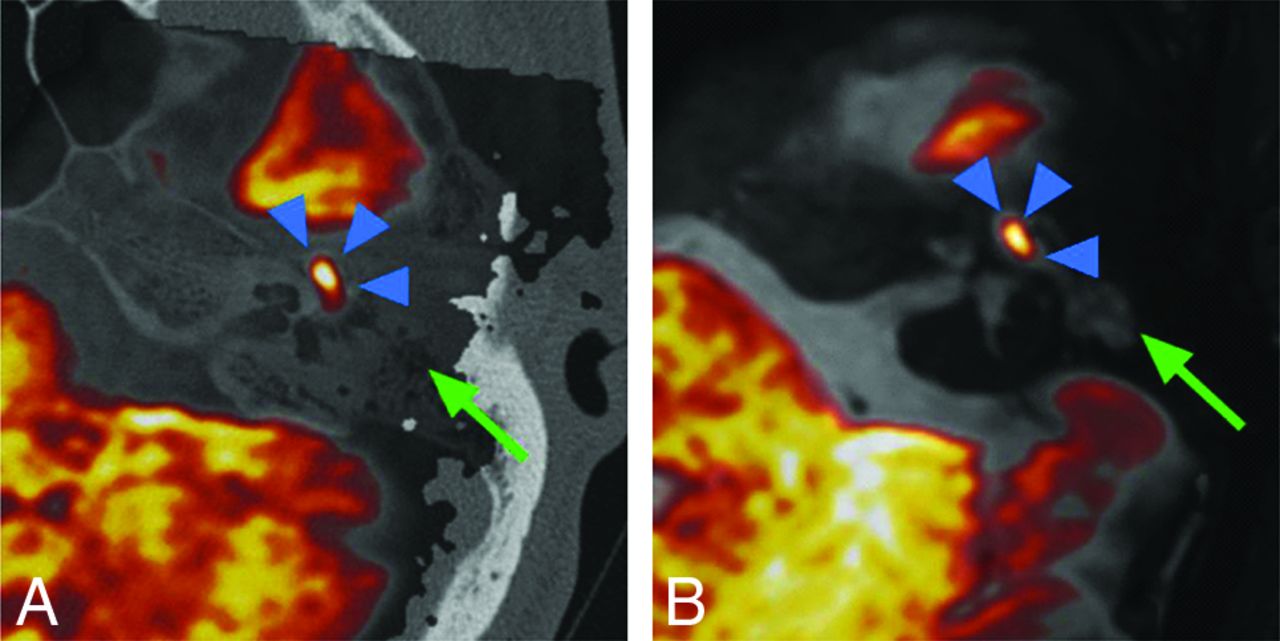
  • FIG 2.
    • Download figure
    • Open in new tab
    • Download powerpoint
    FIG 2.

    Left middle ear mastoid region. A, CT-DWI fusion image of a cholesteatoma in red (blue arrowhead) and inflammatory tissue in gray (green arrow). B, T2WI-DWI fusion image of cholesteatoma in red (blue arrowhead) and inflammatory tissue in gray (green arrow).

  • FIG 3.
    • Download figure
    • Open in new tab
    • Download powerpoint
    FIG 3.

    Subjective evaluation of CT-DWI and T2WI fusion image quality.
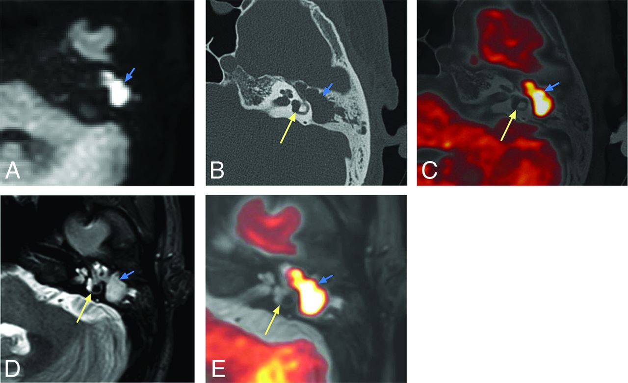
  • FIG 4.
    • Download figure
    • Open in new tab
    • Download powerpoint
    FIG 4.

    Pathologically confirmed cholesteatoma. The attic and mastoid antrum are filled with cholesteatoma during the operation. A, TSE-DWI: a high signal intensity area (blue arrow) is seen in the mastoid of the left middle ear with clear borders and poorly displayed semicircular canal. B, HRCT: a soft-tissue density shadow is seen within the middle ear mastoid cavity (blue arrow), and the anterior and posterior pedicles of the horizontal semicircular canal are clearly displayed (yellow arrow). CT-DWI fusion image (C) shows the cholesteatoma exceeding the posterior branch of the horizontal semicircular canal, involving the mastoid antrum. D, T2WI shows a nonspecific high-intensity-signal shadow in the mastoid process of the left middle ear. E, T2WI-DWI: the horizontal semicircular canal is clear, the cholesteatoma shows yellow changes, and the lesion involves the attic and mastoid antrum.

  • FIG 5.
    • Download figure
    • Open in new tab
    • Download powerpoint
    FIG 5.

    Surgical confirmation of a cholesteatoma in the tympanic cavity. A, TSE-DWI: a clear high-intensity-signal area (blue arrow) with a clear border in the right middle ear. B, HRCT: soft-tissue density shadow (blue arrow) in the middle ear with an unclear border and clear cochlear structures (yellow arrows). CT-DWI fusion image (C) clearly shows the cholesteatoma in the tympanic cavity. D, T2WI shows a nonspecific high-intensity-signal shadow in the mastoid process of the right middle ear. E, T2WI-DWI shows a clear cochlea with the cholesteatoma, localized in the tympanic cavity with reddish-yellow changes.

Tables

  • Figures
    • View popup
    Table 1:

    Subjective evaluation of fusion image quality

    ScoreOverall Quality of the Fusion ImageSemicircular Canal DisplayClarity of the LesionDiagnostic Confidence
    1UnacceptableDifficult to identify edgesSevere blurring of contoursVery poor
    2Poor, evaluation moderately limitedBlurred edges, but identifiableBlurred contoursPoor
    3Moderate, evaluation mildly limitedMargins recognizableContours recognizableModerate
    4Good, evaluation less limitedEdges visible, no distortionContour edges visibleGood
    5Very goodClear edgesClear contoursVery good
    • View popup
    Table 2:

    Comparison of subjective evaluation agreement between CT-DWI and T2WI-DWI fusion imagesa

    CT-DWI, Interobserver κ (95% CI)T2WI-DWI, Interobserver κ (95% CI)
    Overall quality of fusion image0.82 (0.71–0.93)0.87 (0.73–1.00)
    Semicircular canal display0.88 (0.77–0.98)0.81 (0.70–0.92)
    Clarity of the lesion0.94 (0.88–0.99)0.93 (0.88–0.99)
    Diagnostic confidence0.93 (0.86–0.99)0.91 (0.85–0.97)
    • ↵a κ indicates weighted κ coefficients.

    • View popup
    Table 3:

    Diagnostic efficacy of middle ear mastoid localization in CT-DWI fusion and T2WI-DWI fusion images

    SensitivitySpecificityAUCAUC 95% CIP Value
    Attic.32
     CT-DWI0.980.750.870.76–0.94
     T2WI-DWI1.000.750.880.77–0.95
    Tympanic cavity.30
     CT-DWI0.840.860.850.74–0.93
     T2WI-DWI0.840.720.780.66–0.88
    Mastoid antrum.70
     CT-DWI0.760.870.820.70–0.90
     T2WI-DWI0.790.870.830.71–0.91
    Mastoid cavity.16
     CT-DWI0.750.850.800.68–0.89
     T2WI-DWI0.820.820.850.74–0.93
    • Note:—AUC indicates area under the curve.

PreviousNext
Back to top

In this issue

American Journal of Neuroradiology: 43 (7)
American Journal of Neuroradiology
Vol. 43, Issue 7
1 Jul 2022
  • Table of Contents
  • Index by author
  • Complete Issue (PDF)
Advertisement
Print
Download PDF
Email Article

Thank you for your interest in spreading the word on American Journal of Neuroradiology.

NOTE: We only request your email address so that the person you are recommending the page to knows that you wanted them to see it, and that it is not junk mail. We do not capture any email address.

Enter multiple addresses on separate lines or separate them with commas.
Comparison of the Utility of High-Resolution CT-DWI and T2WI-DWI Fusion Images for the Localization of Cholesteatoma
(Your Name) has sent you a message from American Journal of Neuroradiology
(Your Name) thought you would like to see the American Journal of Neuroradiology web site.
CAPTCHA
This question is for testing whether or not you are a human visitor and to prevent automated spam submissions.
Cite this article
X. Fan, C. Ding, Z. Liu
Comparison of the Utility of High-Resolution CT-DWI and T2WI-DWI Fusion Images for the Localization of Cholesteatoma
American Journal of Neuroradiology Jul 2022, 43 (7) 1029-1035; DOI: 10.3174/ajnr.A7538

Citation Manager Formats

  • BibTeX
  • Bookends
  • EasyBib
  • EndNote (tagged)
  • EndNote 8 (xml)
  • Medlars
  • Mendeley
  • Papers
  • RefWorks Tagged
  • Ref Manager
  • RIS
  • Zotero
0 Responses
Respond to this article
Share
Bookmark this article
CT-DWI vs. T2WI-DWI for Cholesteatoma Localization
X. Fan, C. Ding, Z. Liu
American Journal of Neuroradiology Jul 2022, 43 (7) 1029-1035; DOI: 10.3174/ajnr.A7538
del.icio.us logo Twitter logo Facebook logo Mendeley logo
  • Tweet Widget
  • Facebook Like
  • Google Plus One
Purchase

Jump to section

  • Article
    • Abstract
    • ABBREVIATION:
    • MATERIALS AND METHODS
    • RESULTS
    • DISCUSSION
    • CONCLUSIONS
    • Footnotes
    • References
  • Figures & Data
  • Info & Metrics
  • Responses
  • References
  • PDF

Related Articles

  • No related articles found.
  • PubMed
  • Google Scholar

Cited By...

  • No citing articles found.
  • Crossref
  • Google Scholar

This article has not yet been cited by articles in journals that are participating in Crossref Cited-by Linking.

More in this TOC Section

  • MRI of the Parasellar Ligaments
  • ASL Sensitivity for Head and Neck Paraganglioma
  • Post SRS Peritumoral Hyperintense Signal of VSs
Show more Head and Neck Imaging

Similar Articles

Advertisement

Indexed Content

  • Current Issue
  • Accepted Manuscripts
  • Article Preview
  • Past Issues
  • Editorials
  • Editor's Choice
  • Fellows' Journal Club
  • Letters to the Editor
  • Video Articles

Cases

  • Case Collection
  • Archive - Case of the Week
  • Archive - Case of the Month
  • Archive - Classic Case

More from AJNR

  • Trainee Corner
  • Imaging Protocols
  • MRI Safety Corner
  • Book Reviews

Multimedia

  • AJNR Podcasts
  • AJNR Scantastics

Resources

  • Turnaround Time
  • Submit a Manuscript
  • Submit a Video Article
  • Submit an eLetter to the Editor/Response
  • Manuscript Submission Guidelines
  • Statistical Tips
  • Fast Publishing of Accepted Manuscripts
  • Graphical Abstract Preparation
  • Imaging Protocol Submission
  • Evidence-Based Medicine Level Guide
  • Publishing Checklists
  • Author Policies
  • Become a Reviewer/Academy of Reviewers
  • News and Updates

About Us

  • About AJNR
  • Editorial Board
  • Editorial Board Alumni
  • Alerts
  • Permissions
  • Not an AJNR Subscriber? Join Now
  • Advertise with Us
  • Librarian Resources
  • Feedback
  • Terms and Conditions
  • AJNR Editorial Board Alumni

American Society of Neuroradiology

  • Not an ASNR Member? Join Now

© 2025 by the American Society of Neuroradiology All rights, including for text and data mining, AI training, and similar technologies, are reserved.
Print ISSN: 0195-6108 Online ISSN: 1936-959X

Powered by HighWire