Advertisement

Main menu

  • Home
  • Content
    • Current Issue
    • Accepted Manuscripts
    • Article Preview
    • Past Issue Archive
    • Video Articles
    • AJNR Case Collection
    • Case of the Week Archive
    • Case of the Month Archive
    • Classic Case Archive
  • Special Collections
    • AJNR Awards
    • Low-Field MRI
    • Alzheimer Disease
    • ASNR Foundation Special Collection
    • Photon-Counting CT
    • View All
  • Multimedia
    • AJNR Podcasts
    • AJNR SCANtastic
    • Trainee Corner
    • MRI Safety Corner
    • Imaging Protocols
  • For Authors
    • Submit a Manuscript
    • Submit a Video Article
    • Submit an eLetter to the Editor/Response
    • Manuscript Submission Guidelines
    • Statistical Tips
    • Fast Publishing of Accepted Manuscripts
    • Graphical Abstract Preparation
    • Imaging Protocol Submission
    • Author Policies
  • About Us
    • About AJNR
    • Editorial Board
    • Editorial Board Alumni
  • More
    • Become a Reviewer/Academy of Reviewers
    • Subscribers
    • Permissions
    • Alerts
    • Feedback
    • Advertisers
    • ASNR Home

User menu

  • Alerts
  • Log in

Search

  • Advanced search
American Journal of Neuroradiology

American Journal of Neuroradiology

ASHNR American Society of Functional Neuroradiology ASHNR American Society of Pediatric Neuroradiology ASSR
  • Alerts
  • Log in

Advanced Search

  • Home
  • Content
    • Current Issue
    • Accepted Manuscripts
    • Article Preview
    • Past Issue Archive
    • Video Articles
    • AJNR Case Collection
    • Case of the Week Archive
    • Case of the Month Archive
    • Classic Case Archive
  • Special Collections
    • AJNR Awards
    • Low-Field MRI
    • Alzheimer Disease
    • ASNR Foundation Special Collection
    • Photon-Counting CT
    • View All
  • Multimedia
    • AJNR Podcasts
    • AJNR SCANtastic
    • Trainee Corner
    • MRI Safety Corner
    • Imaging Protocols
  • For Authors
    • Submit a Manuscript
    • Submit a Video Article
    • Submit an eLetter to the Editor/Response
    • Manuscript Submission Guidelines
    • Statistical Tips
    • Fast Publishing of Accepted Manuscripts
    • Graphical Abstract Preparation
    • Imaging Protocol Submission
    • Author Policies
  • About Us
    • About AJNR
    • Editorial Board
    • Editorial Board Alumni
  • More
    • Become a Reviewer/Academy of Reviewers
    • Subscribers
    • Permissions
    • Alerts
    • Feedback
    • Advertisers
    • ASNR Home
  • Follow AJNR on Twitter
  • Visit AJNR on Facebook
  • Follow AJNR on Instagram
  • Join AJNR on LinkedIn
  • RSS Feeds

AJNR Awards, New Junior Editors, and more. Read the latest AJNR updates

Research ArticleAdult Brain

Brain Abnormalities and Epilepsy in Patients with Parry-Romberg Syndrome

C. De la Garza-Ramos, A. Jain, S.A. Montazeri, L. Okromelidze, R. McGeary, A.A. Bhatt, S.J.S. Sandhu, S.S. Grewal, A. Feyissa, J.I. Sirven, A.L. Ritaccio, W.O. Tatum, V. Gupta and E.H. Middlebrooks
American Journal of Neuroradiology June 2022, 43 (6) 850-856; DOI: https://doi.org/10.3174/ajnr.A7517
C. De la Garza-Ramos
aFrom the Department of Neuroradiology (C.D.l.G.-R., A.J., S.A.M., L.O., R.M., A.A.B., S.J.S., V.G., E.H.M.), Mayo Clinic, Jacksonville, Florida
  • Find this author on Google Scholar
  • Find this author on PubMed
  • Search for this author on this site
  • ORCID record for C. De la Garza-Ramos
A. Jain
aFrom the Department of Neuroradiology (C.D.l.G.-R., A.J., S.A.M., L.O., R.M., A.A.B., S.J.S., V.G., E.H.M.), Mayo Clinic, Jacksonville, Florida
  • Find this author on Google Scholar
  • Find this author on PubMed
  • Search for this author on this site
  • ORCID record for A. Jain
S.A. Montazeri
aFrom the Department of Neuroradiology (C.D.l.G.-R., A.J., S.A.M., L.O., R.M., A.A.B., S.J.S., V.G., E.H.M.), Mayo Clinic, Jacksonville, Florida
  • Find this author on Google Scholar
  • Find this author on PubMed
  • Search for this author on this site
  • ORCID record for S.A. Montazeri
L. Okromelidze
aFrom the Department of Neuroradiology (C.D.l.G.-R., A.J., S.A.M., L.O., R.M., A.A.B., S.J.S., V.G., E.H.M.), Mayo Clinic, Jacksonville, Florida
  • Find this author on Google Scholar
  • Find this author on PubMed
  • Search for this author on this site
  • ORCID record for L. Okromelidze
R. McGeary
aFrom the Department of Neuroradiology (C.D.l.G.-R., A.J., S.A.M., L.O., R.M., A.A.B., S.J.S., V.G., E.H.M.), Mayo Clinic, Jacksonville, Florida
  • Find this author on Google Scholar
  • Find this author on PubMed
  • Search for this author on this site
  • ORCID record for R. McGeary
A.A. Bhatt
aFrom the Department of Neuroradiology (C.D.l.G.-R., A.J., S.A.M., L.O., R.M., A.A.B., S.J.S., V.G., E.H.M.), Mayo Clinic, Jacksonville, Florida
  • Find this author on Google Scholar
  • Find this author on PubMed
  • Search for this author on this site
  • ORCID record for A.A. Bhatt
S.J.S. Sandhu
aFrom the Department of Neuroradiology (C.D.l.G.-R., A.J., S.A.M., L.O., R.M., A.A.B., S.J.S., V.G., E.H.M.), Mayo Clinic, Jacksonville, Florida
  • Find this author on Google Scholar
  • Find this author on PubMed
  • Search for this author on this site
  • ORCID record for S.J.S. Sandhu
S.S. Grewal
bDepartment of Neurologic Surgery (S.S.G.), Mayo Clinic, Jacksonville, Florida
  • Find this author on Google Scholar
  • Find this author on PubMed
  • Search for this author on this site
  • ORCID record for S.S. Grewal
A. Feyissa
cDepartment of Neurology (A.F., J.I.S., A.L.R., W.O.T.), Mayo Clinic, Jacksonville, Florida
  • Find this author on Google Scholar
  • Find this author on PubMed
  • Search for this author on this site
  • ORCID record for A. Feyissa
J.I. Sirven
cDepartment of Neurology (A.F., J.I.S., A.L.R., W.O.T.), Mayo Clinic, Jacksonville, Florida
  • Find this author on Google Scholar
  • Find this author on PubMed
  • Search for this author on this site
  • ORCID record for J.I. Sirven
A.L. Ritaccio
cDepartment of Neurology (A.F., J.I.S., A.L.R., W.O.T.), Mayo Clinic, Jacksonville, Florida
  • Find this author on Google Scholar
  • Find this author on PubMed
  • Search for this author on this site
  • ORCID record for A.L. Ritaccio
W.O. Tatum
cDepartment of Neurology (A.F., J.I.S., A.L.R., W.O.T.), Mayo Clinic, Jacksonville, Florida
  • Find this author on Google Scholar
  • Find this author on PubMed
  • Search for this author on this site
  • ORCID record for W.O. Tatum
V. Gupta
aFrom the Department of Neuroradiology (C.D.l.G.-R., A.J., S.A.M., L.O., R.M., A.A.B., S.J.S., V.G., E.H.M.), Mayo Clinic, Jacksonville, Florida
  • Find this author on Google Scholar
  • Find this author on PubMed
  • Search for this author on this site
  • ORCID record for V. Gupta
E.H. Middlebrooks
aFrom the Department of Neuroradiology (C.D.l.G.-R., A.J., S.A.M., L.O., R.M., A.A.B., S.J.S., V.G., E.H.M.), Mayo Clinic, Jacksonville, Florida
  • Find this author on Google Scholar
  • Find this author on PubMed
  • Search for this author on this site
  • ORCID record for E.H. Middlebrooks
  • Article
  • Figures & Data
  • Supplemental
  • Info & Metrics
  • Responses
  • References
  • PDF
Loading

Article Figures & Data

Figures

  • Tables
  • FIG 1.
    • Download figure
    • Open in new tab
    • Download powerpoint
    FIG 1.

    Exemplary MR imaging of atrophy and white matter disease severity. Coronal FLAIR MR imaging shows mild left hemisphere atrophy (A), moderate right hemisphere atrophy (B), and severe left hemisphere atrophy (C). Axial FLAIR MR imaging shows mild right hemisphere WMD (D), moderate left hemisphere WMD (E), and severe right hemisphere WMD (F).

  • FIG 2.
    • Download figure
    • Open in new tab
    • Download powerpoint
    FIG 2.

    A, Axial FLAIR MR imaging in a patient with epilepsy shows severe, confluent left hemisphere white matter hyperintensity with extensive areas of punctate susceptibility artifacts throughout the left hemisphere on susceptibility-weighted imaging (B). Most of the hypointense foci on susceptibility-weighted imaging are hypointense on the matching phase image (C), consistent with prior microhemorrhages; however, few show hyperintense signal on the phase image (arrows in B and C), consistent with calcifications (confirmed by CT, not shown).
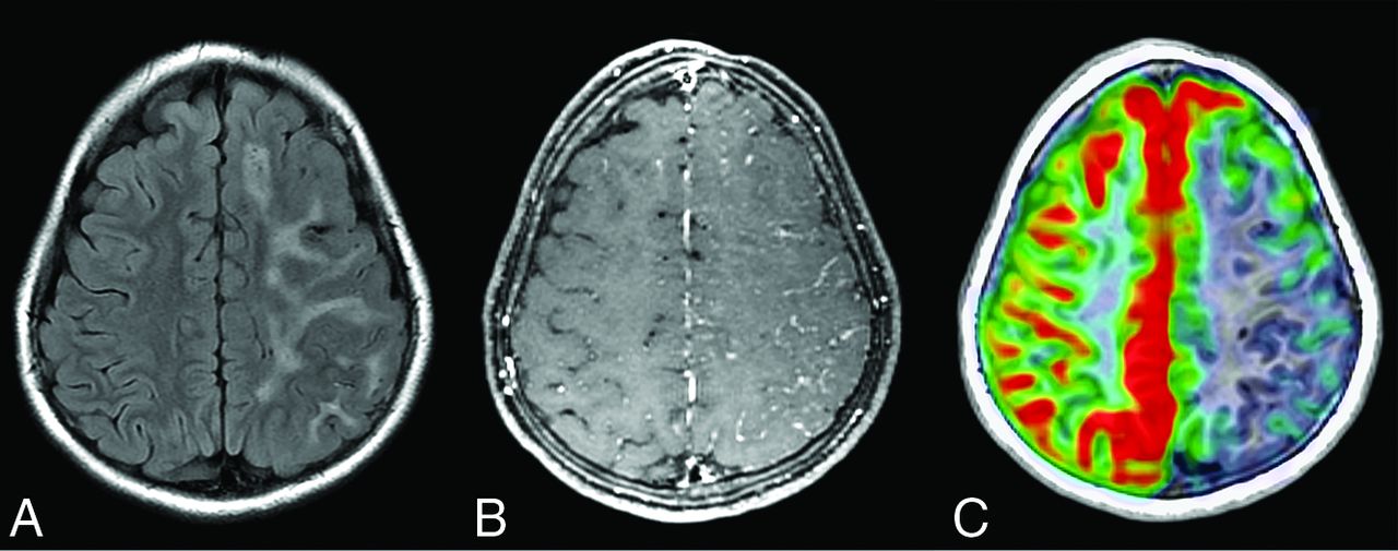
  • FIG 3.
    • Download figure
    • Open in new tab
    • Download powerpoint
    FIG 3.

    A, Axial FLAIR MR imaging in a patient with epilepsy shows severe, confluent left hemisphere white matter hyperintensity. B, Postcontrast T1-weighted MR imaging reveals leptomeningeal enhancement and sulcal effacement with decreased perfusion to the same area on arterial spin-labeling perfusion MR imaging (C).

  • FIG 4.
    • Download figure
    • Open in new tab
    • Download powerpoint
    FIG 4.

    A, Initial head CT in a patient with epilepsy reveals effacement of the sulci over the left parietal high convexity. B, Subsequent T1-weighted MR imaging performed 10 months later shows improvement in sulcal effacement, but with diffuse left hemisphere white matter hyperintensity on double inversion recovery MR imaging (C, arrow). D, Sagittal postcontrast T1-weighted MR imaging shows faint periventricular and perivascular enhancement (arrowheads). E, Concomitant FDG-PET shows left parietal hypometabolism in the affected area.

  • FIG 5.
    • Download figure
    • Open in new tab
    • Download powerpoint
    FIG 5.

    Spectrum of unilateral sulcal effacement. In 1 patient with epilepsy, axial T2-weighted MR imaging (A) shows right hemispheric sulcal effacement affecting the right frontal and parietal lobes, which remained unchanged on MR imaging performed 1 year later (B). C, Coronal FLAIR MR imaging in a second patient with epilepsy shows diffuse right hemisphere sulcal effacement with hyperintensity in the right hippocampal head, with resolution of sulcal effacement and development of right mesial temporal sclerosis on MR imaging approximately 6 months later (D).

  • FIG 6.
    • Download figure
    • Open in new tab
    • Download powerpoint
    FIG 6.

    Proton MRS in a 12-year-old boy with left-sided Parry-Romberg syndrome and epilepsy shows the spectrum from the normal-appearing right hemisphere white matter (A) compared with the area of white matter FLAIR hyperintensity in the left hemisphere (B). There is a relative reduction in Cho and NAA, with a slight elevation of Cr on the abnormal side. PPM indicates parts per million.

Tables

  • Figures
    • View popup
    Table 1:

    Parry-Romberg syndrome patient characteristicsa

    Variables
    Total80
    Age at work-up and imaging (yr)37 (26–56)
    Age at hemifacial atrophy identification (yr)20 (13–43)
    Sex
        Male24 (30%)
        Female56 (70%)
    Laterality of hemifacial atrophy
        Right37 (46%)
        Left43 (54%)
        Bilateral0 (0%)
    Brain abnormalities on MR imaging
        Normal32 (40%)
        Abnormal48 (60%)
    Overall laterality of brain MR imaging findings
        Unilateral34 (42%)
        Bilateral14 (17%)
    Laterality of unilateral brain MR imaging findings in relation to hemifacial atrophy
        Ipsilateral26 (35%)
        Contralateral6 (7%)
    Epilepsy
        Present16 (20%)
        Absent64 (80%)
    • ↵a Data are No. (%) or median (IQR, 1–3).

    • View popup
    Table 2:

    Frequency and severity of unilateral brain abnormalities in patients with Parry-Romberg syndrome and epilepsy compared with cases without epilepsya

    Nonepilepsy (n = 64)Epilepsy (n = 16)P Value CategoriesP Value Overall
    Hemispheric atrophy
        None61 (95%)8 (50%)<.001<.001
        Mild1 (1.6%)1 (6%)
        Moderate1 (1.6%)3 (19%)
        Severe1 (1.6%)4 (25%)
    WMD
        None49 (77%)2 (12%)<.001<.001
        Mild10 (15%)7 (44%)
        Moderate2 (3%)4 (25%)
        Severe3 (5%)3 (19%)
    Hemispheric microhemorrhage
        None61 (95%)13 (81%).015.091
        Mild3 (5%)1 (6%)
        Moderate00
        Severe02 (12%)
    Leptomeningeal enhancement
        Absent63 (98%)15 (94%).362
        Present1 (1.6%)1 (6%)
    • ↵a Some patients had more than 1 finding.

PreviousNext
Back to top

In this issue

American Journal of Neuroradiology: 43 (6)
American Journal of Neuroradiology
Vol. 43, Issue 6
1 Jun 2022
  • Table of Contents
  • Index by author
  • Complete Issue (PDF)
Advertisement
Print
Download PDF
Email Article

Thank you for your interest in spreading the word on American Journal of Neuroradiology.

NOTE: We only request your email address so that the person you are recommending the page to knows that you wanted them to see it, and that it is not junk mail. We do not capture any email address.

Enter multiple addresses on separate lines or separate them with commas.
Brain Abnormalities and Epilepsy in Patients with Parry-Romberg Syndrome
(Your Name) has sent you a message from American Journal of Neuroradiology
(Your Name) thought you would like to see the American Journal of Neuroradiology web site.
CAPTCHA
This question is for testing whether or not you are a human visitor and to prevent automated spam submissions.
Cite this article
C. De la Garza-Ramos, A. Jain, S.A. Montazeri, L. Okromelidze, R. McGeary, A.A. Bhatt, S.J.S. Sandhu, S.S. Grewal, A. Feyissa, J.I. Sirven, A.L. Ritaccio, W.O. Tatum, V. Gupta, E.H. Middlebrooks
Brain Abnormalities and Epilepsy in Patients with Parry-Romberg Syndrome
American Journal of Neuroradiology Jun 2022, 43 (6) 850-856; DOI: 10.3174/ajnr.A7517

Citation Manager Formats

  • BibTeX
  • Bookends
  • EasyBib
  • EndNote (tagged)
  • EndNote 8 (xml)
  • Medlars
  • Mendeley
  • Papers
  • RefWorks Tagged
  • Ref Manager
  • RIS
  • Zotero
0 Responses
Respond to this article
Share
Bookmark this article
Brain Abnormalities in Parry-Romberg Syndrome
C. De la Garza-Ramos, A. Jain, S.A. Montazeri, L. Okromelidze, R. McGeary, A.A. Bhatt, S.J.S. Sandhu, S.S. Grewal, A. Feyissa, J.I. Sirven, A.L. Ritaccio, W.O. Tatum, V. Gupta, E.H. Middlebrooks
American Journal of Neuroradiology Jun 2022, 43 (6) 850-856; DOI: 10.3174/ajnr.A7517
del.icio.us logo Twitter logo Facebook logo Mendeley logo
  • Tweet Widget
  • Facebook Like
  • Google Plus One
Purchase

Jump to section

  • Article
    • Abstract
    • ABBREVIATIONS:
    • MATERIALS AND METHODS
    • RESULTS
    • DISCUSSION
    • CONCLUSIONS
    • Footnotes
    • References
  • Figures & Data
  • Supplemental
  • Info & Metrics
  • Responses
  • References
  • PDF

Related Articles

  • PubMed
  • Google Scholar

Cited By...

  • No citing articles found.
  • Crossref
  • Google Scholar

This article has not yet been cited by articles in journals that are participating in Crossref Cited-by Linking.

More in this TOC Section

  • Diagnostic Neuroradiology of Monoclonal Antibodies
  • Clinical Outcomes After Chiari I Decompression
  • Segmentation of Brain Metastases with BLAST
Show more Adult Brain

Similar Articles

Advertisement

Indexed Content

  • Current Issue
  • Accepted Manuscripts
  • Article Preview
  • Past Issues
  • Editorials
  • Editor's Choice
  • Fellows' Journal Club
  • Letters to the Editor
  • Video Articles

Cases

  • Case Collection
  • Archive - Case of the Week
  • Archive - Case of the Month
  • Archive - Classic Case

More from AJNR

  • Trainee Corner
  • Imaging Protocols
  • MRI Safety Corner
  • Book Reviews

Multimedia

  • AJNR Podcasts
  • AJNR Scantastics

Resources

  • Turnaround Time
  • Submit a Manuscript
  • Submit a Video Article
  • Submit an eLetter to the Editor/Response
  • Manuscript Submission Guidelines
  • Statistical Tips
  • Fast Publishing of Accepted Manuscripts
  • Graphical Abstract Preparation
  • Imaging Protocol Submission
  • Evidence-Based Medicine Level Guide
  • Publishing Checklists
  • Author Policies
  • Become a Reviewer/Academy of Reviewers
  • News and Updates

About Us

  • About AJNR
  • Editorial Board
  • Editorial Board Alumni
  • Alerts
  • Permissions
  • Not an AJNR Subscriber? Join Now
  • Advertise with Us
  • Librarian Resources
  • Feedback
  • Terms and Conditions
  • AJNR Editorial Board Alumni

American Society of Neuroradiology

  • Not an ASNR Member? Join Now

© 2025 by the American Society of Neuroradiology All rights, including for text and data mining, AI training, and similar technologies, are reserved.
Print ISSN: 0195-6108 Online ISSN: 1936-959X

Powered by HighWire