Advertisement

Main menu

  • Home
  • Content
    • Current Issue
    • Accepted Manuscripts
    • Article Preview
    • Past Issue Archive
    • Video Articles
    • AJNR Case Collection
    • Case of the Week Archive
    • Case of the Month Archive
    • Classic Case Archive
  • Special Collections
    • AJNR Awards
    • Low-Field MRI
    • Alzheimer Disease
    • ASNR Foundation Special Collection
    • Photon-Counting CT
    • View All
  • Multimedia
    • AJNR Podcasts
    • AJNR SCANtastic
    • Trainee Corner
    • MRI Safety Corner
    • Imaging Protocols
  • For Authors
    • Submit a Manuscript
    • Submit a Video Article
    • Submit an eLetter to the Editor/Response
    • Manuscript Submission Guidelines
    • Statistical Tips
    • Fast Publishing of Accepted Manuscripts
    • Graphical Abstract Preparation
    • Imaging Protocol Submission
    • Author Policies
  • About Us
    • About AJNR
    • Editorial Board
    • Editorial Board Alumni
  • More
    • Become a Reviewer/Academy of Reviewers
    • Subscribers
    • Permissions
    • Alerts
    • Feedback
    • Advertisers
    • ASNR Home

User menu

  • Alerts
  • Log in

Search

  • Advanced search
American Journal of Neuroradiology

American Journal of Neuroradiology

ASHNR American Society of Functional Neuroradiology ASHNR American Society of Pediatric Neuroradiology ASSR
  • Alerts
  • Log in

Advanced Search

  • Home
  • Content
    • Current Issue
    • Accepted Manuscripts
    • Article Preview
    • Past Issue Archive
    • Video Articles
    • AJNR Case Collection
    • Case of the Week Archive
    • Case of the Month Archive
    • Classic Case Archive
  • Special Collections
    • AJNR Awards
    • Low-Field MRI
    • Alzheimer Disease
    • ASNR Foundation Special Collection
    • Photon-Counting CT
    • View All
  • Multimedia
    • AJNR Podcasts
    • AJNR SCANtastic
    • Trainee Corner
    • MRI Safety Corner
    • Imaging Protocols
  • For Authors
    • Submit a Manuscript
    • Submit a Video Article
    • Submit an eLetter to the Editor/Response
    • Manuscript Submission Guidelines
    • Statistical Tips
    • Fast Publishing of Accepted Manuscripts
    • Graphical Abstract Preparation
    • Imaging Protocol Submission
    • Author Policies
  • About Us
    • About AJNR
    • Editorial Board
    • Editorial Board Alumni
  • More
    • Become a Reviewer/Academy of Reviewers
    • Subscribers
    • Permissions
    • Alerts
    • Feedback
    • Advertisers
    • ASNR Home
  • Follow AJNR on Twitter
  • Visit AJNR on Facebook
  • Follow AJNR on Instagram
  • Join AJNR on LinkedIn
  • RSS Feeds

AJNR Awards, New Junior Editors, and more. Read the latest AJNR updates

Research ArticleHead and Neck Imaging

A New Frontier in Temporal Bone Imaging: Photon-Counting Detector CT Demonstrates Superior Visualization of Critical Anatomic Structures at Reduced Radiation Dose

J.C. Benson, K. Rajendran, J.I. Lane, F.E. Diehn, N.M. Weber, J.E. Thorne, N.B. Larson, J.G. Fletcher, C.H. McCollough and S. Leng
American Journal of Neuroradiology April 2022, 43 (4) 579-584; DOI: https://doi.org/10.3174/ajnr.A7452
J.C. Benson
aFrom the Departments of Radiology (J.C.B., K.R., J.I.L., F.E.D., N.M.W., J.E.T., J.G.F., C.H.M., S.L.)
  • Find this author on Google Scholar
  • Find this author on PubMed
  • Search for this author on this site
  • ORCID record for J.C. Benson
K. Rajendran
aFrom the Departments of Radiology (J.C.B., K.R., J.I.L., F.E.D., N.M.W., J.E.T., J.G.F., C.H.M., S.L.)
  • Find this author on Google Scholar
  • Find this author on PubMed
  • Search for this author on this site
  • ORCID record for K. Rajendran
J.I. Lane
aFrom the Departments of Radiology (J.C.B., K.R., J.I.L., F.E.D., N.M.W., J.E.T., J.G.F., C.H.M., S.L.)
  • Find this author on Google Scholar
  • Find this author on PubMed
  • Search for this author on this site
  • ORCID record for J.I. Lane
F.E. Diehn
aFrom the Departments of Radiology (J.C.B., K.R., J.I.L., F.E.D., N.M.W., J.E.T., J.G.F., C.H.M., S.L.)
  • Find this author on Google Scholar
  • Find this author on PubMed
  • Search for this author on this site
  • ORCID record for F.E. Diehn
N.M. Weber
aFrom the Departments of Radiology (J.C.B., K.R., J.I.L., F.E.D., N.M.W., J.E.T., J.G.F., C.H.M., S.L.)
  • Find this author on Google Scholar
  • Find this author on PubMed
  • Search for this author on this site
  • ORCID record for N.M. Weber
J.E. Thorne
aFrom the Departments of Radiology (J.C.B., K.R., J.I.L., F.E.D., N.M.W., J.E.T., J.G.F., C.H.M., S.L.)
  • Find this author on Google Scholar
  • Find this author on PubMed
  • Search for this author on this site
  • ORCID record for J.E. Thorne
N.B. Larson
bQuantitative Health Sciences (N.B.L.), Mayo Clinic, Rochester, Minnesota
  • Find this author on Google Scholar
  • Find this author on PubMed
  • Search for this author on this site
  • ORCID record for N.B. Larson
J.G. Fletcher
aFrom the Departments of Radiology (J.C.B., K.R., J.I.L., F.E.D., N.M.W., J.E.T., J.G.F., C.H.M., S.L.)
  • Find this author on Google Scholar
  • Find this author on PubMed
  • Search for this author on this site
  • ORCID record for J.G. Fletcher
C.H. McCollough
aFrom the Departments of Radiology (J.C.B., K.R., J.I.L., F.E.D., N.M.W., J.E.T., J.G.F., C.H.M., S.L.)
  • Find this author on Google Scholar
  • Find this author on PubMed
  • Search for this author on this site
  • ORCID record for C.H. McCollough
S. Leng
aFrom the Departments of Radiology (J.C.B., K.R., J.I.L., F.E.D., N.M.W., J.E.T., J.G.F., C.H.M., S.L.)
  • Find this author on Google Scholar
  • Find this author on PubMed
  • Search for this author on this site
  • ORCID record for S. Leng
  • Article
  • Figures & Data
  • Supplemental
  • Info & Metrics
  • Responses
  • References
  • PDF
Loading

Article Figures & Data

Figures

  • FIG 1.
    • Download figure
    • Open in new tab
    • Download powerpoint
    FIG 1.

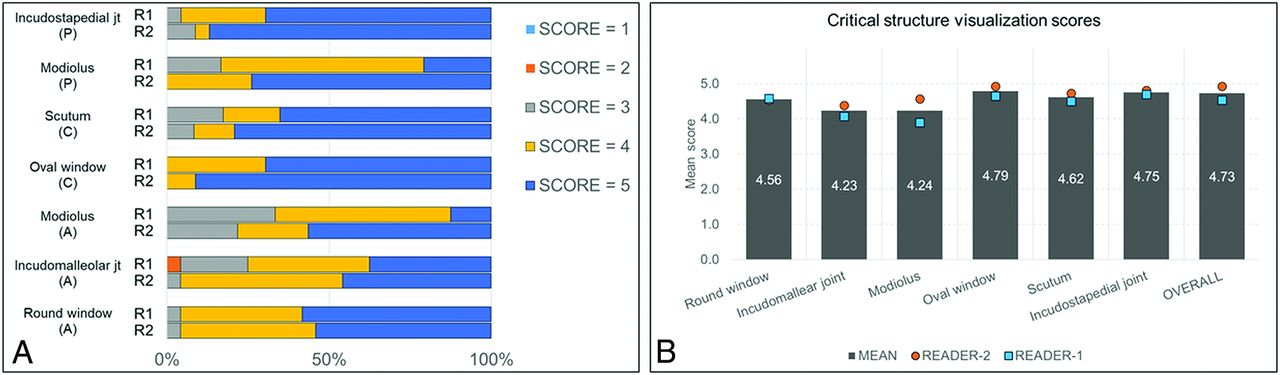
    Readers’ score distribution for spatial resolution and visualization of critical anatomic structures in different reformatted planes (A) and mean readers’ scores for individual anatomic structures and overall image quality (B). All scores were based on a 5-point Likert scale, comparing PCD-CT with EID-CT: 1 = inferior resolution with degraded visualization, 2 = slightly inferior resolution without affecting visualization, 3 = equivalent resolution and visualization, 4 = slightly superior resolution without affecting visualization, and 5 = superior spatial resolution with improved visualization.

  • FIG 2.
    • Download figure
    • Open in new tab
    • Download powerpoint
    FIG 2.

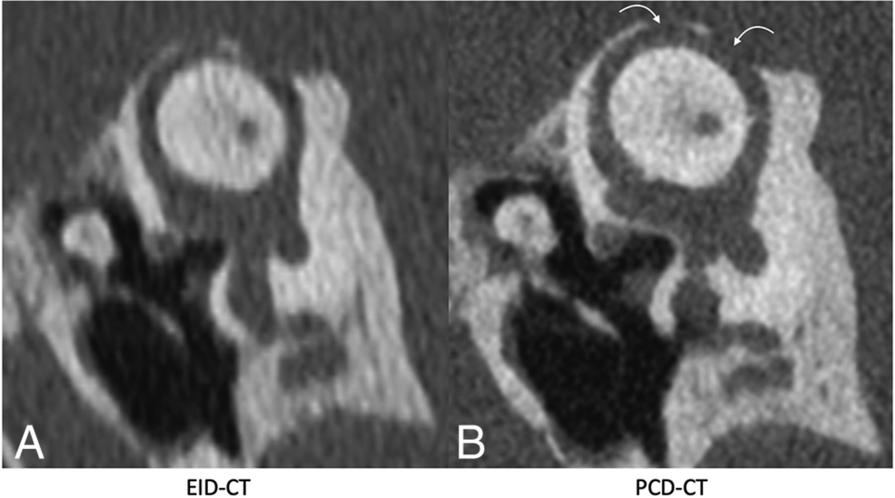
    Pöschl reformatted images in a patient with superior semicircular canal dehiscence, shown on EID-CT (left) and PCD-CT (right) images. The PCD-CT image (B) clearly demonstrates 2 discrete regions of dehiscence (curved arrows). These regions are also identifiable on conventional EID-CT (A), though the intact adjacent bone is less well-visualized. The integrity of the roof of the superior semicircular canal was not formally evaluated in the readers’ study but is shown for illustrative purposes.

  • FIG 3.
    • Download figure
    • Open in new tab
    • Download powerpoint
    FIG 3.

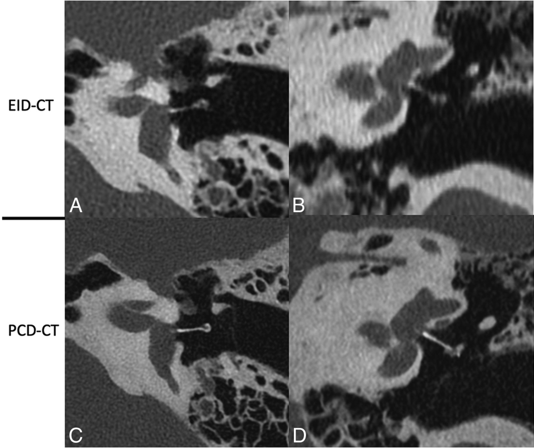
    Ossicular anatomy, shown on conventional EID-CT (upper row) and PCD-CT (lower row). Reformatted images along the plane of the tensor tympani (A and E) demonstrate the tensor tympani (TT) extending to the upper handle of the malleus (HA); the lateral process (L) of the malleus is also clearly visible. An image reformatted along the long plane of the malleus (B and F) shows its handle (HA), lateral process (L), neck (N), and head (H). An image reformatted along the length of the stapes (C and G) clearly demonstrates the suprastructure (SS) and both crura. A “molar tooth” reformatted image (D and H) shows the HA of the malleus, as well as the body (B) and long process (LP) of the incus. These additional reformatted images were generated by a nonreviewer radiologist to demonstrate certain anatomic features but were not used by the readers to score image quality.

  • FIG 4.
    • Download figure
    • Open in new tab
    • Download powerpoint
    FIG 4.

    The incudostapedial joint (arrows), shown on EID-CT (left) and PCD-CT (right) images. The joint was one of several anatomic structures specifically graded using a 5-point Likert score, with higher scores favoring the quality of the PCD-CT images. The images reformatted in this plane were generated by a nonreviewer radiologist to demonstrate certain anatomic features but were not used by the readers to score image quality.

  • FIG 5.
    • Download figure
    • Open in new tab
    • Download powerpoint
    FIG 5.

    A stapes piston prosthesis shown on EID-CT (upper row) and PCD-CT (lower row) images. The prothesis is shown on oblique axial (A and C) and oblique coronal (B and D) reformats along the plane of the piston. In both planes, the stapes prosthesis was better delineated on PCD-CT due to reduced partial volume averaging from 0.2 mm sections. These additional reformatted images were generated by a nonreviewer radiologist to demonstrate certain anatomic features but were not used by the readers to score image quality.

  • FIG 6.
    • Download figure
    • Open in new tab
    • Download powerpoint
    FIG 6.

    Images reformatted along the long axis of the stapes in a patient with fenestral otosclerosis shown on EID-CT (left) and conventional PCD-CT (right) images. The arrow points to the insertion of the anterior crus of the stapes into the region involved by otosclerosis. These additional reformatted images were generated by a nonreviewer radiologist to demonstrate certain anatomic features but were not used by the readers to score image quality.

PreviousNext
Back to top

In this issue

American Journal of Neuroradiology: 43 (4)
American Journal of Neuroradiology
Vol. 43, Issue 4
1 Apr 2022
  • Table of Contents
  • Index by author
  • Complete Issue (PDF)
Advertisement
Print
Download PDF
Email Article

Thank you for your interest in spreading the word on American Journal of Neuroradiology.

NOTE: We only request your email address so that the person you are recommending the page to knows that you wanted them to see it, and that it is not junk mail. We do not capture any email address.

Enter multiple addresses on separate lines or separate them with commas.
A New Frontier in Temporal Bone Imaging: Photon-Counting Detector CT Demonstrates Superior Visualization of Critical Anatomic Structures at Reduced Radiation Dose
(Your Name) has sent you a message from American Journal of Neuroradiology
(Your Name) thought you would like to see the American Journal of Neuroradiology web site.
CAPTCHA
This question is for testing whether or not you are a human visitor and to prevent automated spam submissions.
Cite this article
J.C. Benson, K. Rajendran, J.I. Lane, F.E. Diehn, N.M. Weber, J.E. Thorne, N.B. Larson, J.G. Fletcher, C.H. McCollough, S. Leng
A New Frontier in Temporal Bone Imaging: Photon-Counting Detector CT Demonstrates Superior Visualization of Critical Anatomic Structures at Reduced Radiation Dose
American Journal of Neuroradiology Apr 2022, 43 (4) 579-584; DOI: 10.3174/ajnr.A7452

Citation Manager Formats

  • BibTeX
  • Bookends
  • EasyBib
  • EndNote (tagged)
  • EndNote 8 (xml)
  • Medlars
  • Mendeley
  • Papers
  • RefWorks Tagged
  • Ref Manager
  • RIS
  • Zotero
0 Responses
Respond to this article
Share
Bookmark this article
A New Frontier in Temporal Bone Imaging: Photon-Counting Detector CT Demonstrates Superior Visualization of Critical Anatomic Structures at Reduced Radiation Dose
J.C. Benson, K. Rajendran, J.I. Lane, F.E. Diehn, N.M. Weber, J.E. Thorne, N.B. Larson, J.G. Fletcher, C.H. McCollough, S. Leng
American Journal of Neuroradiology Apr 2022, 43 (4) 579-584; DOI: 10.3174/ajnr.A7452
del.icio.us logo Twitter logo Facebook logo Mendeley logo
  • Tweet Widget
  • Facebook Like
  • Google Plus One
Purchase

Jump to section

  • Article
    • Abstract
    • ABBREVIATIONS:
    • MATERIALS AND METHODS
    • RESULTS
    • DISCUSSION
    • CONCLUSIONS
    • Acknowledgments
    • Footnotes
    • References
  • Figures & Data
  • Supplemental
  • Info & Metrics
  • Responses
  • References
  • PDF

Related Articles

  • PubMed
  • Google Scholar

Cited By...

  • Systematic Review and Meta-Analysis of Radiation Dose Reduction Studies in Pediatric Head CT
  • Ultra-High-Resolution Photon-Counting-Detector CT with a Dedicated Denoising Convolutional Neural Network for Enhanced Temporal Bone Imaging
  • Pre- and Postoperative Imaging of Cochlear Implantation in Cadaveric Specimens Using Low-Dose Photon-Counting Detector CT
  • Comprehensive Review of External and Middle Ear Anatomy on Photon-Counting CT
  • Neurovascular Imaging with Ultra-High-Resolution Photon-Counting CT: Preliminary Findings on Image-Quality Evaluation
  • High-Resolution Head CTA: A Prospective Patient Study Comparing Image Quality of Photon-Counting Detector CT and Energy-Integrating Detector CT
  • Photon-Counting CT in the Head and Neck: Current Applications and Future Prospects
  • Back to the Future: Dynamic Contrast-Enhanced Photon-Counting Detector CT for the Detection of Pituitary Adenoma in Cushing Disease
  • Automatic analysis of skull thickness, scalp-to-cortex distance and association with age and sex in cognitively normal elderly
  • Recalling the Usefulness of Conebeam CT in Temporal Bone Imaging: Higher Resolution with Lower Radiation Dose
  • Reply:
  • Crossref
  • Google Scholar

This article has not yet been cited by articles in journals that are participating in Crossref Cited-by Linking.

More in this TOC Section

  • MRI of the Parasellar Ligaments
  • ASL Sensitivity for Head and Neck Paraganglioma
  • Post SRS Peritumoral Hyperintense Signal of VSs
Show more Head and Neck Imaging

Similar Articles

Advertisement

Indexed Content

  • Current Issue
  • Accepted Manuscripts
  • Article Preview
  • Past Issues
  • Editorials
  • Editor's Choice
  • Fellows' Journal Club
  • Letters to the Editor
  • Video Articles

Cases

  • Case Collection
  • Archive - Case of the Week
  • Archive - Case of the Month
  • Archive - Classic Case

More from AJNR

  • Trainee Corner
  • Imaging Protocols
  • MRI Safety Corner
  • Book Reviews

Multimedia

  • AJNR Podcasts
  • AJNR Scantastics

Resources

  • Turnaround Time
  • Submit a Manuscript
  • Submit a Video Article
  • Submit an eLetter to the Editor/Response
  • Manuscript Submission Guidelines
  • Statistical Tips
  • Fast Publishing of Accepted Manuscripts
  • Graphical Abstract Preparation
  • Imaging Protocol Submission
  • Evidence-Based Medicine Level Guide
  • Publishing Checklists
  • Author Policies
  • Become a Reviewer/Academy of Reviewers
  • News and Updates

About Us

  • About AJNR
  • Editorial Board
  • Editorial Board Alumni
  • Alerts
  • Permissions
  • Not an AJNR Subscriber? Join Now
  • Advertise with Us
  • Librarian Resources
  • Feedback
  • Terms and Conditions
  • AJNR Editorial Board Alumni

American Society of Neuroradiology

  • Not an ASNR Member? Join Now

© 2025 by the American Society of Neuroradiology All rights, including for text and data mining, AI training, and similar technologies, are reserved.
Print ISSN: 0195-6108 Online ISSN: 1936-959X

Powered by HighWire