
Article Figures & Data
Figures
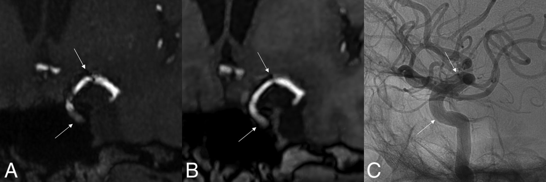
- FIG 1.
3D-TOF with HyperSense MRA (A), LAVA-Flex MRA (B), and DSA (C) of a 61-year-old woman treated with a 4 × 25 Surpass Evolve FD located from the left ICA to the left MCA. There is a significant reduction in metal artifacts on the LAVA-Flex (OutPhase) sequence, which shows the absence of in-stent stenosis, confirmed by DSA. Note that there is not exactly the same projection between MRA and DSA. White arrows indicate the proximal and distal ends of the FDs.
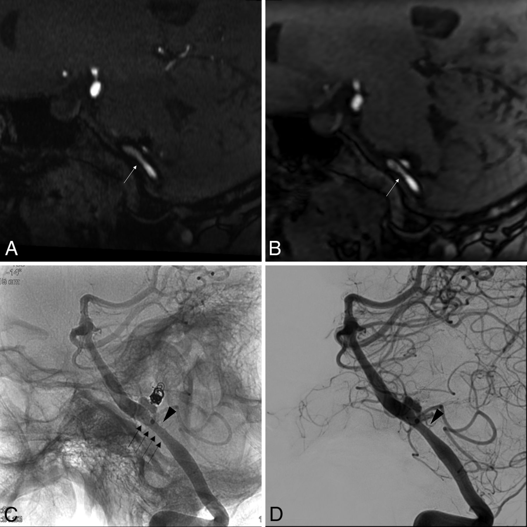
- FIG 2.
In-stent stenosis in a 50-year-old woman treated with a 5 × 15 Surpass Evolve FD placed in the left VA. This was a complementary embolization of a left PICA aneurysm, initially revealed by a subarachnoid hemorrhage 10 years earlier and treated by coiling at that time. 3D-TOF with HyperSense MRA (A) and LAVA-Flex MRA (Outphase) (B) demonstrates moderate in-stent stenosis (white arrows) predominating a few millimeters upstream of the neck of the still patent aneurysm, 6 months after placement of the FD. Confirmation of a moderate in-stent stenosis (black arrowhead) on angiography without (C) and with (D) digital subtraction. The black arrows show the limits of the FD metallic mesh.
- FIG 3.
3D-TOF with HyperSense MRA comparison of artifacts created by the 3 types of FDs used in our study. A, A 68-year-old woman treated with a Surpass Evolve FD (3.25 × 17 mm) located in the right VA. B, A 61-year-old woman treated with a Pipeline Shield FD (3.5 × 18 mm) located from the left VA to the basilar artery. C, A 52-year-old woman treated with a Silk Vista Baby FD (2.5 × 20 mm) located in the right anterior cerebral artery (A1–A2). The extent of the metallic artifacts is large for the Surpass Evolve FD, small for the Pipeline Shield FD, and minimal for the Silk Vista Baby FD. White arrows indicate the proximal and distal ends of the FDs.


Tables
NC-MRA CE-MRA DSA Three-grade scale Patent 36 (64.3%) 24 (42.9%) 42 (75.0%) Stenosis 19 (33.9%) 31 (55.4%) 13 (23.2%) Occlusion 1 (1.8%) 1 (1.8%) 1 (1.8%) Simplified 2-grade scale Patent 36 (64.3%) 24 (42.9%) 42 (75.0%) Stenosis or occlusion 20 (35.7%) 32 (57.1%) 14 (25.0%) Consensus Evaluation for DSA Pathologic (Stenosis or Occlusion) Normal (Patent) Consensus Evaluation for NC-MRA Pathologic (stenosis or occlusion) 13 7 Normal (patent) 1 35 Consensus Evaluation for DSA Pathologic (Stenosis or Occlusion) Normal (Patent) Consensus Evaluation for CE-MRA Pathologic (stenosis or occlusion) 13 19 Normal (patent) 1 23 - Table 4:
Diagnostic accuracies for parent artery patency of NC-MRA for each separate device, for patients treated with FD exclusively and those treated with additional device
Pipeline Shield (n = 28) Surpass Evolve (n = 19) Silk Vista Baby (n = 9) FD Exclusively (n = 22) Additional Device (n = 34) Specificity (95% CI) 0.96 (0.78–1.00) 0.57 (0.29–0.82) 1 (0.48–1.00) 0.87 (0.60–0.98) 0.81 (0.62–0.94) PPV (95% CI) 0.80 (0.28–0.99) 0.45 (0.17–0.77) 1 (0.40–1.00) 0.78 (0.40–0.97) 0.55 (0.23–0.83) Sensitivity (95% CI) 0.80 (0.28–0.99) 1 (0.48–1.00) 1 (0.40–1.00) 1 (0.59–1.00) 0.86 (0.42–1.00) NPV (95% CI) 0.96 (0.78–1.00) 1 (0.63–1.00) 1 (0.48–1.00) 1 (0.75–1.00) 0.96 (0.78–1.00)


