Advertisement

Main menu

  • Home
  • Content
    • Current Issue
    • Accepted Manuscripts
    • Article Preview
    • Past Issue Archive
    • Video Articles
    • AJNR Case Collection
    • Case of the Week Archive
    • Case of the Month Archive
    • Classic Case Archive
  • Special Collections
    • AJNR Awards
    • Low-Field MRI
    • Alzheimer Disease
    • ASNR Foundation Special Collection
    • Photon-Counting CT
    • View All
  • Multimedia
    • AJNR Podcasts
    • AJNR SCANtastic
    • Trainee Corner
    • MRI Safety Corner
    • Imaging Protocols
  • For Authors
    • Submit a Manuscript
    • Submit a Video Article
    • Submit an eLetter to the Editor/Response
    • Manuscript Submission Guidelines
    • Statistical Tips
    • Fast Publishing of Accepted Manuscripts
    • Graphical Abstract Preparation
    • Imaging Protocol Submission
    • Author Policies
  • About Us
    • About AJNR
    • Editorial Board
    • Editorial Board Alumni
  • More
    • Become a Reviewer/Academy of Reviewers
    • Subscribers
    • Permissions
    • Alerts
    • Feedback
    • Advertisers
    • ASNR Home

User menu

  • Alerts
  • Log in

Search

  • Advanced search
American Journal of Neuroradiology

American Journal of Neuroradiology

ASHNR American Society of Functional Neuroradiology ASHNR American Society of Pediatric Neuroradiology ASSR
  • Alerts
  • Log in

Advanced Search

  • Home
  • Content
    • Current Issue
    • Accepted Manuscripts
    • Article Preview
    • Past Issue Archive
    • Video Articles
    • AJNR Case Collection
    • Case of the Week Archive
    • Case of the Month Archive
    • Classic Case Archive
  • Special Collections
    • AJNR Awards
    • Low-Field MRI
    • Alzheimer Disease
    • ASNR Foundation Special Collection
    • Photon-Counting CT
    • View All
  • Multimedia
    • AJNR Podcasts
    • AJNR SCANtastic
    • Trainee Corner
    • MRI Safety Corner
    • Imaging Protocols
  • For Authors
    • Submit a Manuscript
    • Submit a Video Article
    • Submit an eLetter to the Editor/Response
    • Manuscript Submission Guidelines
    • Statistical Tips
    • Fast Publishing of Accepted Manuscripts
    • Graphical Abstract Preparation
    • Imaging Protocol Submission
    • Author Policies
  • About Us
    • About AJNR
    • Editorial Board
    • Editorial Board Alumni
  • More
    • Become a Reviewer/Academy of Reviewers
    • Subscribers
    • Permissions
    • Alerts
    • Feedback
    • Advertisers
    • ASNR Home
  • Follow AJNR on Twitter
  • Visit AJNR on Facebook
  • Follow AJNR on Instagram
  • Join AJNR on LinkedIn
  • RSS Feeds

AJNR Awards, New Junior Editors, and more. Read the latest AJNR updates

Research ArticlePediatric Neuroimaging

DWI Hyperintensity in the Fornix Fimbria on MRI in Children

M.S. Rootman, L. Kornreich, A.N. Osherov and O. Konen
American Journal of Neuroradiology March 2022, 43 (3) 480-485; DOI: https://doi.org/10.3174/ajnr.A7437
M.S. Rootman
aFrom the Department of Radiology (M.S.R., L.K., A.N.O., O.K.), Schneider Children’s Medical Center of Israel, Petach Tikvah, Israel
bThe Sackler Faculty of Medicine (M.S.R., L.K., A.N.O., O.K.), Tel Aviv University, Tel Aviv, Israel
  • Find this author on Google Scholar
  • Find this author on PubMed
  • Search for this author on this site
  • ORCID record for M.S. Rootman
L. Kornreich
aFrom the Department of Radiology (M.S.R., L.K., A.N.O., O.K.), Schneider Children’s Medical Center of Israel, Petach Tikvah, Israel
bThe Sackler Faculty of Medicine (M.S.R., L.K., A.N.O., O.K.), Tel Aviv University, Tel Aviv, Israel
  • Find this author on Google Scholar
  • Find this author on PubMed
  • Search for this author on this site
  • ORCID record for L. Kornreich
A.N. Osherov
aFrom the Department of Radiology (M.S.R., L.K., A.N.O., O.K.), Schneider Children’s Medical Center of Israel, Petach Tikvah, Israel
bThe Sackler Faculty of Medicine (M.S.R., L.K., A.N.O., O.K.), Tel Aviv University, Tel Aviv, Israel
  • Find this author on Google Scholar
  • Find this author on PubMed
  • Search for this author on this site
  • ORCID record for A.N. Osherov
O. Konen
aFrom the Department of Radiology (M.S.R., L.K., A.N.O., O.K.), Schneider Children’s Medical Center of Israel, Petach Tikvah, Israel
bThe Sackler Faculty of Medicine (M.S.R., L.K., A.N.O., O.K.), Tel Aviv University, Tel Aviv, Israel
  • Find this author on Google Scholar
  • Find this author on PubMed
  • Search for this author on this site
  • ORCID record for O. Konen
  • Article
  • Figures & Data
  • Info & Metrics
  • Responses
  • References
  • PDF
Loading

Article Figures & Data

Figures

  • Tables
  • FIG 1.
    • Download figure
    • Open in new tab
    • Download powerpoint
    FIG 1.

    DWI signal hyperintensity in the fornix-fimbria. MR imaging study of a 7.5-month-old girl with hypsarrhythmia. Consecutive diffusion-weighted images from top (A) to bottom (E) show signal hyperintensity of the fornices (arrows) and fimbriae (arrowheads). F, A magnified view shows DWI hyperintensity involving the fornix-fimbria complex. Also notable is mild volume loss.
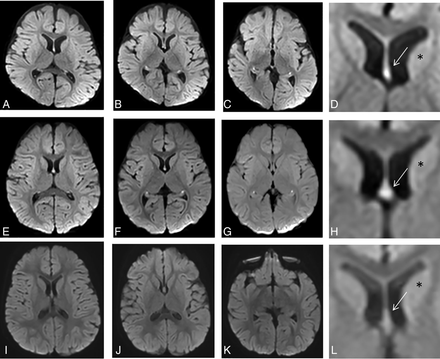
  • FIG 2.
    • Download figure
    • Open in new tab
    • Download powerpoint
    FIG 2.

    DWI signal intensity in the fornix-fimbria in a control group versus patients with high signal intensity. Two examples of children (patient 1, A–D; patient, 2 E–H) with high signal intensity detected in the fornix-fimbria on DWI, in axial slices from top to bottom. D and H, Magnified view at the level of the forniceal body (arrow) shows high signal intensity (compared with the adjacent caudate, asterisk). A third patient (I–L) with normal signal intensity of the fornix-fimbria on DWI. L, An enlarged image of the forniceal body (arrow). Usually, the DWI signal intensity of the fornix is approximately the same as that of the adjacent caudate nucleus (asterisk).
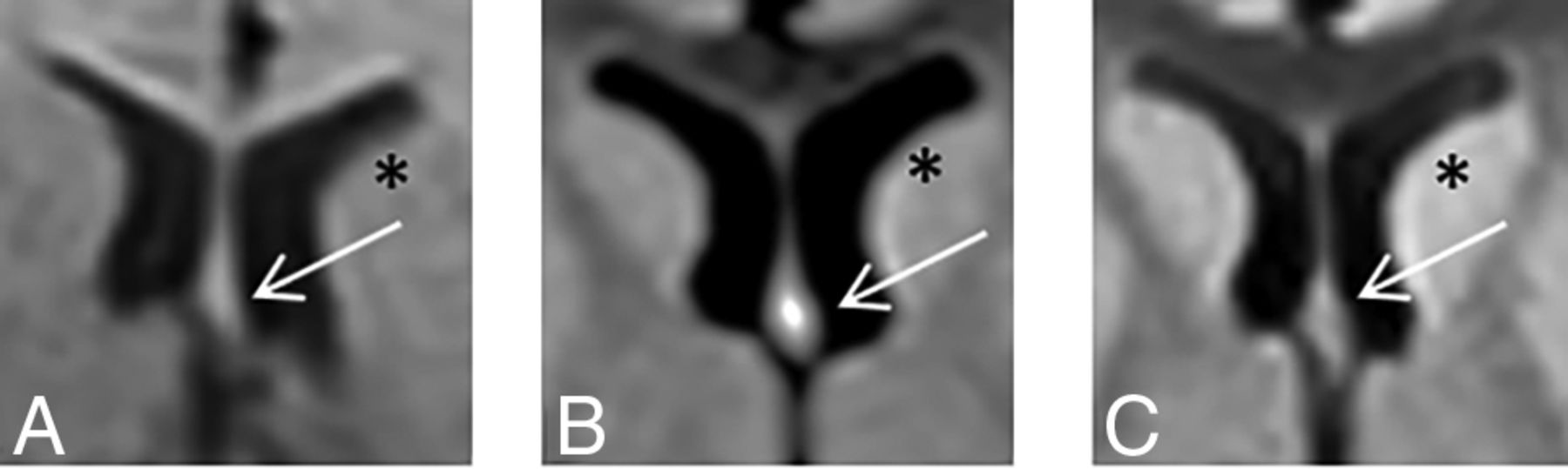
  • FIG 3.
    • Download figure
    • Open in new tab
    • Download powerpoint
    FIG 3.

    Changes in DWI signal intensity of the fornix-fimbria with time. A brain MR imaging study of a child with a low-grade cervicomedullary tumor on a DWI sequence. The body of the fornix (arrows) is shown in 3 follow-up studies performed at 3 months of age (A), 13 months of age (B), and 24 months of age (C). Fornix DWI hyperintensity is noted only at 13 months of age. Comparing the signal intensity of the head of the caudate nucleus (asterisk) shows similar signals at 3 and 24 months of age and a hypersignal at 13 months of age.
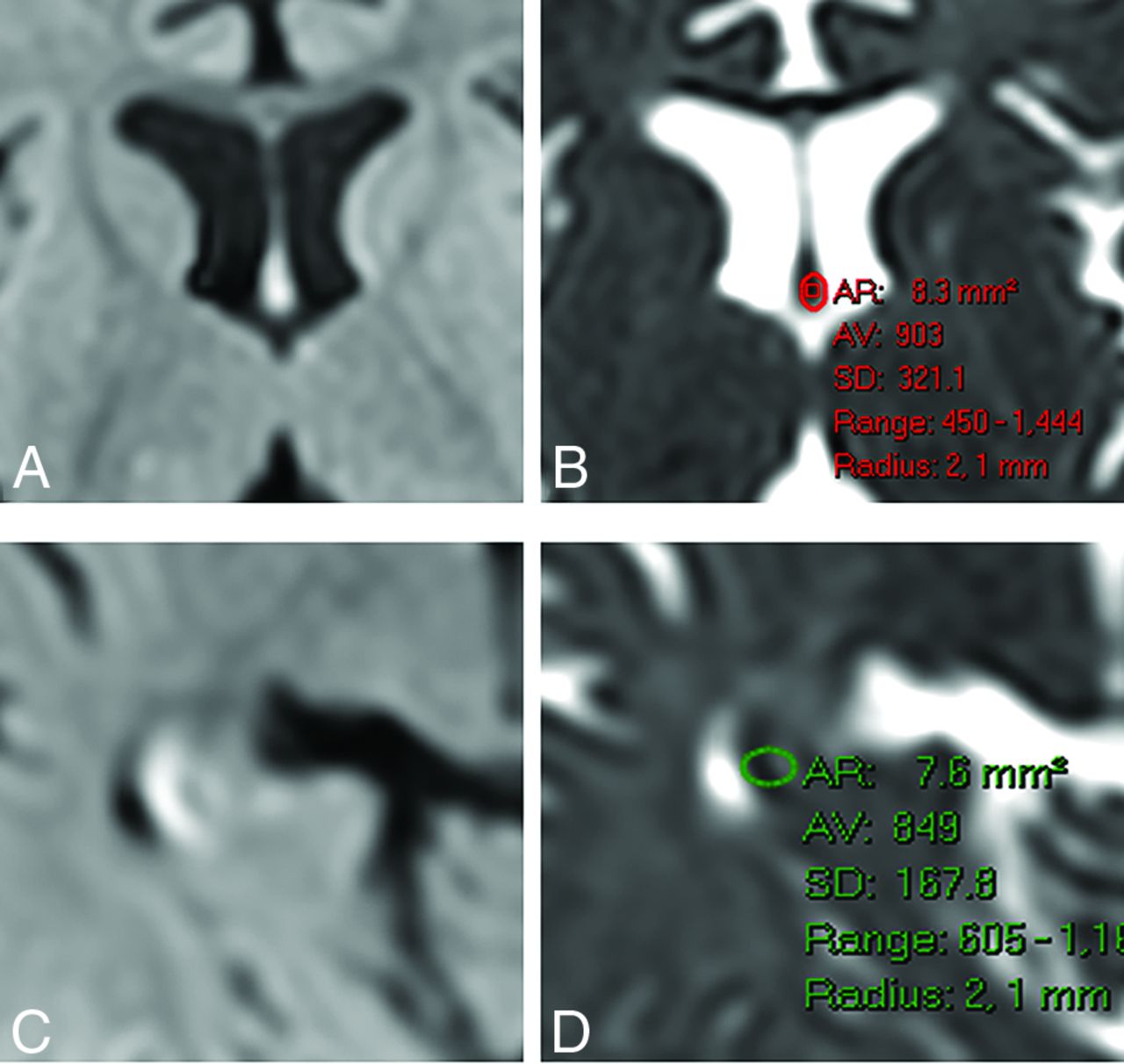
  • FIG 4.
    • Download figure
    • Open in new tab
    • Download powerpoint
    FIG 4.

    ADC measurement in the fornix (A and B) and fimbria (C and D). After detecting DWI hyperintensity on the fornix (A) or fimbria (C), we placed an ROI manually on the corresponding ADC image while trying to avoid CSF contamination and obtained the measurements. Similar measurements were performed for the control group using anatomic guidance. AR indicates area; AV, average.

Tables

  • Figures
    • View popup
    Table 1:

    Characteristics of the 53 patients with fornix-fimbria DWI signal changes

    CharacteristicsNo.(%)
    Age
     Median10 months
     Range12 days to 22 months
    Sex
     Female3770
     Male1630
    MR imaging magnet
     1.5T917
     3T4483
    Follow-up studies
     Patients with a follow-up MR imaging47.5
    Common findings on MR imaginga
     Normal scan findings2047
     Parenchymal volume loss815
    • ↵a Additional imaging findings were variable and included old infarcts, subdural collections, hydrocephalus, tuberous-sclerosis stigmata, and heterotopia.

    • View popup
    Table 2:

    Indications for performing brain MR imaging in 53 children with DWI hyperintensity in the fornix-fimbria complexa

    IndicationsNo.%
    Seizures1426.5
    Developmental delay/FTT815
    Sensorineural hearing loss47.5
    Hemiparesis24
    Nystagmus24
    Torticollis35.5
    Acute lymphocytic leukemia35.5
    Macrocephaly24
    Microcephaly12
    Hypoxic-ischemic insult12
    Suspected nonaccidental injury12
    Suspected Sturge-Weber syndrome12
    Strabismus12
    Brain tumor12
    Suspected Miller Fisher syndrome12
    Drowsiness12
    Microtia12
    Tuberous sclerosis12
    Ventriculomegaly35.5
    Meningitis24
    • Note:—FTT indicates failure to thrive.

    • ↵a Children 0–2 years of age; mean, 10 months of age.

PreviousNext
Back to top

In this issue

American Journal of Neuroradiology: 43 (3)
American Journal of Neuroradiology
Vol. 43, Issue 3
1 Mar 2022
  • Table of Contents
  • Index by author
  • Complete Issue (PDF)
Advertisement
Print
Download PDF
Email Article

Thank you for your interest in spreading the word on American Journal of Neuroradiology.

NOTE: We only request your email address so that the person you are recommending the page to knows that you wanted them to see it, and that it is not junk mail. We do not capture any email address.

Enter multiple addresses on separate lines or separate them with commas.
DWI Hyperintensity in the Fornix Fimbria on MRI in Children
(Your Name) has sent you a message from American Journal of Neuroradiology
(Your Name) thought you would like to see the American Journal of Neuroradiology web site.
CAPTCHA
This question is for testing whether or not you are a human visitor and to prevent automated spam submissions.
Cite this article
M.S. Rootman, L. Kornreich, A.N. Osherov, O. Konen
DWI Hyperintensity in the Fornix Fimbria on MRI in Children
American Journal of Neuroradiology Mar 2022, 43 (3) 480-485; DOI: 10.3174/ajnr.A7437

Citation Manager Formats

  • BibTeX
  • Bookends
  • EasyBib
  • EndNote (tagged)
  • EndNote 8 (xml)
  • Medlars
  • Mendeley
  • Papers
  • RefWorks Tagged
  • Ref Manager
  • RIS
  • Zotero
0 Responses
Respond to this article
Share
Bookmark this article
DWI Hyperintensity in the Fornix Fimbria on MRI in Children
M.S. Rootman, L. Kornreich, A.N. Osherov, O. Konen
American Journal of Neuroradiology Mar 2022, 43 (3) 480-485; DOI: 10.3174/ajnr.A7437
del.icio.us logo Twitter logo Facebook logo Mendeley logo
  • Tweet Widget
  • Facebook Like
  • Google Plus One
Purchase

Jump to section

  • Article
    • Abstract
    • MATERIALS AND METHODS
    • RESULTS
    • DISCUSSION
    • CONCLUSIONS
    • Footnotes
    • References
  • Figures & Data
  • Info & Metrics
  • Responses
  • References
  • PDF

Related Articles

  • No related articles found.
  • PubMed
  • Google Scholar

Cited By...

  • Neuroimaging Features of Biotinidase Deficiency
  • Crossref
  • Google Scholar

This article has not yet been cited by articles in journals that are participating in Crossref Cited-by Linking.

More in this TOC Section

  • Frontal Paraventricular Cysts
  • Sodium MRI in Pediatric Brain Tumors
  • FRACTURE MR in Congenital Vertebral Anomalies
Show more Pediatric Neuroimaging

Similar Articles

Advertisement

Indexed Content

  • Current Issue
  • Accepted Manuscripts
  • Article Preview
  • Past Issues
  • Editorials
  • Editor's Choice
  • Fellows' Journal Club
  • Letters to the Editor
  • Video Articles

Cases

  • Case Collection
  • Archive - Case of the Week
  • Archive - Case of the Month
  • Archive - Classic Case

More from AJNR

  • Trainee Corner
  • Imaging Protocols
  • MRI Safety Corner
  • Book Reviews

Multimedia

  • AJNR Podcasts
  • AJNR Scantastics

Resources

  • Turnaround Time
  • Submit a Manuscript
  • Submit a Video Article
  • Submit an eLetter to the Editor/Response
  • Manuscript Submission Guidelines
  • Statistical Tips
  • Fast Publishing of Accepted Manuscripts
  • Graphical Abstract Preparation
  • Imaging Protocol Submission
  • Evidence-Based Medicine Level Guide
  • Publishing Checklists
  • Author Policies
  • Become a Reviewer/Academy of Reviewers
  • News and Updates

About Us

  • About AJNR
  • Editorial Board
  • Editorial Board Alumni
  • Alerts
  • Permissions
  • Not an AJNR Subscriber? Join Now
  • Advertise with Us
  • Librarian Resources
  • Feedback
  • Terms and Conditions
  • AJNR Editorial Board Alumni

American Society of Neuroradiology

  • Not an ASNR Member? Join Now

© 2025 by the American Society of Neuroradiology All rights, including for text and data mining, AI training, and similar technologies, are reserved.
Print ISSN: 0195-6108 Online ISSN: 1936-959X

Powered by HighWire