
Article Figures & Data
Figures
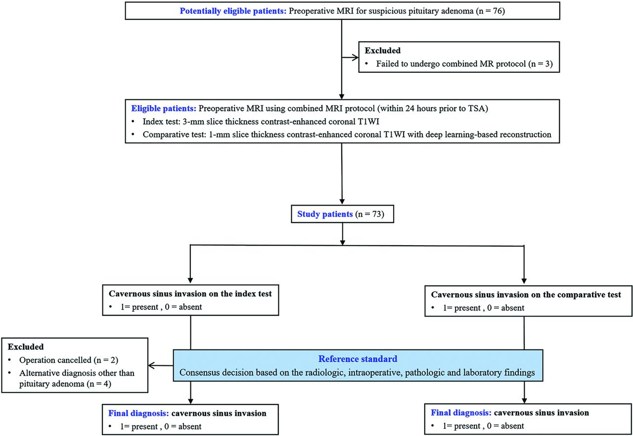
- FIG 1.
Patient inclusion process and image analysis.
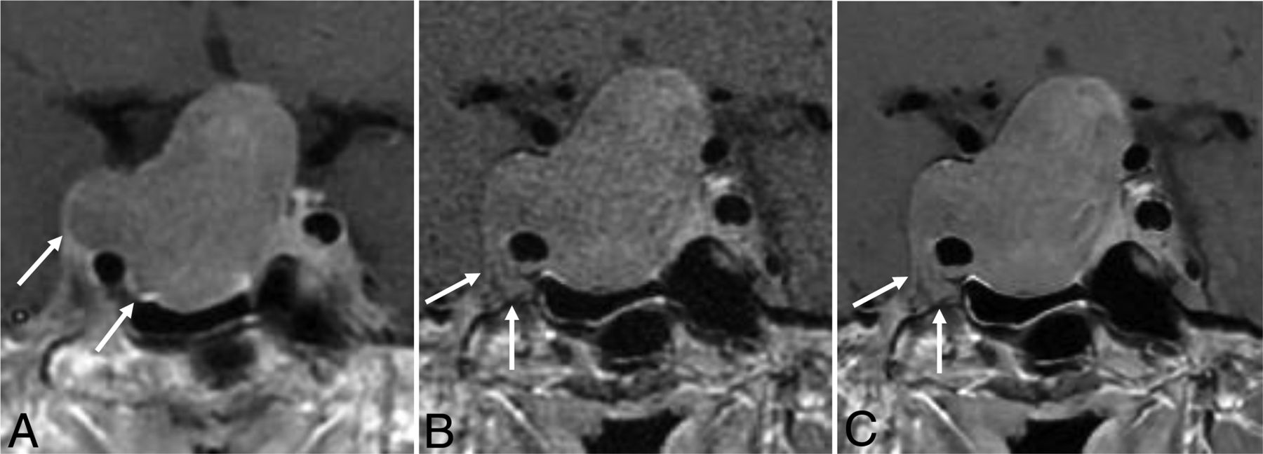
- FIG 2.
A 55-year-old man who underwent a combined MR imaging protocol for preoperative evaluation of pituitary adenoma. On a 3-mmMRI, coronal, contrast-enhanced T1-weighted image (A), the tumor is present beyond the lateral intercarotid line (modified Knosp classification 3A) and is about 180° in contact with the intracavernous ICA. One-mmMRI, coronal, contrast-enhanced T1-weighted image (B) was reconstructed using DLR (C), and the tumor is shown to encase the intracavernous ICA (arrows, modified Knosp classification 4).
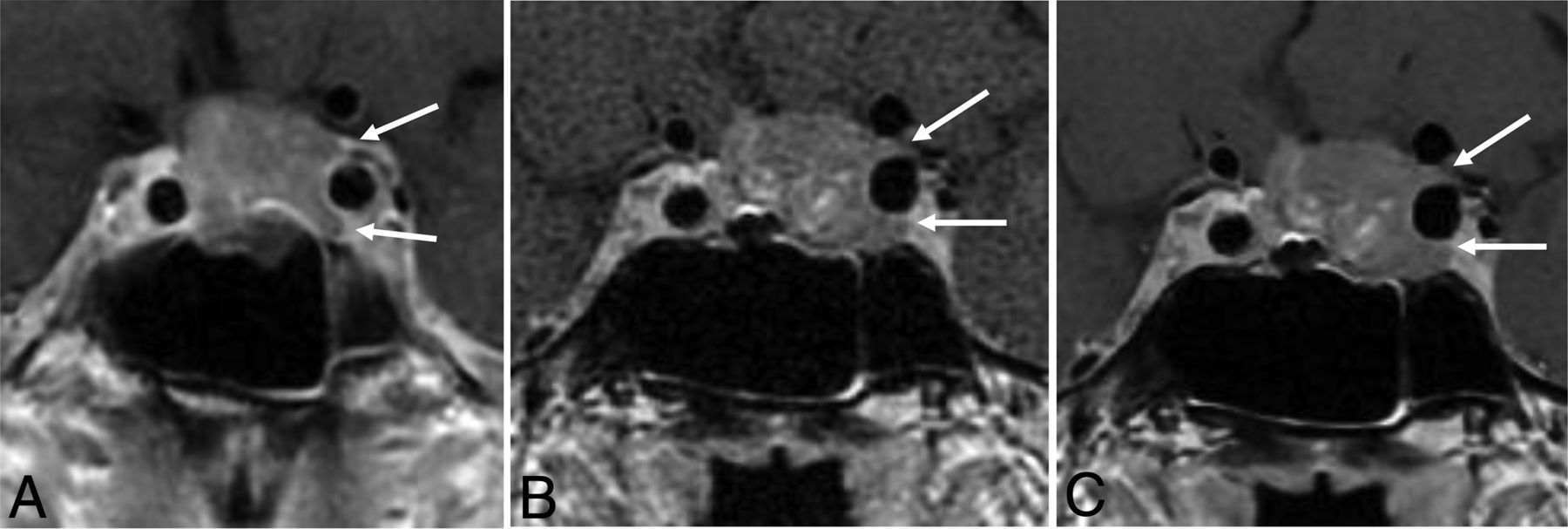
- FIG 3.
A 60-year-old woman who underwent a combined MR imaging protocol for preoperative evaluation of pituitary adenoma. On a 3-mmMRI, coronal, contrast-enhanced T1-weighted image (A), the tumor is present beyond the medial intercarotid line but not beyond the median intercarotid line (modified Knosp classification 1) and is <180° in contact with the intracavernous ICA. One-mmMRI, coronal, contrast-enhanced T1-weighted image (B) was reconstructed with DLR (C), and the tumor is shown to extend beyond the median intercarotid line (modified Knosp classification 2) and is <270° in contact with the intracavernous ICA (arrows).


Tables
Characteristics Total 67 Age (yr) 53 (SD, 12) No. of female patients 28 (42) Functioning adenoma 14 (21) Prolactin 5 (7.5) Growth hormone 7 (10) Adrenocorticotropic hormone 1 (1.5) Thyroid stimulating hormone 1 (1.5) CS invasion by reference standard 29 (43) Residual tumor following operation determined by follow-up MR imaging 11 (16) Postoperative stereotactic radiation therapy 7 (10) Note:—CS indicates cavernous sinus.
↵a Data are expressed as the mean (SD) or numbers with percentages in parenthesis.
- Table 2:
Depth of invasion and degree of contact with the intracavernous ICA by pituitary adenoma on 3-mmMRI, 1-mmMRI and 1-mmMRI with DLRa
3-mmMRI P Value 1-mmMRI P Value 1-mmMRI+DLR Depth of invasion (mm) 3.12 (2.44–3.81) <.001 3.82 (3.10–4.55) .004 4.07 (3.35–4.80) Degree of contact with intracavernous ICA Total encasement 9 (13.4%) <.001 13 (19.4%) .02 25 (37.3%) >270° 11 (16.4%) <.001 15 (22.4%) .04 26 (38.8%) >180° 17 (25.4%) .001 21 (31.3%) .11 30 (44.8%) ↵a P value was calculated by using paired t test for depth of invasion and McNemar test for degree of contact with the intracavernous ICA. Data are mean or number of patients, and data in parentheses are 95% confidence intervals unless otherwise specified.


Jump to section
Related Articles
Cited By...
- No citing articles found.