
Article Figures & Data
Figures
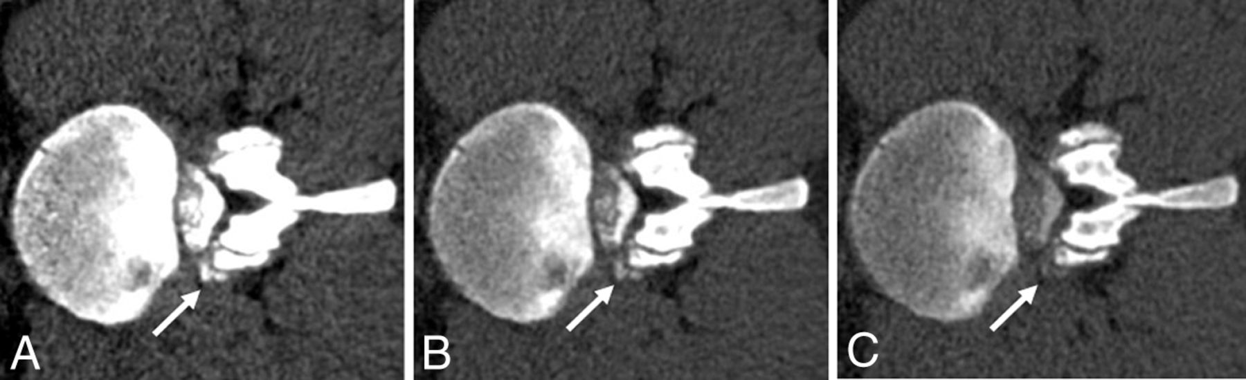
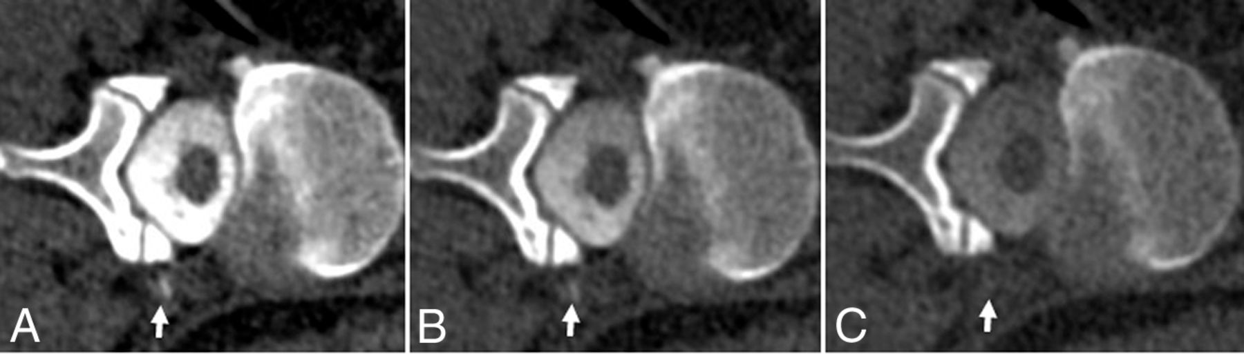
- FIG 1.
A, 50-keV VMI. B, 100 kV. C, 140 kV. Case 1: Left-side-down CTM with linear contrast at the left T12–L1 neural foramen, thought to reflect a CVF (arrows). Contrast is also noted in the renal collecting system. ROI Hounsfield units: A, 50-keV VMI Hounsfield unit maximum (max): 283 HU; mean, 76 HU. B, 100 -kV Hounsfield unit max: 147 HU; mean, 68 HU. C, 140-kV Hounsfield unit max: 116 HU; mean, 46 HU. D, 50-keV VMI. E, 100 kV. F, 140 kV. Case 1: Right-side-down CTM with paraspinal contrast at the level of T11–12, representing a second CVF (arrows). ROI Hounsfield units: D, 50-keV VMI Hounsfield unit max: 981 HU; mean, 693 HU. E, 100-kV Hounsfield unit max: 542 HU; mean, 369 HU. F, 140-kV Hounsfield unit max: 233 HU; mean, 107 HU.
- FIG 2.
A, 50-keV VMI. B, 100 kV. C, 140 kV. Case 2: Left-side-down CTM with a suspected distal nerve root sleeve tear at the left L2–3 (arrows). The patient also had contrast in the renal collecting system (not shown). ROI Hounsfield units: A, 50-keV VMI Hounsfield unit maximum (max): 806 HU; mean, 271 HU. B, 100-kV Hounsfield unit max: 435 HU; mean, 133 HU. C, 140-kV Hounsfield unit max: 210 HU; mean, 68 HU.
- FIG 3.
A, 50 -keV VMI. B, 100 kV. C, 150 kV. Case 3: Right-side-down CTM with linear contrast at the right L2–3 neuroforamen (arrows), extending into the paraspinal soft tissues, thought to reflect a vessel associated with contrast leakage at a higher right-T10 distal nerve root sleeve tear (not shown). ROI Hounsfield units: A, 50-keV VMI Hounsfield unit maximum (max): 485 HU; mean, 118 HU. B, 100-kV Hounsfield unit max: 271 HU; mean, 51 HU. C, 150-kV Hounsfield unit max: 151 HU; mean, 16 HU.
- FIG 4.
A, 50-keV VMI. B, 100 kV. C, 140 kV. Case 4: Right-side-down CTM with a small focus of extradural contrast at the right aspect of the thecal sac at T7–8 (arrows). ROI Hounsfield units: A, 50-keV VMI Hounsfield unit maximum (max): 441 HU; mean, 275 HU). B, 100-kV Hounsfield unit max: 277 HU; mean, 165 HU. C, 140-kV Hounsfield unit max: 163 HU; mean, 102 HU.
- FIG 5.
A, 50-keV VMI. B, 100 kV. C, 140 kV. Case 5: CTM right-side-down at the level of T11–12 shows linear focus most consistent with a CSF venous fistula (arrows). ROI Hounsfield units: A, 50-keV Hounsfield unit maximum (max): 436 HU; mean, 154 HU. B, 100-kV Hounsfield unit max: 251 HU; mean, 88 HU. C, 140-kV Hounsfield unit max: 125 HU; mean, 33 HU.


Tables
- Table 1:
Parameters for CT cervical, thoracic, and lumbar spine myelogram (Siemens dual-source, dual-energy CT models)
DECT Model Somatom Definition Flash Somatom Force Description 128-Section dual-source, dual-energy CT 192-Section dual-source, dual-energy CT kV(p) A: 100 B: Sn 140 A: 100 B: Sn 150 Quality reference mAs A: 230 B: 178 A: 260 B: 130 Scan FOV (mm) A: 500 B: 332 A: 500 B: 356 Rotation time (sec) 1.0 1.0 Pitch 0.9 0.9 Collimation (mm) 32 × 0.6 128 × .06 Patient Age Sex Other Imaging Findings CTM Findings Symptoms Treatment Outcome 1 28 F High-probability brain MR imaging T12-L1 CVF on left; T11-12 on right; contrast in the renal collecting system Orthostatic headache, multifocal pain T11 nerve root ligation Initial relief with subsequent recurrence of symptoms in the setting of Marfan syndrome 2 50 M Intermediate-probability brain MR imaging CVF at left L2-3 and contrast in the renal collecting system Orthostatic headache Blood patch, transvenous embolization of the left L2 paraspinal vein Dramatic symptom improvement following embolization 3 52 M High-probability brain MR imaging, positive cisternogram findings Right T10 distal nerve root sleeve tear with extradural contrast first detected at L2-3 Orthostatic headache, vision changes, pulsatile tinnitus Hemi-laminectomy right T10-11, repair of CSF leak Complete symptom resolution sustained for at least 1 year 4 34 F Intermediate-probability brain MR imaging Focus of CSF leak arising from the right lateral thecal sac at T7-8 thought to represent dural tear; contrast in the renal collecting system Orthostatic headache, facial and hand paresthesia Three targeted blood patches Improvement after 3 targeted blood patches with recurrence of symptoms 5 50 F Multiple nerve root diverticula on spine MR imaging Faint linear hyperattenuation extending from a right T11-12 nerve sleeve diverticulum thought to represent CVF Orthostatic headache, vision changes Two targeted and 1 multifocal blood patch Transient improvement after 2 targeted and 1 multifocal blood patch Note:—M indicates male; F, female.
Case 50 keV VMI Mean 50 keV VMI Max 100 kV Mean 100 kV Max 140/150 kV Mean 140/150 kV Max Case 1L 76 283 68 147 46 116 Case 1R 693 981 369 542 107 233 Case 2 271 806 133 435 68 210 Case 3 118 485 51 271 16 150 Case 4 275 441 165 277 102 163 Case 5 154 436 88 251 33 125 Note:—1L indicates left side down CTM; 1R, right side down CTM


