Advertisement

Main menu

  • Home
  • Content
    • Current Issue
    • Accepted Manuscripts
    • Article Preview
    • Past Issue Archive
    • Video Articles
    • AJNR Case Collection
    • Case of the Week Archive
    • Case of the Month Archive
    • Classic Case Archive
  • Special Collections
    • AJNR Awards
    • Low-Field MRI
    • Alzheimer Disease
    • ASNR Foundation Special Collection
    • Photon-Counting CT
    • View All
  • Multimedia
    • AJNR Podcasts
    • AJNR SCANtastic
    • Trainee Corner
    • MRI Safety Corner
    • Imaging Protocols
  • For Authors
    • Submit a Manuscript
    • Submit a Video Article
    • Submit an eLetter to the Editor/Response
    • Manuscript Submission Guidelines
    • Statistical Tips
    • Fast Publishing of Accepted Manuscripts
    • Graphical Abstract Preparation
    • Imaging Protocol Submission
    • Author Policies
  • About Us
    • About AJNR
    • Editorial Board
    • Editorial Board Alumni
  • More
    • Become a Reviewer/Academy of Reviewers
    • Subscribers
    • Permissions
    • Alerts
    • Feedback
    • Advertisers
    • ASNR Home

User menu

  • Alerts
  • Log in

Search

  • Advanced search
American Journal of Neuroradiology

American Journal of Neuroradiology

ASHNR American Society of Functional Neuroradiology ASHNR American Society of Pediatric Neuroradiology ASSR
  • Alerts
  • Log in

Advanced Search

  • Home
  • Content
    • Current Issue
    • Accepted Manuscripts
    • Article Preview
    • Past Issue Archive
    • Video Articles
    • AJNR Case Collection
    • Case of the Week Archive
    • Case of the Month Archive
    • Classic Case Archive
  • Special Collections
    • AJNR Awards
    • Low-Field MRI
    • Alzheimer Disease
    • ASNR Foundation Special Collection
    • Photon-Counting CT
    • View All
  • Multimedia
    • AJNR Podcasts
    • AJNR SCANtastic
    • Trainee Corner
    • MRI Safety Corner
    • Imaging Protocols
  • For Authors
    • Submit a Manuscript
    • Submit a Video Article
    • Submit an eLetter to the Editor/Response
    • Manuscript Submission Guidelines
    • Statistical Tips
    • Fast Publishing of Accepted Manuscripts
    • Graphical Abstract Preparation
    • Imaging Protocol Submission
    • Author Policies
  • About Us
    • About AJNR
    • Editorial Board
    • Editorial Board Alumni
  • More
    • Become a Reviewer/Academy of Reviewers
    • Subscribers
    • Permissions
    • Alerts
    • Feedback
    • Advertisers
    • ASNR Home
  • Follow AJNR on Twitter
  • Visit AJNR on Facebook
  • Follow AJNR on Instagram
  • Join AJNR on LinkedIn
  • RSS Feeds

AJNR Awards, New Junior Editors, and more. Read the latest AJNR updates

Research ArticleNeurointervention

Parent Artery Straightening after Flow-Diverter Stenting Improves the Odds of Aneurysm Occlusion

K. Janot, R. Fahed, A. Rouchaud, K. Zuber, G. Boulouis, G. Forestier, C. Mounayer and M. Piotin
American Journal of Neuroradiology January 2022, 43 (1) 87-92; DOI: https://doi.org/10.3174/ajnr.A7350
K. Janot
aFrom the Department of Interventional Neuroradiology (K.J., G.B.), University Hospital of Tours, Tours, France
  • Find this author on Google Scholar
  • Find this author on PubMed
  • Search for this author on this site
  • ORCID record for K. Janot
R. Fahed
bDepartment of Interventional Neuroradiology (R.F., M.P.)
  • Find this author on Google Scholar
  • Find this author on PubMed
  • Search for this author on this site
  • ORCID record for R. Fahed
A. Rouchaud
dDepartment of Interventional Neuroradiology (A.R., G.F., C.M.), Dupuytren University Hospital, Limoges, Franc
  • Find this author on Google Scholar
  • Find this author on PubMed
  • Search for this author on this site
  • ORCID record for A. Rouchaud
K. Zuber
cResearch and Biostatistics Unit (K.Z.), Rothschild Foundation Hospital, Paris, France
  • Find this author on Google Scholar
  • Find this author on PubMed
  • Search for this author on this site
  • ORCID record for K. Zuber
G. Boulouis
aFrom the Department of Interventional Neuroradiology (K.J., G.B.), University Hospital of Tours, Tours, France
  • Find this author on Google Scholar
  • Find this author on PubMed
  • Search for this author on this site
  • ORCID record for G. Boulouis
G. Forestier
dDepartment of Interventional Neuroradiology (A.R., G.F., C.M.), Dupuytren University Hospital, Limoges, Franc
  • Find this author on Google Scholar
  • Find this author on PubMed
  • Search for this author on this site
  • ORCID record for G. Forestier
C. Mounayer
dDepartment of Interventional Neuroradiology (A.R., G.F., C.M.), Dupuytren University Hospital, Limoges, Franc
  • Find this author on Google Scholar
  • Find this author on PubMed
  • Search for this author on this site
  • ORCID record for C. Mounayer
M. Piotin
bDepartment of Interventional Neuroradiology (R.F., M.P.)
  • Find this author on Google Scholar
  • Find this author on PubMed
  • Search for this author on this site
  • ORCID record for M. Piotin
  • Article
  • Figures & Data
  • Info & Metrics
  • Responses
  • References
  • PDF
Loading

Article Figures & Data

Figures

  • Tables
  • FIG 1.
    • Download figure
    • Open in new tab
    • Download powerpoint
    FIG 1.

    Measurement of parent artery straightening. Angles were measured at the intersection of 2 lines drawn from the neck center in the axis of the proximal and distal segments of the parent artery. α is the angle measured before stent placement, and β is the angle measured after stent deployment. Preoperative (1) and postoperative (2) drawings.

  • FIG 2.
    • Download figure
    • Open in new tab
    • Download powerpoint
    FIG 2.

    Distribution of angles of parent artery straightening and association with aneurysmal occlusion. A, Shadowgram distribution of parent artery straightening. B, Stacked histograms of the proportion of patients with complete occlusion in bins of increasing parent artery straightening (offset = 0, width = 25°).
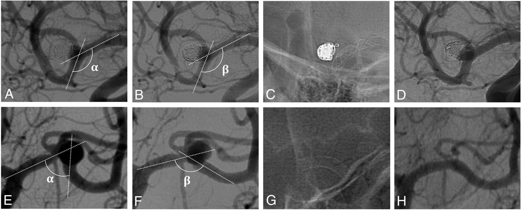
  • FIG 3.
    • Download figure
    • Open in new tab
    • Download powerpoint
    FIG 3.

    Comparison of vascular geometry modifications in 2 treatments of MCA aneurysms. The second flow-diverter stent deployment appears to modify the vascular anatomy (E–G) more than the first flow-diverter stent (A–C). Angiogram controls show complete aneurysm occlusion with the second flow-diverter stent (H) compared with the first-flow diverter (D) stent. A and E, Preoperative angiogram. B and F, Angiogram after stent placement. C and G, Unsubtracted view after stent deployment. D and H, First angiogram follow-up. α indicates the angle measured before stent placement; β, the angle measured after stent placement.

Tables

  • Figures
    • View popup
    Table 1:

    Comparison of demographic, angiographic, and treatment characteristics between complete occluded and incomplete occluded aneurysmsa

    All Aneurysms (n = 99)Complete Occlusion (n = 64)Incomplete Occlusion (n = 20)P Value
    Age (yr) (mean)54.1 (SD, 11.2)53.8 (SD, 10.8)54.3 (SD, 10.4).76
    Sex, M/F31 (31.3%)/68 (68.7%)18 (28.1%)/46 (71.9%)9 (45%)/11 (55%).26
    Follow-up (mo) (n = 84)23.9 (SD, 16.3)24.2 (SD, 15.0)22.9 (SD, 20.2).27
    Previous treatment45 (45.5%)28 (43.8%)13 (65%).16
    Aneurysm location.62
     MCA44 (44.4%)25 (39.1%)12 (60%)
     AcomA27 (27.3%)20 (31.3%)5 (25%)
     A12 (2.0%)2 (3.1%)0
     A22 (2.0%)2 (3.1%)0
     Pericallosal24 (24.2%)15 (23.4%)3 (15%)
    Aneurysm size (mm)
     Neck3.9 (SD, 2.1)3.6 (SD, 2.1)4.3 (SD, 2.0).06
     Diameter6.1 (SD, 5.0)6.1 (SD, 5.7)6.5 (SD, 3.5).14
    Concomitant coiling15 (15.2%)9 (14.1%)2 (10%)1
    Flow-diverter stent
     Length (mm)16.9 (SD, 3.9)16.5 (SD, 3.8)18.2 (SD, 4.3).42
     Type.004
      Nitinol47 (47.5%)26 (40.6%)16 (80%)
      Cobalt chromium52 (52.5%)38 (59.4%)4 (20%)
    Angle of parent artery
     Initial angle Embedded Image 124.1° (SD, 64.0°)118.3° (SD, 29.8°)122.7° (SD, 28.1°).58
     Postdeployment angle Embedded Image 141.6° (SD, 24.8°)146.0° (SD, 21.8°)135.9° (SD, 24°).12
     Parent artery straightening Embedded Image 3.8° (SD, 21.6°)27.7  (SD, 22.6)13.3° (SD, 12.3°)<.001
    • Note:—AcomA indicates anterior communicating artery; A1, proximal segment of the anterior cerebral artery; A2, distal segment of the anterior cerebral artery.

    • ↵a Data are presented as mean (standard deviation, SD) for continuous variables and absolute number (percentage of column total) for discrete variables.

    • View popup
    Table 2:

    Multivariable analysis of aneurysm occlusion determinants

    VariablesMultivariate Analysis, aOR (95% CI)P Value
    Aneurysm neck (mm)0.57 (0.38–0.87)<.001
    Stent type: chromium cobalt2.13(1.26–11.93).020
    Parent artery straightening1.04° (1.01°–1.17°).036
    Initial angle Embedded Image 0.95°(0.91°–1.02°.401
    Parent artery straightening × stent typea0.99 (0.94–1.02).055
    • Note:—aOR indicates adjusted odds ratio.

    • ↵a Interaction term.

PreviousNext
Back to top

In this issue

American Journal of Neuroradiology: 43 (1)
American Journal of Neuroradiology
Vol. 43, Issue 1
1 Jan 2022
  • Table of Contents
  • Index by author
  • Complete Issue (PDF)
Advertisement
Print
Download PDF
Email Article

Thank you for your interest in spreading the word on American Journal of Neuroradiology.

NOTE: We only request your email address so that the person you are recommending the page to knows that you wanted them to see it, and that it is not junk mail. We do not capture any email address.

Enter multiple addresses on separate lines or separate them with commas.
Parent Artery Straightening after Flow-Diverter Stenting Improves the Odds of Aneurysm Occlusion
(Your Name) has sent you a message from American Journal of Neuroradiology
(Your Name) thought you would like to see the American Journal of Neuroradiology web site.
CAPTCHA
This question is for testing whether or not you are a human visitor and to prevent automated spam submissions.
Cite this article
K. Janot, R. Fahed, A. Rouchaud, K. Zuber, G. Boulouis, G. Forestier, C. Mounayer, M. Piotin
Parent Artery Straightening after Flow-Diverter Stenting Improves the Odds of Aneurysm Occlusion
American Journal of Neuroradiology Jan 2022, 43 (1) 87-92; DOI: 10.3174/ajnr.A7350

Citation Manager Formats

  • BibTeX
  • Bookends
  • EasyBib
  • EndNote (tagged)
  • EndNote 8 (xml)
  • Medlars
  • Mendeley
  • Papers
  • RefWorks Tagged
  • Ref Manager
  • RIS
  • Zotero
0 Responses
Respond to this article
Share
Bookmark this article
Parent Artery Straightening after Flow-Diverter Stenting Improves the Odds of Aneurysm Occlusion
K. Janot, R. Fahed, A. Rouchaud, K. Zuber, G. Boulouis, G. Forestier, C. Mounayer, M. Piotin
American Journal of Neuroradiology Jan 2022, 43 (1) 87-92; DOI: 10.3174/ajnr.A7350
del.icio.us logo Twitter logo Facebook logo Mendeley logo
  • Tweet Widget
  • Facebook Like
  • Google Plus One
Purchase

Jump to section

  • Article
    • Abstract
    • MATERIALS AND METHODS
    • RESULTS
    • DISCUSSION
    • CONCLUSIONS
    • Footnotes
    • References
  • Figures & Data
  • Info & Metrics
  • Responses
  • References
  • PDF

Related Articles

  • No related articles found.
  • PubMed
  • Google Scholar

Cited By...

  • In vitro flow diversion effect of the ReSolv stent with the shelf technique in a bifurcation aneurysm model
  • Correlation of Flow Diverter Malapposition at the Aneurysm Neck with Incomplete Aneurysm Occlusion in Patients with Small Intracranial Aneurysms: A Single-Center Experience
  • In vitro flow diversion effect of the ReSolv stent with the shelf technique in a bifurcation aneurysm model
  • Crossref
  • Google Scholar

This article has not yet been cited by articles in journals that are participating in Crossref Cited-by Linking.

More in this TOC Section

  • Rescue Reentry in Carotid Near-Occlusion
  • Contour Neurovascular System: Five Year Follow Up
  • Effect of SARS-CoV2 on Endovascular Thrombectomy
Show more Neurointervention

Similar Articles

Advertisement

Indexed Content

  • Current Issue
  • Accepted Manuscripts
  • Article Preview
  • Past Issues
  • Editorials
  • Editor's Choice
  • Fellows' Journal Club
  • Letters to the Editor
  • Video Articles

Cases

  • Case Collection
  • Archive - Case of the Week
  • Archive - Case of the Month
  • Archive - Classic Case

More from AJNR

  • Trainee Corner
  • Imaging Protocols
  • MRI Safety Corner
  • Book Reviews

Multimedia

  • AJNR Podcasts
  • AJNR Scantastics

Resources

  • Turnaround Time
  • Submit a Manuscript
  • Submit a Video Article
  • Submit an eLetter to the Editor/Response
  • Manuscript Submission Guidelines
  • Statistical Tips
  • Fast Publishing of Accepted Manuscripts
  • Graphical Abstract Preparation
  • Imaging Protocol Submission
  • Evidence-Based Medicine Level Guide
  • Publishing Checklists
  • Author Policies
  • Become a Reviewer/Academy of Reviewers
  • News and Updates

About Us

  • About AJNR
  • Editorial Board
  • Editorial Board Alumni
  • Alerts
  • Permissions
  • Not an AJNR Subscriber? Join Now
  • Advertise with Us
  • Librarian Resources
  • Feedback
  • Terms and Conditions
  • AJNR Editorial Board Alumni

American Society of Neuroradiology

  • Not an ASNR Member? Join Now

© 2025 by the American Society of Neuroradiology All rights, including for text and data mining, AI training, and similar technologies, are reserved.
Print ISSN: 0195-6108 Online ISSN: 1936-959X

Powered by HighWire