Advertisement

Main menu

  • Home
  • Content
    • Current Issue
    • Accepted Manuscripts
    • Article Preview
    • Past Issue Archive
    • Video Articles
    • AJNR Case Collection
    • Case of the Week Archive
    • Case of the Month Archive
    • Classic Case Archive
  • Special Collections
    • AJNR Awards
    • Low-Field MRI
    • Alzheimer Disease
    • ASNR Foundation Special Collection
    • Photon-Counting CT
    • View All
  • Multimedia
    • AJNR Podcasts
    • AJNR SCANtastic
    • Trainee Corner
    • MRI Safety Corner
    • Imaging Protocols
  • For Authors
    • Submit a Manuscript
    • Submit a Video Article
    • Submit an eLetter to the Editor/Response
    • Manuscript Submission Guidelines
    • Statistical Tips
    • Fast Publishing of Accepted Manuscripts
    • Graphical Abstract Preparation
    • Imaging Protocol Submission
    • Author Policies
  • About Us
    • About AJNR
    • Editorial Board
    • Editorial Board Alumni
  • More
    • Become a Reviewer/Academy of Reviewers
    • Subscribers
    • Permissions
    • Alerts
    • Feedback
    • Advertisers
    • ASNR Home

User menu

  • Alerts
  • Log in

Search

  • Advanced search
American Journal of Neuroradiology

American Journal of Neuroradiology

ASHNR American Society of Functional Neuroradiology ASHNR American Society of Pediatric Neuroradiology ASSR
  • Alerts
  • Log in

Advanced Search

  • Home
  • Content
    • Current Issue
    • Accepted Manuscripts
    • Article Preview
    • Past Issue Archive
    • Video Articles
    • AJNR Case Collection
    • Case of the Week Archive
    • Case of the Month Archive
    • Classic Case Archive
  • Special Collections
    • AJNR Awards
    • Low-Field MRI
    • Alzheimer Disease
    • ASNR Foundation Special Collection
    • Photon-Counting CT
    • View All
  • Multimedia
    • AJNR Podcasts
    • AJNR SCANtastic
    • Trainee Corner
    • MRI Safety Corner
    • Imaging Protocols
  • For Authors
    • Submit a Manuscript
    • Submit a Video Article
    • Submit an eLetter to the Editor/Response
    • Manuscript Submission Guidelines
    • Statistical Tips
    • Fast Publishing of Accepted Manuscripts
    • Graphical Abstract Preparation
    • Imaging Protocol Submission
    • Author Policies
  • About Us
    • About AJNR
    • Editorial Board
    • Editorial Board Alumni
  • More
    • Become a Reviewer/Academy of Reviewers
    • Subscribers
    • Permissions
    • Alerts
    • Feedback
    • Advertisers
    • ASNR Home
  • Follow AJNR on Twitter
  • Visit AJNR on Facebook
  • Follow AJNR on Instagram
  • Join AJNR on LinkedIn
  • RSS Feeds

AJNR Awards, New Junior Editors, and more. Read the latest AJNR updates

Research ArticleSpine Imaging and Spine Image-Guided Interventions

Diagnostic Accuracy and Failure Mode Analysis of a Deep Learning Algorithm for the Detection of Cervical Spine Fractures

A.F. Voter, M.E. Larson, J.W. Garrett and J.-P.J. Yu
American Journal of Neuroradiology August 2021, 42 (8) 1550-1556; DOI: https://doi.org/10.3174/ajnr.A7179
A.F. Voter
aSchool of Medicine and Public Health (A.F.V.), University of Wisconsin-Madison, Madison, Wisconsin
  • Find this author on Google Scholar
  • Find this author on PubMed
  • Search for this author on this site
  • ORCID record for A.F. Voter
M.E. Larson
bDepartment of Radiology (M.E.L., J.W.G., J.-P.J.Y.), University of Wisconsin-Madison, Madison, Wisconsin
  • Find this author on Google Scholar
  • Find this author on PubMed
  • Search for this author on this site
  • ORCID record for M.E. Larson
J.W. Garrett
bDepartment of Radiology (M.E.L., J.W.G., J.-P.J.Y.), University of Wisconsin-Madison, Madison, Wisconsin
  • Find this author on Google Scholar
  • Find this author on PubMed
  • Search for this author on this site
  • ORCID record for J.W. Garrett
J.-P.J. Yu
bDepartment of Radiology (M.E.L., J.W.G., J.-P.J.Y.), University of Wisconsin-Madison, Madison, Wisconsin
cDepartment of Biomedical Engineering (J.-P.J.Y.), College of Engineering, University of Wisconsin-Madison, Madison, Wisconsin
dDepartment of Psychiatry (J.-P.J.Y.), University of Wisconsin School of Medicine and Public Health, Madison, Wisconsin
  • Find this author on Google Scholar
  • Find this author on PubMed
  • Search for this author on this site
  • ORCID record for J.-P.J. Yu
  • Article
  • Figures & Data
  • Supplemental
  • Info & Metrics
  • Responses
  • References
  • PDF
Loading

Article Figures & Data

Figures

  • Tables
  • FIG 1.
    • Download figure
    • Open in new tab
    • Download powerpoint
    FIG 1.

    Standards for reporting diagnostic accuracy studies (STARD) patient flow diagram.
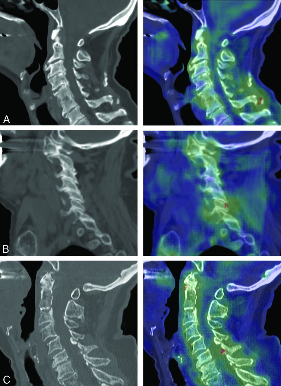
  • FIG 2.
    • Download figure
    • Open in new tab
    • Download powerpoint
    FIG 2.

    Examples of degenerative findings falsely flagged by Aidoc. Each panel shows the sagittal noncontrast cervical spine CT (left) and the Aidoc key image indicating the flagged pathology in red (right). A, A chronic ossicle falsely flagged by Aidoc. B, False-positive findings triggered by facet degeneration. C, Ossification of the ligamentum flavum incorrectly identified as a fracture by Aidoc.
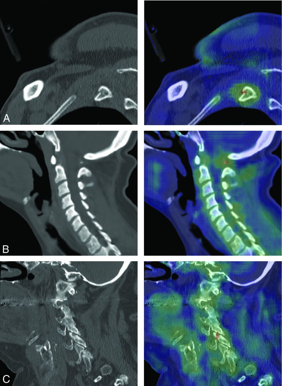
  • FIG 3.
    • Download figure
    • Open in new tab
    • Download powerpoint
    FIG 3.

    Examples of nondegenerative findings falsely flagged by Aidoc. Sagittal noncontrast cervical spine CT (left) and the Aidoc key image indicating the flagged pathology in red (right). A, Rib fracture outside of the cervical spine incorrectly flagged by Aidoc. B, Congenital hypoplasia of the posterior arch of the atlas flagged as a fracture. C, A nonpathologic nutrient foramen with degenerative changes identified as a fracture by Aidoc.

Tables

  • Figures
    • View popup
    Table 1:

    The impact of patient characteristics on Aidoc performance

    FactorAidoc Incorrect (No.)%P Value
    Total (No.) (%)1904, 100161100
    Indication (No.) (%).97
        Trauma1796, 9415596
        Critical511, 274528
        Minor888, 477446
        Not specified397, 213622
        Neck pain27, 111
        Neurologic deficit33, 221
        Postoperative10, 111
        Other38, 221
    Sex (No.) (%).08
        Male958, 509257
        Female946, 506943
    Imaging location (No.) (%).86
        Academic center1659. 8714188
        Outreach center245, 132012
    History of cervical spine surgery (No.) (%).57
        Prior surgery67, 474
        No prior surgery1837, 9615496
    Age (mean) (yr).03
        Overall60 (SD, 22)
        Aidoc incorrect64 (SD, 21)
        Aidoc correct60 (SD, 22)
    • View popup
    Table 2:

    Etiology of the false-positive flagged studies

    False-Positive EtiologyCountPercentage of All Flagged Studies (n = 173)
    Degeneration5531.8
        Degenerative ossicle1810.4
        Facet degeneration148.1
        Calcified ligament63.5
        Cortical irregularity74.0
        Osteopenia42.3
        Cystic degeneration42.3
        Atlantodental joint10.6
        Osteophyte10.6
    Noncervical pathology158.7
        Rib fracture84.6
        Degeneration, thoracic42.3
        Skull fracture21.2
        Carotid calcification10.6
    Anatomic variant105.8
        Nonunion vertebrae42.3
        Transitional anatomy21.2
        Limbus21.2
        Bifid spinous process10.6
        Secondary transverse foramen10.6
    Nutrient foramen95.2
    Artifact74.0
    Unknown84.6
    Other21.7
        DISH10.6
        Occipital suture10.6
    Total10661.3
    • Note:—DISH indicates diffuse idiopathic skeletal hyperostosis.

PreviousNext
Back to top

In this issue

American Journal of Neuroradiology: 42 (8)
American Journal of Neuroradiology
Vol. 42, Issue 8
1 Aug 2021
  • Table of Contents
  • Index by author
  • Complete Issue (PDF)
Advertisement
Print
Download PDF
Email Article

Thank you for your interest in spreading the word on American Journal of Neuroradiology.

NOTE: We only request your email address so that the person you are recommending the page to knows that you wanted them to see it, and that it is not junk mail. We do not capture any email address.

Enter multiple addresses on separate lines or separate them with commas.
Diagnostic Accuracy and Failure Mode Analysis of a Deep Learning Algorithm for the Detection of Cervical Spine Fractures
(Your Name) has sent you a message from American Journal of Neuroradiology
(Your Name) thought you would like to see the American Journal of Neuroradiology web site.
CAPTCHA
This question is for testing whether or not you are a human visitor and to prevent automated spam submissions.
Cite this article
A.F. Voter, M.E. Larson, J.W. Garrett, J.-P.J. Yu
Diagnostic Accuracy and Failure Mode Analysis of a Deep Learning Algorithm for the Detection of Cervical Spine Fractures
American Journal of Neuroradiology Aug 2021, 42 (8) 1550-1556; DOI: 10.3174/ajnr.A7179

Citation Manager Formats

  • BibTeX
  • Bookends
  • EasyBib
  • EndNote (tagged)
  • EndNote 8 (xml)
  • Medlars
  • Mendeley
  • Papers
  • RefWorks Tagged
  • Ref Manager
  • RIS
  • Zotero
0 Responses
Respond to this article
Share
Bookmark this article
Diagnostic Accuracy and Failure Mode Analysis of a Deep Learning Algorithm for the Detection of Cervical Spine Fractures
A.F. Voter, M.E. Larson, J.W. Garrett, J.-P.J. Yu
American Journal of Neuroradiology Aug 2021, 42 (8) 1550-1556; DOI: 10.3174/ajnr.A7179
del.icio.us logo Twitter logo Facebook logo Mendeley logo
  • Tweet Widget
  • Facebook Like
  • Google Plus One
Purchase

Jump to section

  • Article
    • Abstract
    • ABBREVIATIONS:
    • MATERIALS AND METHODS
    • RESULTS
    • DISCUSSION
    • CONCLUSIONS
    • Footnotes
    • References
  • Figures & Data
  • Supplemental
  • Info & Metrics
  • Responses
  • References
  • PDF

Related Articles

  • PubMed
  • Google Scholar

Cited By...

  • The Future of Artificial Intelligence in Clinical Radiology: Savior or False Hope?
  • Automated Detection of Cervical Spinal Stenosis and Cord Compression via Vision Transformer and Rules-Based Classification
  • Crossref
  • Google Scholar

This article has not yet been cited by articles in journals that are participating in Crossref Cited-by Linking.

More in this TOC Section

  • Optimization of Photon Counting CT Myelography
  • Characteristics of SIH Type I Culprit Lesions
  • Management Outcomes For VO Spine Biopsy
Show more Spine Imaging and Spine Image-Guided Interventions

Similar Articles

Advertisement

Indexed Content

  • Current Issue
  • Accepted Manuscripts
  • Article Preview
  • Past Issues
  • Editorials
  • Editor's Choice
  • Fellows' Journal Club
  • Letters to the Editor
  • Video Articles

Cases

  • Case Collection
  • Archive - Case of the Week
  • Archive - Case of the Month
  • Archive - Classic Case

More from AJNR

  • Trainee Corner
  • Imaging Protocols
  • MRI Safety Corner
  • Book Reviews

Multimedia

  • AJNR Podcasts
  • AJNR Scantastics

Resources

  • Turnaround Time
  • Submit a Manuscript
  • Submit a Video Article
  • Submit an eLetter to the Editor/Response
  • Manuscript Submission Guidelines
  • Statistical Tips
  • Fast Publishing of Accepted Manuscripts
  • Graphical Abstract Preparation
  • Imaging Protocol Submission
  • Evidence-Based Medicine Level Guide
  • Publishing Checklists
  • Author Policies
  • Become a Reviewer/Academy of Reviewers
  • News and Updates

About Us

  • About AJNR
  • Editorial Board
  • Editorial Board Alumni
  • Alerts
  • Permissions
  • Not an AJNR Subscriber? Join Now
  • Advertise with Us
  • Librarian Resources
  • Feedback
  • Terms and Conditions
  • AJNR Editorial Board Alumni

American Society of Neuroradiology

  • Not an ASNR Member? Join Now

© 2025 by the American Society of Neuroradiology All rights, including for text and data mining, AI training, and similar technologies, are reserved.
Print ISSN: 0195-6108 Online ISSN: 1936-959X

Powered by HighWire