Advertisement

Main menu

  • Home
  • Content
    • Current Issue
    • Accepted Manuscripts
    • Article Preview
    • Past Issue Archive
    • Video Articles
    • AJNR Case Collection
    • Case of the Week Archive
    • Case of the Month Archive
    • Classic Case Archive
  • Special Collections
    • AJNR Awards
    • Low-Field MRI
    • Alzheimer Disease
    • ASNR Foundation Special Collection
    • Photon-Counting CT
    • View All
  • Multimedia
    • AJNR Podcasts
    • AJNR SCANtastic
    • Trainee Corner
    • MRI Safety Corner
    • Imaging Protocols
  • For Authors
    • Submit a Manuscript
    • Submit a Video Article
    • Submit an eLetter to the Editor/Response
    • Manuscript Submission Guidelines
    • Statistical Tips
    • Fast Publishing of Accepted Manuscripts
    • Graphical Abstract Preparation
    • Imaging Protocol Submission
    • Author Policies
  • About Us
    • About AJNR
    • Editorial Board
    • Editorial Board Alumni
  • More
    • Become a Reviewer/Academy of Reviewers
    • Subscribers
    • Permissions
    • Alerts
    • Feedback
    • Advertisers
    • ASNR Home

User menu

  • Alerts
  • Log in

Search

  • Advanced search
American Journal of Neuroradiology

American Journal of Neuroradiology

ASHNR American Society of Functional Neuroradiology ASHNR American Society of Pediatric Neuroradiology ASSR
  • Alerts
  • Log in

Advanced Search

  • Home
  • Content
    • Current Issue
    • Accepted Manuscripts
    • Article Preview
    • Past Issue Archive
    • Video Articles
    • AJNR Case Collection
    • Case of the Week Archive
    • Case of the Month Archive
    • Classic Case Archive
  • Special Collections
    • AJNR Awards
    • Low-Field MRI
    • Alzheimer Disease
    • ASNR Foundation Special Collection
    • Photon-Counting CT
    • View All
  • Multimedia
    • AJNR Podcasts
    • AJNR SCANtastic
    • Trainee Corner
    • MRI Safety Corner
    • Imaging Protocols
  • For Authors
    • Submit a Manuscript
    • Submit a Video Article
    • Submit an eLetter to the Editor/Response
    • Manuscript Submission Guidelines
    • Statistical Tips
    • Fast Publishing of Accepted Manuscripts
    • Graphical Abstract Preparation
    • Imaging Protocol Submission
    • Author Policies
  • About Us
    • About AJNR
    • Editorial Board
    • Editorial Board Alumni
  • More
    • Become a Reviewer/Academy of Reviewers
    • Subscribers
    • Permissions
    • Alerts
    • Feedback
    • Advertisers
    • ASNR Home
  • Follow AJNR on Twitter
  • Visit AJNR on Facebook
  • Follow AJNR on Instagram
  • Join AJNR on LinkedIn
  • RSS Feeds

AJNR Awards, New Junior Editors, and more. Read the latest AJNR updates

Research ArticleAdult Brain

Is Contrast Medium Really Needed for Follow-up MRI of Untreated Intracranial Meningiomas?

J. Boto, R. Guatta, A. Fitsiori, J. Hofmeister, T.R. Meling and M.I. Vargas
American Journal of Neuroradiology August 2021, 42 (8) 1421-1428; DOI: https://doi.org/10.3174/ajnr.A7170
J. Boto
aFrom the Division of Neuroradiology (J.B., A.F., M.I.V.), Geneva University Hospital and Faculty of Medicine of Geneva, Geneva, Switzerland
  • Find this author on Google Scholar
  • Find this author on PubMed
  • Search for this author on this site
  • ORCID record for J. Boto
R. Guatta
bDivision of Neurosurgery (R.G., T.R.M.), Lugano Regional Hospital (Civic), Lugano, Switzerland
  • Find this author on Google Scholar
  • Find this author on PubMed
  • Search for this author on this site
  • ORCID record for R. Guatta
A. Fitsiori
aFrom the Division of Neuroradiology (J.B., A.F., M.I.V.), Geneva University Hospital and Faculty of Medicine of Geneva, Geneva, Switzerland
  • Find this author on Google Scholar
  • Find this author on PubMed
  • Search for this author on this site
  • ORCID record for A. Fitsiori
J. Hofmeister
cDivision of Radiology (J.H.), Geneva University Hospital and Faculty of Medicine of Geneva, Geneva, Switzerland.
  • Find this author on Google Scholar
  • Find this author on PubMed
  • Search for this author on this site
  • ORCID record for J. Hofmeister
T.R. Meling
bDivision of Neurosurgery (R.G., T.R.M.), Lugano Regional Hospital (Civic), Lugano, Switzerland
  • Find this author on Google Scholar
  • Find this author on PubMed
  • Search for this author on this site
  • ORCID record for T.R. Meling
M.I. Vargas
aFrom the Division of Neuroradiology (J.B., A.F., M.I.V.), Geneva University Hospital and Faculty of Medicine of Geneva, Geneva, Switzerland
  • Find this author on Google Scholar
  • Find this author on PubMed
  • Search for this author on this site
  • ORCID record for M.I. Vargas
  • Article
  • Figures & Data
  • Supplemental
  • Info & Metrics
  • Responses
  • References
  • PDF
Loading

Article Figures & Data

Figures

  • FIG 1.
    • Download figure
    • Open in new tab
    • Download powerpoint
    FIG 1.

    Flow diagram showing the steps in patient selection. NF2 indicates neurofibromatosis type 2.
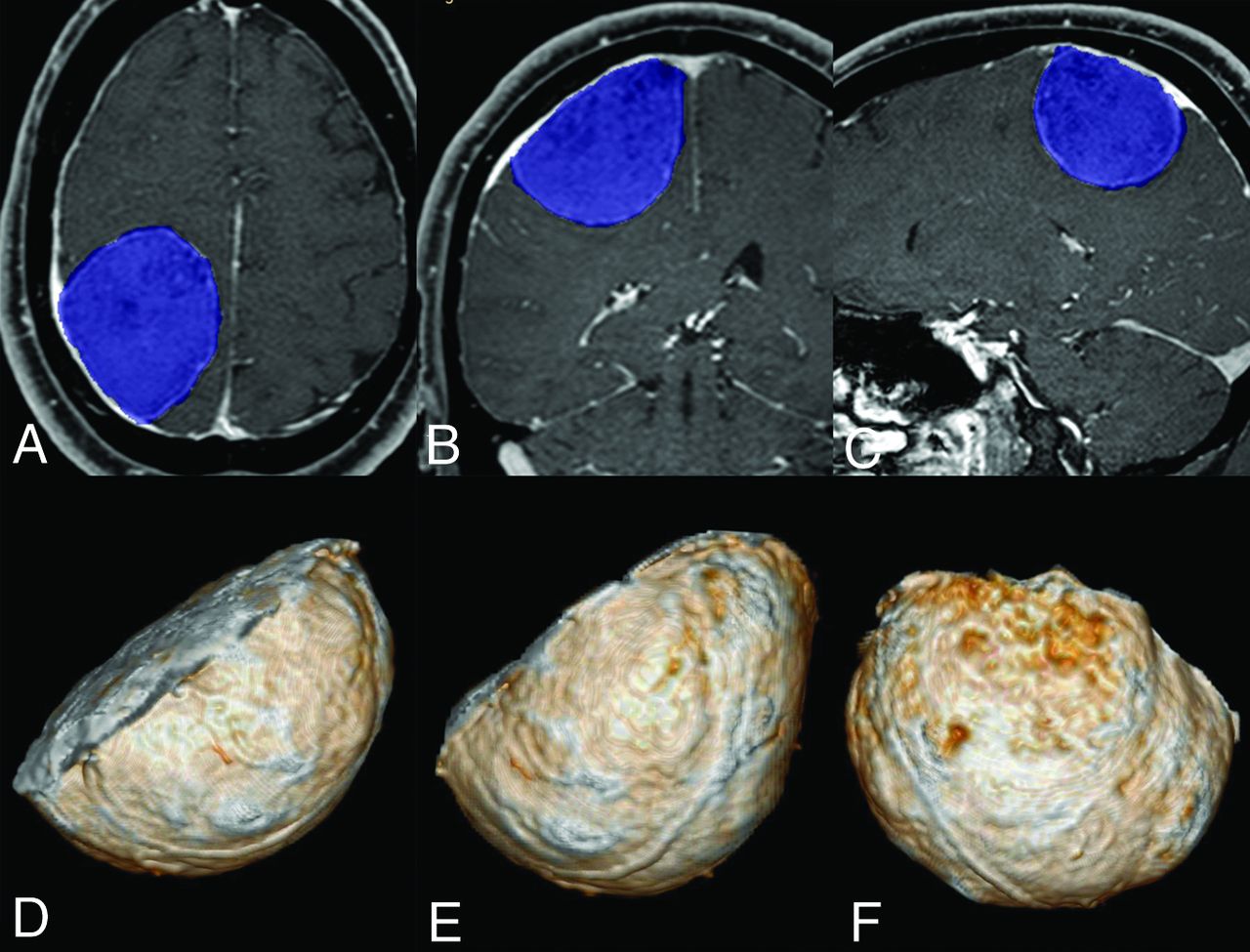
  • FIG 2.
    • Download figure
    • Open in new tab
    • Download powerpoint
    FIG 2.

    A 41-year-old man with a meningioma of the right convexity adjacent to the parietal lobe. Segmentation of the meningioma is achieved by shading the lesion in blue using a semi-automated software (A–C). The voxels outside the shaded volume can be discarded, and the voxels within the volume (shown as 3D-rendering in D, E, and F) can be isolated and exported to the PACS or any other software for further analysis.

  • FIG 3.
    • Download figure
    • Open in new tab
    • Download powerpoint
    FIG 3.

    Chart showing the number of included intracranial meningiomas by sex and specific location. Cerebral convexity parasagittal, falcine, intraventricular, and cerebellar convexity meningiomas are considered non-skull base. All other locations are considered skull base. Post. indicates posterior.
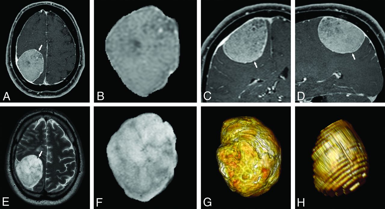
  • FIG 4.
    • Download figure
    • Open in new tab
    • Download powerpoint
    FIG 4.

    Same patient as in Fig 2 with a meningioma of the right convexity adjacent to the parietal lobe. Axial (A), coronal (C) and sagittal (D) THRIVE Gd and axial T2-weighted (E) images depict a meningioma of the right convexity exerting mass effect on the adjacent brain parenchyma of the parietal lobe (arrow, A, C, D, and E). Axial THRIVE Gd (B) and T2-weighted (F) images of the isolated lesion obtained with the semi-automated software, from which 3D-rendering can easily be performed with any DICOM viewer (G, THRIVE Gd. H, T2WI).

  • FIG 5.
    • Download figure
    • Open in new tab
    • Download powerpoint
    FIG 5.

    A 52-year-old woman. Axial (A), coronal (C) and sagittal (D) THRIVE Gd and axial T2-weighted (E) images show a meningioma arising from the planum sphenoidale and orbital roof on the right (white arrow, A, C, D, and E). The lesion crosses the midline and is in close contact with the cisternal segment of the right optic nerve (white arrowhead in D), the supraclinoid right internal carotid artery (black arrow, C), and cranial aspect of the right cavernous sinus (black arrowhead, C). Axial THRIVE Gd (B) and T2-weighted (F) images of the isolated lesion obtained with the semi-automated software, from which 3D-rendering was performed with our DICOM viewer (G, T1 3D-fat-saturated Gd; H, T2WI). This case illustrates a particularly challenging meningioma for which the skull base location, close contact with other structures, and irregular shape made the segmentation process more difficult.

PreviousNext
Back to top

In this issue

American Journal of Neuroradiology: 42 (8)
American Journal of Neuroradiology
Vol. 42, Issue 8
1 Aug 2021
  • Table of Contents
  • Index by author
  • Complete Issue (PDF)
Advertisement
Print
Download PDF
Email Article

Thank you for your interest in spreading the word on American Journal of Neuroradiology.

NOTE: We only request your email address so that the person you are recommending the page to knows that you wanted them to see it, and that it is not junk mail. We do not capture any email address.

Enter multiple addresses on separate lines or separate them with commas.
Is Contrast Medium Really Needed for Follow-up MRI of Untreated Intracranial Meningiomas?
(Your Name) has sent you a message from American Journal of Neuroradiology
(Your Name) thought you would like to see the American Journal of Neuroradiology web site.
CAPTCHA
This question is for testing whether or not you are a human visitor and to prevent automated spam submissions.
Cite this article
J. Boto, R. Guatta, A. Fitsiori, J. Hofmeister, T.R. Meling, M.I. Vargas
Is Contrast Medium Really Needed for Follow-up MRI of Untreated Intracranial Meningiomas?
American Journal of Neuroradiology Aug 2021, 42 (8) 1421-1428; DOI: 10.3174/ajnr.A7170

Citation Manager Formats

  • BibTeX
  • Bookends
  • EasyBib
  • EndNote (tagged)
  • EndNote 8 (xml)
  • Medlars
  • Mendeley
  • Papers
  • RefWorks Tagged
  • Ref Manager
  • RIS
  • Zotero
0 Responses
Respond to this article
Share
Bookmark this article
Is Contrast Medium Really Needed for Follow-up MRI of Untreated Intracranial Meningiomas?
J. Boto, R. Guatta, A. Fitsiori, J. Hofmeister, T.R. Meling, M.I. Vargas
American Journal of Neuroradiology Aug 2021, 42 (8) 1421-1428; DOI: 10.3174/ajnr.A7170
del.icio.us logo Twitter logo Facebook logo Mendeley logo
  • Tweet Widget
  • Facebook Like
  • Google Plus One
Purchase

Jump to section

  • Article
    • Abstract
    • ABBREVIATIONS:
    • MATERIALS AND METHODS
    • RESULTS
    • DISCUSSION
    • CONCLUSIONS
    • References
  • Figures & Data
  • Supplemental
  • Info & Metrics
  • Responses
  • References
  • PDF

Related Articles

  • PubMed
  • Google Scholar

Cited By...

  • Reply:
  • Regarding "Comparative Evaluation of Lower Gadolinium Doses for MR Imaging of Meningiomas: How Low Can We Go?"
  • Comparative Evaluation of Lower Gadolinium Doses for MR Imaging of Meningiomas: How Low Can We Go?
  • Do We Need Gadolinium-Based Contrast Agents for Routine MRI Surveillance of Unoperated Pituitary Macroadenoma?
  • Crossref
  • Google Scholar

This article has not yet been cited by articles in journals that are participating in Crossref Cited-by Linking.

More in this TOC Section

  • Diagnostic Neuroradiology of Monoclonal Antibodies
  • Clinical Outcomes After Chiari I Decompression
  • Segmentation of Brain Metastases with BLAST
Show more Adult Brain

Similar Articles

Advertisement

Indexed Content

  • Current Issue
  • Accepted Manuscripts
  • Article Preview
  • Past Issues
  • Editorials
  • Editor's Choice
  • Fellows' Journal Club
  • Letters to the Editor
  • Video Articles

Cases

  • Case Collection
  • Archive - Case of the Week
  • Archive - Case of the Month
  • Archive - Classic Case

More from AJNR

  • Trainee Corner
  • Imaging Protocols
  • MRI Safety Corner
  • Book Reviews

Multimedia

  • AJNR Podcasts
  • AJNR Scantastics

Resources

  • Turnaround Time
  • Submit a Manuscript
  • Submit a Video Article
  • Submit an eLetter to the Editor/Response
  • Manuscript Submission Guidelines
  • Statistical Tips
  • Fast Publishing of Accepted Manuscripts
  • Graphical Abstract Preparation
  • Imaging Protocol Submission
  • Evidence-Based Medicine Level Guide
  • Publishing Checklists
  • Author Policies
  • Become a Reviewer/Academy of Reviewers
  • News and Updates

About Us

  • About AJNR
  • Editorial Board
  • Editorial Board Alumni
  • Alerts
  • Permissions
  • Not an AJNR Subscriber? Join Now
  • Advertise with Us
  • Librarian Resources
  • Feedback
  • Terms and Conditions
  • AJNR Editorial Board Alumni

American Society of Neuroradiology

  • Not an ASNR Member? Join Now

© 2025 by the American Society of Neuroradiology All rights, including for text and data mining, AI training, and similar technologies, are reserved.
Print ISSN: 0195-6108 Online ISSN: 1936-959X

Powered by HighWire