Advertisement

Main menu

  • Home
  • Content
    • Current Issue
    • Accepted Manuscripts
    • Article Preview
    • Past Issue Archive
    • Video Articles
    • AJNR Case Collection
    • Case of the Week Archive
    • Case of the Month Archive
    • Classic Case Archive
  • Special Collections
    • AJNR Awards
    • Low-Field MRI
    • Alzheimer Disease
    • ASNR Foundation Special Collection
    • Photon-Counting CT
    • View All
  • Multimedia
    • AJNR Podcasts
    • AJNR SCANtastic
    • Trainee Corner
    • MRI Safety Corner
    • Imaging Protocols
  • For Authors
    • Submit a Manuscript
    • Submit a Video Article
    • Submit an eLetter to the Editor/Response
    • Manuscript Submission Guidelines
    • Statistical Tips
    • Fast Publishing of Accepted Manuscripts
    • Graphical Abstract Preparation
    • Imaging Protocol Submission
    • Author Policies
  • About Us
    • About AJNR
    • Editorial Board
    • Editorial Board Alumni
  • More
    • Become a Reviewer/Academy of Reviewers
    • Subscribers
    • Permissions
    • Alerts
    • Feedback
    • Advertisers
    • ASNR Home

User menu

  • Alerts
  • Log in

Search

  • Advanced search
American Journal of Neuroradiology

American Journal of Neuroradiology

ASHNR American Society of Functional Neuroradiology ASHNR American Society of Pediatric Neuroradiology ASSR
  • Alerts
  • Log in

Advanced Search

  • Home
  • Content
    • Current Issue
    • Accepted Manuscripts
    • Article Preview
    • Past Issue Archive
    • Video Articles
    • AJNR Case Collection
    • Case of the Week Archive
    • Case of the Month Archive
    • Classic Case Archive
  • Special Collections
    • AJNR Awards
    • Low-Field MRI
    • Alzheimer Disease
    • ASNR Foundation Special Collection
    • Photon-Counting CT
    • View All
  • Multimedia
    • AJNR Podcasts
    • AJNR SCANtastic
    • Trainee Corner
    • MRI Safety Corner
    • Imaging Protocols
  • For Authors
    • Submit a Manuscript
    • Submit a Video Article
    • Submit an eLetter to the Editor/Response
    • Manuscript Submission Guidelines
    • Statistical Tips
    • Fast Publishing of Accepted Manuscripts
    • Graphical Abstract Preparation
    • Imaging Protocol Submission
    • Author Policies
  • About Us
    • About AJNR
    • Editorial Board
    • Editorial Board Alumni
  • More
    • Become a Reviewer/Academy of Reviewers
    • Subscribers
    • Permissions
    • Alerts
    • Feedback
    • Advertisers
    • ASNR Home
  • Follow AJNR on Twitter
  • Visit AJNR on Facebook
  • Follow AJNR on Instagram
  • Join AJNR on LinkedIn
  • RSS Feeds

AJNR Awards, New Junior Editors, and more. Read the latest AJNR updates

Research ArticleRadiology-Pathology Correlation

Hypothalamic Pilomyxoid Astrocytoma in a Child with Lipodystrophy

J.C. Benson, J. Trejo-Lopez, S.E. Bach, J. Schwartz, T.J. Kaufmann, L. Eckel and J. Guerin
American Journal of Neuroradiology August 2021, 42 (8) 1370-1374; DOI: https://doi.org/10.3174/ajnr.A7136
J.C. Benson
aDepartment of Radiology (J.C.B., T.J.K., L.E., J.G.), Mayo Clinic, Rochester, Minnesota
  • Find this author on Google Scholar
  • Find this author on PubMed
  • Search for this author on this site
  • ORCID record for J.C. Benson
J. Trejo-Lopez
bDepartment of Laboratory Medicine and Pathology (J.T.-L.), Mayo Clinic, Rochester, Minnesota
  • Find this author on Google Scholar
  • Find this author on PubMed
  • Search for this author on this site
  • ORCID record for J. Trejo-Lopez
S.E. Bach
cDepartment of Laboratory Medicine and Pathology (S.E.B.), OSF Healthcare, Peoria, Illinois
  • Find this author on Google Scholar
  • Find this author on PubMed
  • Search for this author on this site
  • ORCID record for S.E. Bach
J. Schwartz
dDepartments of Pediatric and Adolescent Medicine (J.S.), Mayo Clinic, Rochester, Minnesota.
  • Find this author on Google Scholar
  • Find this author on PubMed
  • Search for this author on this site
  • ORCID record for J. Schwartz
T.J. Kaufmann
aDepartment of Radiology (J.C.B., T.J.K., L.E., J.G.), Mayo Clinic, Rochester, Minnesota
  • Find this author on Google Scholar
  • Find this author on PubMed
  • Search for this author on this site
  • ORCID record for T.J. Kaufmann
L. Eckel
aDepartment of Radiology (J.C.B., T.J.K., L.E., J.G.), Mayo Clinic, Rochester, Minnesota
  • Find this author on Google Scholar
  • Find this author on PubMed
  • Search for this author on this site
  • ORCID record for L. Eckel
J. Guerin
aDepartment of Radiology (J.C.B., T.J.K., L.E., J.G.), Mayo Clinic, Rochester, Minnesota
  • Find this author on Google Scholar
  • Find this author on PubMed
  • Search for this author on this site
  • ORCID record for J. Guerin
  • Article
  • Figures & Data
  • Info & Metrics
  • Responses
  • References
  • PDF
Loading

Article Figures & Data

Figures

  • Tables
  • FIG 1.
    • Download figure
    • Open in new tab
    • Download powerpoint
    FIG 1.

    Photographs of the patient demonstrate clinical sequelae of lipodystrophy, with profound loss of subcutaneous fat. Printed with authorization from the patient's parents.

  • FIG 2.
    • Download figure
    • Open in new tab
    • Download powerpoint
    FIG 2.

    Tumor appearance on axial MPRAGE (A) and T2WI (B), coronal postgadolinium T1-weighted Cube (GE Healthcare) (C), and sagittal FIESTA (D) and postgadolinium T1-weighted Cube (E). Images demonstrate an enhancing lobulated mass arising from the hypothalamic/chiasmatic region, with indistinct borders between the tumor and adjacent left hypothalamic parenchyma (arrow).

  • FIG 3.
    • Download figure
    • Open in new tab
    • Download powerpoint
    FIG 3.

    Leptomeningeal seeding seen on axial postgadolinium T1 Cube (A and B) and sagittal postgadolinium T1 images of the lower spine (C). Multiple enhancing foci are noted, including along the right temporal lobe (long straight arrow in A), ventral medulla (curved arrow in B), and ventral conus medullaris (short straight arrows in C). There is near-complete absence of subcutaneous fat, compatible with the patient's known lipodystrophy.

  • FIG 4.
    • Download figure
    • Open in new tab
    • Download powerpoint
    FIG 4.

    Photomicrographs of the tumor reveal a glioma composed of overall monotonous neoplastic cells embedded in a myxoid background (A, H&E, ×100 magnification) and exhibiting prominent angiocentric arrangements (B, H&E, ×200 magnification). Neoplastic cells demonstrate immunoreactivity for glial fibrillary acidic protein. C, Glial fibrillary acidic protein immunohistochemistry, ×200 magnification. Neurofilament reveals, overall, few background axons, compatible with a predominantly expansile growth pattern (D, neurofilament immunohistochemistry, ×100 magnification).
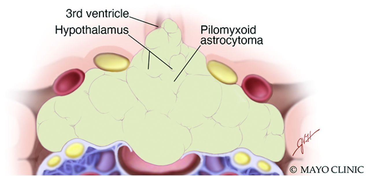
  • FIG 5.
    • Download figure
    • Open in new tab
    • Download powerpoint
    FIG 5.

    Artistic illustration of a PMA extending from the hypothalamus, with involvement of the suprasellar region and adjacent structures. PMAs tend to be solid and are typically devoid of both calcifications and intratumoral cysts. Used with permission of Mayo Foundation for Medical Education and Research. All rights reserved.

Tables

  • Figures
  • Comparison of typical PMA and pilocytic astrocytoma clinical and imaging features

    PMAPilocytic Astrocytoma
    AgeYounger (mean age, 1.5 yr)Older (mean age, 4.8 yr)
    LocationHypothalamic/chiasmatic regionCerebellum > hypothalamic/chiasmatic region
    EnhancementHomogeneousHeterogeneous
    Intratumoral contentsMost solid, with minimal tumoral cystsMost have cystic content
    Intratumoral hemorrhage12%–25%1%–8%
    Evidence of leptomeningeal seedingMore frequent (up to 20%)Exceedingly rare
PreviousNext
Back to top

In this issue

American Journal of Neuroradiology: 42 (8)
American Journal of Neuroradiology
Vol. 42, Issue 8
1 Aug 2021
  • Table of Contents
  • Index by author
  • Complete Issue (PDF)
Advertisement
Print
Download PDF
Email Article

Thank you for your interest in spreading the word on American Journal of Neuroradiology.

NOTE: We only request your email address so that the person you are recommending the page to knows that you wanted them to see it, and that it is not junk mail. We do not capture any email address.

Enter multiple addresses on separate lines or separate them with commas.
Hypothalamic Pilomyxoid Astrocytoma in a Child with Lipodystrophy
(Your Name) has sent you a message from American Journal of Neuroradiology
(Your Name) thought you would like to see the American Journal of Neuroradiology web site.
CAPTCHA
This question is for testing whether or not you are a human visitor and to prevent automated spam submissions.
Cite this article
J.C. Benson, J. Trejo-Lopez, S.E. Bach, J. Schwartz, T.J. Kaufmann, L. Eckel, J. Guerin
Hypothalamic Pilomyxoid Astrocytoma in a Child with Lipodystrophy
American Journal of Neuroradiology Aug 2021, 42 (8) 1370-1374; DOI: 10.3174/ajnr.A7136

Citation Manager Formats

  • BibTeX
  • Bookends
  • EasyBib
  • EndNote (tagged)
  • EndNote 8 (xml)
  • Medlars
  • Mendeley
  • Papers
  • RefWorks Tagged
  • Ref Manager
  • RIS
  • Zotero
0 Responses
Respond to this article
Share
Bookmark this article
Hypothalamic Pilomyxoid Astrocytoma in a Child with Lipodystrophy
J.C. Benson, J. Trejo-Lopez, S.E. Bach, J. Schwartz, T.J. Kaufmann, L. Eckel, J. Guerin
American Journal of Neuroradiology Aug 2021, 42 (8) 1370-1374; DOI: 10.3174/ajnr.A7136
del.icio.us logo Twitter logo Facebook logo Mendeley logo
  • Tweet Widget
  • Facebook Like
  • Google Plus One
Purchase

Jump to section

  • Article
    • Abstract
    • ABBREVIATION:
    • DISCUSSION
    • References
  • Figures & Data
  • Info & Metrics
  • Responses
  • References
  • PDF

Related Articles

  • No related articles found.
  • PubMed
  • Google Scholar

Cited By...

  • No citing articles found.
  • Crossref
  • Google Scholar

This article has not yet been cited by articles in journals that are participating in Crossref Cited-by Linking.

More in this TOC Section

Radiology-Pathology Correlation

  • Histopathological Analysis of Five Patients with CSF-Venous Fistulas After Surgical Nerve Root Ligation and Resection
  • Calcified Pseudoneoplasm of the Neuraxis
Show more Radiology-Pathology Correlation

Pediatric Neuroimaging

  • Histopathological Analysis of Five Patients with CSF-Venous Fistulas After Surgical Nerve Root Ligation and Resection
  • Calcified Pseudoneoplasm of the Neuraxis
  • Frontal Paraventricular Cysts
Show more Pediatric Neuroimaging

Similar Articles

Advertisement

Indexed Content

  • Current Issue
  • Accepted Manuscripts
  • Article Preview
  • Past Issues
  • Editorials
  • Editor's Choice
  • Fellows' Journal Club
  • Letters to the Editor
  • Video Articles

Cases

  • Case Collection
  • Archive - Case of the Week
  • Archive - Case of the Month
  • Archive - Classic Case

More from AJNR

  • Trainee Corner
  • Imaging Protocols
  • MRI Safety Corner
  • Book Reviews

Multimedia

  • AJNR Podcasts
  • AJNR Scantastics

Resources

  • Turnaround Time
  • Submit a Manuscript
  • Submit a Video Article
  • Submit an eLetter to the Editor/Response
  • Manuscript Submission Guidelines
  • Statistical Tips
  • Fast Publishing of Accepted Manuscripts
  • Graphical Abstract Preparation
  • Imaging Protocol Submission
  • Evidence-Based Medicine Level Guide
  • Publishing Checklists
  • Author Policies
  • Become a Reviewer/Academy of Reviewers
  • News and Updates

About Us

  • About AJNR
  • Editorial Board
  • Editorial Board Alumni
  • Alerts
  • Permissions
  • Not an AJNR Subscriber? Join Now
  • Advertise with Us
  • Librarian Resources
  • Feedback
  • Terms and Conditions
  • AJNR Editorial Board Alumni

American Society of Neuroradiology

  • Not an ASNR Member? Join Now

© 2025 by the American Society of Neuroradiology All rights, including for text and data mining, AI training, and similar technologies, are reserved.
Print ISSN: 0195-6108 Online ISSN: 1936-959X

Powered by HighWire