Advertisement

Main menu

  • Home
  • Content
    • Current Issue
    • Accepted Manuscripts
    • Article Preview
    • Past Issue Archive
    • Video Articles
    • AJNR Case Collection
    • Case of the Week Archive
    • Case of the Month Archive
    • Classic Case Archive
  • Special Collections
    • AJNR Awards
    • Low-Field MRI
    • Alzheimer Disease
    • ASNR Foundation Special Collection
    • Photon-Counting CT
    • View All
  • Multimedia
    • AJNR Podcasts
    • AJNR SCANtastic
    • Trainee Corner
    • MRI Safety Corner
    • Imaging Protocols
  • For Authors
    • Submit a Manuscript
    • Submit a Video Article
    • Submit an eLetter to the Editor/Response
    • Manuscript Submission Guidelines
    • Statistical Tips
    • Fast Publishing of Accepted Manuscripts
    • Graphical Abstract Preparation
    • Imaging Protocol Submission
    • Author Policies
  • About Us
    • About AJNR
    • Editorial Board
    • Editorial Board Alumni
  • More
    • Become a Reviewer/Academy of Reviewers
    • Subscribers
    • Permissions
    • Alerts
    • Feedback
    • Advertisers
    • ASNR Home

User menu

  • Alerts
  • Log in

Search

  • Advanced search
American Journal of Neuroradiology

American Journal of Neuroradiology

ASHNR American Society of Functional Neuroradiology ASHNR American Society of Pediatric Neuroradiology ASSR
  • Alerts
  • Log in

Advanced Search

  • Home
  • Content
    • Current Issue
    • Accepted Manuscripts
    • Article Preview
    • Past Issue Archive
    • Video Articles
    • AJNR Case Collection
    • Case of the Week Archive
    • Case of the Month Archive
    • Classic Case Archive
  • Special Collections
    • AJNR Awards
    • Low-Field MRI
    • Alzheimer Disease
    • ASNR Foundation Special Collection
    • Photon-Counting CT
    • View All
  • Multimedia
    • AJNR Podcasts
    • AJNR SCANtastic
    • Trainee Corner
    • MRI Safety Corner
    • Imaging Protocols
  • For Authors
    • Submit a Manuscript
    • Submit a Video Article
    • Submit an eLetter to the Editor/Response
    • Manuscript Submission Guidelines
    • Statistical Tips
    • Fast Publishing of Accepted Manuscripts
    • Graphical Abstract Preparation
    • Imaging Protocol Submission
    • Author Policies
  • About Us
    • About AJNR
    • Editorial Board
    • Editorial Board Alumni
  • More
    • Become a Reviewer/Academy of Reviewers
    • Subscribers
    • Permissions
    • Alerts
    • Feedback
    • Advertisers
    • ASNR Home
  • Follow AJNR on Twitter
  • Visit AJNR on Facebook
  • Follow AJNR on Instagram
  • Join AJNR on LinkedIn
  • RSS Feeds

AJNR Awards, New Junior Editors, and more. Read the latest AJNR updates

Research ArticleSpine Imaging and Spine Image-Guided Interventions
Open Access

T1 Mapping for Microstructural Assessment of the Cervical Spinal Cord in the Evaluation of Patients with Degenerative Cervical Myelopathy

G. Baucher, H. Rasoanandrianina, S. Levy, L. Pini, L. Troude, P.-H. Roche and V. Callot
American Journal of Neuroradiology July 2021, 42 (7) 1348-1357; DOI: https://doi.org/10.3174/ajnr.A7157
G. Baucher
aFrom the Neurochirurgie adulte (G.B., L.T., P.-H.R.), Assistance Publique-Hôpitaux de Marseille, Hôpital Universitaire Nord, Marseille, France
bCenter for Magnetic Resonance in Biology and Medicine (G.B., H.R., L.P., S.L., V.C.), Assistance Publique-Hôpitaux de Marseille, Hôpital Universitaire Timone, Marseille, France
diLab-Spine International Associated Laboratory (G.B., H.R., S.L., P.-H.R., V.C.), Marseille-Montreal, France-Canada
  • Find this author on Google Scholar
  • Find this author on PubMed
  • Search for this author on this site
  • ORCID record for G. Baucher
H. Rasoanandrianina
bCenter for Magnetic Resonance in Biology and Medicine (G.B., H.R., L.P., S.L., V.C.), Assistance Publique-Hôpitaux de Marseille, Hôpital Universitaire Timone, Marseille, France
cCenter for Magnetic Resonance in Biology and Medicine (H.R., L.P., S.L., V.C.), Aix-Marseille Université, Center National de la Recherche Scientifique, Marseille, France
diLab-Spine International Associated Laboratory (G.B., H.R., S.L., P.-H.R., V.C.), Marseille-Montreal, France-Canada
  • Find this author on Google Scholar
  • Find this author on PubMed
  • Search for this author on this site
  • ORCID record for H. Rasoanandrianina
S. Levy
bCenter for Magnetic Resonance in Biology and Medicine (G.B., H.R., L.P., S.L., V.C.), Assistance Publique-Hôpitaux de Marseille, Hôpital Universitaire Timone, Marseille, France
cCenter for Magnetic Resonance in Biology and Medicine (H.R., L.P., S.L., V.C.), Aix-Marseille Université, Center National de la Recherche Scientifique, Marseille, France
diLab-Spine International Associated Laboratory (G.B., H.R., S.L., P.-H.R., V.C.), Marseille-Montreal, France-Canada
  • Find this author on Google Scholar
  • Find this author on PubMed
  • Search for this author on this site
  • ORCID record for S. Levy
L. Pini
bCenter for Magnetic Resonance in Biology and Medicine (G.B., H.R., L.P., S.L., V.C.), Assistance Publique-Hôpitaux de Marseille, Hôpital Universitaire Timone, Marseille, France
cCenter for Magnetic Resonance in Biology and Medicine (H.R., L.P., S.L., V.C.), Aix-Marseille Université, Center National de la Recherche Scientifique, Marseille, France
  • Find this author on Google Scholar
  • Find this author on PubMed
  • Search for this author on this site
  • ORCID record for L. Pini
L. Troude
aFrom the Neurochirurgie adulte (G.B., L.T., P.-H.R.), Assistance Publique-Hôpitaux de Marseille, Hôpital Universitaire Nord, Marseille, France
  • Find this author on Google Scholar
  • Find this author on PubMed
  • Search for this author on this site
  • ORCID record for L. Troude
P.-H. Roche
aFrom the Neurochirurgie adulte (G.B., L.T., P.-H.R.), Assistance Publique-Hôpitaux de Marseille, Hôpital Universitaire Nord, Marseille, France
diLab-Spine International Associated Laboratory (G.B., H.R., S.L., P.-H.R., V.C.), Marseille-Montreal, France-Canada
  • Find this author on Google Scholar
  • Find this author on PubMed
  • Search for this author on this site
  • ORCID record for P.-H. Roche
V. Callot
bCenter for Magnetic Resonance in Biology and Medicine (G.B., H.R., L.P., S.L., V.C.), Assistance Publique-Hôpitaux de Marseille, Hôpital Universitaire Timone, Marseille, France
cCenter for Magnetic Resonance in Biology and Medicine (H.R., L.P., S.L., V.C.), Aix-Marseille Université, Center National de la Recherche Scientifique, Marseille, France
diLab-Spine International Associated Laboratory (G.B., H.R., S.L., P.-H.R., V.C.), Marseille-Montreal, France-Canada
  • Find this author on Google Scholar
  • Find this author on PubMed
  • Search for this author on this site
  • ORCID record for V. Callot
  • Article
  • Figures & Data
  • Supplemental
  • Info & Metrics
  • Responses
  • References
  • PDF
Loading

Article Figures & Data

Figures

  • Tables
  • FIG 1.
    • Download figure
    • Open in new tab
    • Download powerpoint
    FIG 1.

    Illustrative cases of cervical sagittal MP2RAGE views (T1inv2 and T1Q) acquired in healthy controls (upper row), patients with mild DCM (middle row), and moderate-to-severe DCM (lower row). M indicates mild; M&S, moderate and severe.

  • FIG 2.
    • Download figure
    • Open in new tab
    • Download powerpoint
    FIG 2.

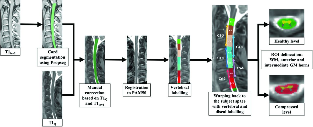
    Postprocessing of the T1 data from cord segmentation to WM/GM ROI analysis, per vertebral and disc levels. A T1Q threshold of 2 seconds was used to check the cord segmentation. Segmentation was performed using the PAM50 atlas.34,35 Quantifications were performed in the subject space.

  • FIG 3.
    • Download figure
    • Open in new tab
    • Download powerpoint
    FIG 3.

    Boxplots of cervical spinal cord T1 values (in milliseconds) in noncompressed disc levels for the 10 healthy controls (HC-0 grade 0; mean T1, 920 [SD, 27] ms) compared with noncompressed (P-0 grade 0; mean T1, 958 [SD, 39] ms), mildly (P-1 grade I; mean T1, 966 [SD, 51] ms), moderately (P-2 grade II; mean T1, 959 [SD, 40] ms), and severely (P-3 grade III; mean T1, 995 [SD, 60]  ms) compressed disc levels for the 20 patients. C1 and C2–C3 to C6–C7 disc levels were considered for each subject (6 levels in total). Grade 0 in patients and healthy controls were considered separately because they present statistical different values. The horizontal blue lines represent the mean value in each group. The horizontal lines within and at the ends of the boxes represent the median value and the first and third quartiles, respectively. The whiskers illustrate the minimum and maximum values. The horizontal green lines demonstrate statistically significant differences between the stenosis grades.

  • FIG 4.
    • Download figure
    • Open in new tab
    • Download powerpoint
    FIG 4.

    Distribution of the cervical spinal cord T1 values (milliseconds) according to the vertebral levels in the healthy subjects (green, no statistical difference between the different cervical levels) and patients with mild (orange) and moderate-to-severe (red) DCM. The horizontal bars represent the SD for each level.

  • FIG 5.
    • Download figure
    • Open in new tab
    • Download powerpoint
    FIG 5.

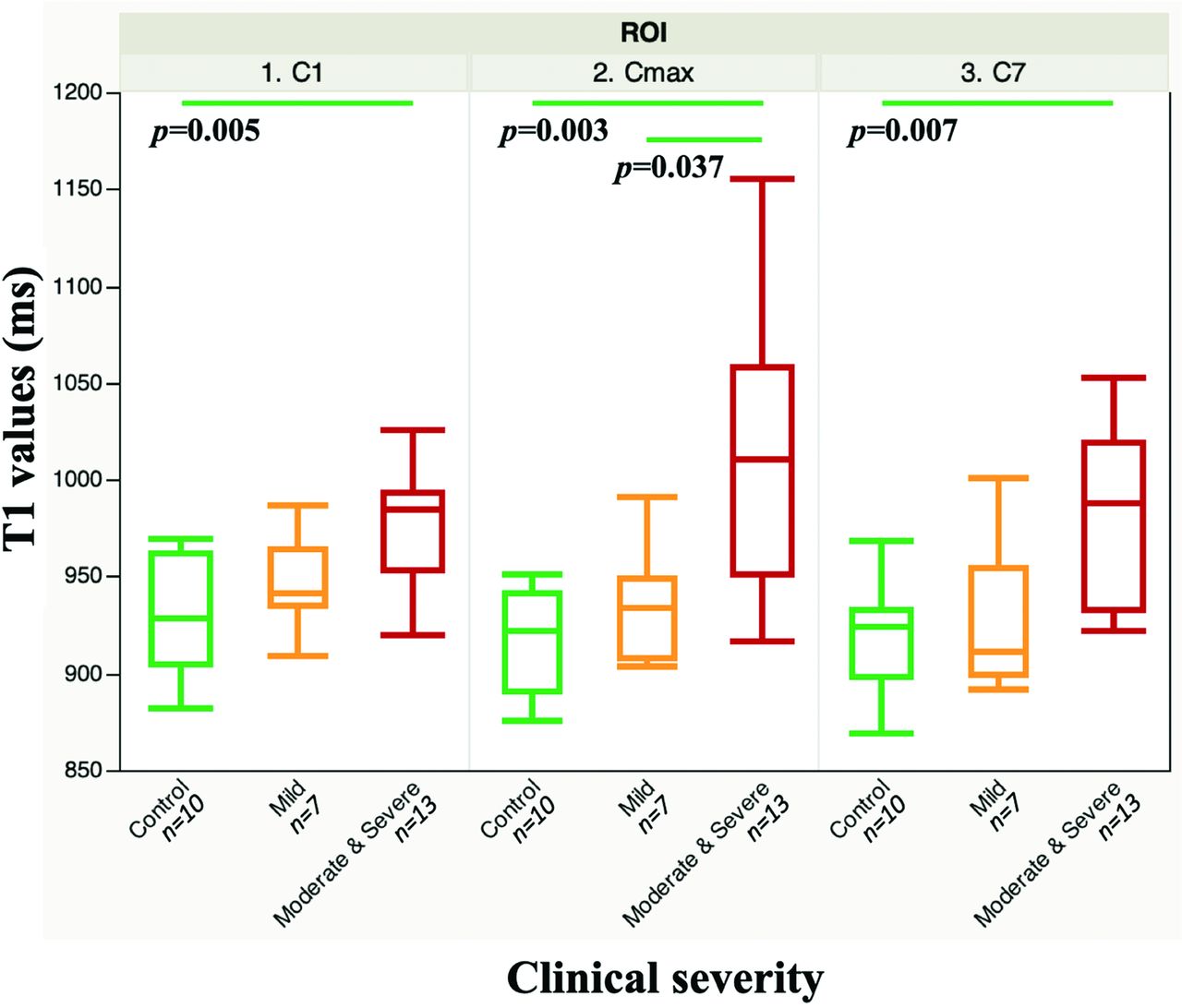
    Boxplots of the T1 values at C1, Cmax, and C7 in the control (green), mild (orange), and moderate-to-severe (red) DCM groups. Horizontal green lines represent statistically significant differences among the clinical groups (P < .05).

  • FIG 6.
    • Download figure
    • Open in new tab
    • Download powerpoint
    FIG 6.

    Distribution of the T1 values (milliseconds) for each clinical group according to the vertebral levels, in the entire spinal cord (blue), white matter (green), and anterior gray matter (yellow). Vertical bars represent the SD for each level.

Tables

  • Figures
  • Summary of the global observations and statistical analyses according to radiologic/clinical settings and regionalized T1-mapping

    Radiologic and Clinical SettingsRegionalized T1 MappingGlobal ObservationsStatistical Significance
    Clinical onsetSC at C1, Cmax, C7T1 values acute > chronicNo
    Cervical spinal canal stenosisSC at disc levelsT1 values increase according to the severity of the stenosis (T1grade0 < T1grade1 < T1grade3)Grades 3/0; 3/1 (considering all subjects)Grades 3/0_HC, 2/0_HC, 1/0_HC, 0/0_HC (differentiating patients and HC grade 0)
    OSSEntire cervical SCLower T1 values in the mild-stenosis groupMild-vs-moderate OSS
    Clinical severityEntire cervical SCT1 values increase according to the clinical severity(T1control < T1mild < T1moderate-to-severe)HC and patients with moderate-to-severe DCM
    Inverse relationship between global T1 values and preoperative mJOA scoresYes
    SC at C1, Cmax, C7T1 values increase at C1, Cmax, C7 according to the clinical severityHC and patients with moderate-to-severe DCM (C1, Cmax, and C7)Patients with mild and moderate-to-severe DCM (Cmax)No statistical difference between controls and patients with mild DCM
    WM, anterior and intermediate GM at C1, Cmax, C7Global T1-value increase at C1, Cmax, C7 in the WM and GM according to the clinical severityHC and patients with moderate-to-severe DCM (Cmax and C7 in WM and Cmax in GM)Patients with mild and moderate-to-severe DCM (C1 and Cmax in WM, Cmax in GM)No statistical difference between controls and patients with mild DCM
PreviousNext
Back to top

In this issue

American Journal of Neuroradiology: 42 (7)
American Journal of Neuroradiology
Vol. 42, Issue 7
1 Jul 2021
  • Table of Contents
  • Index by author
  • Complete Issue (PDF)
Advertisement
Print
Download PDF
Email Article

Thank you for your interest in spreading the word on American Journal of Neuroradiology.

NOTE: We only request your email address so that the person you are recommending the page to knows that you wanted them to see it, and that it is not junk mail. We do not capture any email address.

Enter multiple addresses on separate lines or separate them with commas.
T1 Mapping for Microstructural Assessment of the Cervical Spinal Cord in the Evaluation of Patients with Degenerative Cervical Myelopathy
(Your Name) has sent you a message from American Journal of Neuroradiology
(Your Name) thought you would like to see the American Journal of Neuroradiology web site.
CAPTCHA
This question is for testing whether or not you are a human visitor and to prevent automated spam submissions.
Cite this article
G. Baucher, H. Rasoanandrianina, S. Levy, L. Pini, L. Troude, P.-H. Roche, V. Callot
T1 Mapping for Microstructural Assessment of the Cervical Spinal Cord in the Evaluation of Patients with Degenerative Cervical Myelopathy
American Journal of Neuroradiology Jul 2021, 42 (7) 1348-1357; DOI: 10.3174/ajnr.A7157

Citation Manager Formats

  • BibTeX
  • Bookends
  • EasyBib
  • EndNote (tagged)
  • EndNote 8 (xml)
  • Medlars
  • Mendeley
  • Papers
  • RefWorks Tagged
  • Ref Manager
  • RIS
  • Zotero
0 Responses
Respond to this article
Share
Bookmark this article
T1 Mapping for Microstructural Assessment of the Cervical Spinal Cord in the Evaluation of Patients with Degenerative Cervical Myelopathy
G. Baucher, H. Rasoanandrianina, S. Levy, L. Pini, L. Troude, P.-H. Roche, V. Callot
American Journal of Neuroradiology Jul 2021, 42 (7) 1348-1357; DOI: 10.3174/ajnr.A7157
del.icio.us logo Twitter logo Facebook logo Mendeley logo
  • Tweet Widget
  • Facebook Like
  • Google Plus One
Purchase

Jump to section

  • Article
    • Abstract
    • ABBREVIATIONS:
    • MATERIALS AND METHODS
    • RESULTS
    • DISCUSSION
    • CONCLUSIONS
    • Acknowledgments
    • Footnotes
    • References
  • Figures & Data
  • Supplemental
  • Info & Metrics
  • Responses
  • References
  • PDF

Related Articles

  • PubMed
  • Google Scholar

Cited By...

  • No citing articles found.
  • Crossref
  • Google Scholar

This article has not yet been cited by articles in journals that are participating in Crossref Cited-by Linking.

More in this TOC Section

Spine Imaging and Spine Image-Guided Interventions

  • Optimization of Photon Counting CT Myelography
  • Characteristics of SIH Type I Culprit Lesions
  • Management Outcomes For VO Spine Biopsy
Show more Spine Imaging and Spine Image-Guided Interventions

Functional

  • Kurtosis and Epileptogenic Tubers: A Pilot Study
  • Glutaric Aciduria Type 1: DK vs. Conventional MRI
  • Multiparametric MRI in PEDS Pontine Glioma
Show more Functional

Similar Articles

Advertisement

Indexed Content

  • Current Issue
  • Accepted Manuscripts
  • Article Preview
  • Past Issues
  • Editorials
  • Editor's Choice
  • Fellows' Journal Club
  • Letters to the Editor
  • Video Articles

Cases

  • Case Collection
  • Archive - Case of the Week
  • Archive - Case of the Month
  • Archive - Classic Case

More from AJNR

  • Trainee Corner
  • Imaging Protocols
  • MRI Safety Corner
  • Book Reviews

Multimedia

  • AJNR Podcasts
  • AJNR Scantastics

Resources

  • Turnaround Time
  • Submit a Manuscript
  • Submit a Video Article
  • Submit an eLetter to the Editor/Response
  • Manuscript Submission Guidelines
  • Statistical Tips
  • Fast Publishing of Accepted Manuscripts
  • Graphical Abstract Preparation
  • Imaging Protocol Submission
  • Evidence-Based Medicine Level Guide
  • Publishing Checklists
  • Author Policies
  • Become a Reviewer/Academy of Reviewers
  • News and Updates

About Us

  • About AJNR
  • Editorial Board
  • Editorial Board Alumni
  • Alerts
  • Permissions
  • Not an AJNR Subscriber? Join Now
  • Advertise with Us
  • Librarian Resources
  • Feedback
  • Terms and Conditions
  • AJNR Editorial Board Alumni

American Society of Neuroradiology

  • Not an ASNR Member? Join Now

© 2025 by the American Society of Neuroradiology All rights, including for text and data mining, AI training, and similar technologies, are reserved.
Print ISSN: 0195-6108 Online ISSN: 1936-959X

Powered by HighWire