
Article Figures & Data
Figures
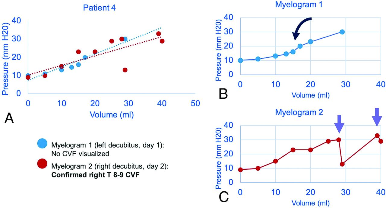
- FIG 1.
Sample craniospinal compliance curves in a 51-year-old man confirmed to have CVF at operation. Combined plot of 2 CTMs with dashed lines showing a linear approximation of compliance (A). Initial nondiagnostic CTM (B) shows a roughly sigmoid pattern with an inflection point seen after a ∼15-mL normal saline bolus was administered (black curved arrow). Repeat CTM with more aggressive positive-pressure augmentation shows an abrupt loss of pressure (purple arrows) after infusion volume of 27 mL of normal saline (C). The CVF became apparent only after pressure was increased beyond this threshold point.
- FIG 2.
A craniospinal compliance curve in a 70-year-old woman with confirmed CVF (A) shows high compliance (ie, smaller change in pressure per increase in volume) with multiple abrupt pressure drops (arrows) above 10 cm H20. Recent brain MR imaging shows characteristic features of SIH (B), including reduced mamillopontine distance, low cerebellar tonsils, and pachymeningeal enhancement. A right T7–8 CVF was identified on decubitus CT myelography (C and D). Hyperdensity of the paraspinal vein and azygous vein (dashed circle) aids in identification of the fistula (white arrows). The patient was treated with percutaneous fibrin glue injection.
- FIG 3.
Craniospinal compliance curve in a 63-year-old woman suspected but not radiographically confirmed to have CVF. Estimated linear compliance for each CTM is shown as a dashed line, approximating the shape of the pressure-volume curve (A). The second CTM was performed after the blood patch showed an increase in opening pressure (B and C). The effect of the blood patch is diminished on delayed repeat CTM (D), in which the left side of the curve, including opening pressure, more closely matches the prepatch curve.
- FIG 4.
Schematic of the components of craniospinal compliance and hypothesized physiology of CVF (CSF = green, arterial blood = red, venous blood = blue). A normal CSC encompasses both intraventricular and subarachnoid CSF and is defined by cranial and spinal compartments (larger and smaller boxes, respectively) as well as the arteriovenous vascular bed (A). In dural tear CSF leak or CVF at low pressure, an equilibrium state (B) may exist in which the leak is occult by CTM. With special maneuvers (dynamic CTM, respiratory-phase variation, jugular pressure, and bolus-pressure augmentation), pressure gradients may open the leak, allowing detection on CTM (C).


Tables
- Table 1:
Demographic and CSF pressure-volume parameters for patients with clinical and imaging features of SIH, all of whom were suspected of having CVFa
Total (n = 22) Definite or Probable CVF (n = 8) No CVF Identified (n = 14) P Value Age (yr) 57.3 60.0 55.8 .5 Sex 59.1% F 37.5% F 71.4% F .12 Increase in relative pressure during normal saline infusion (cm H20) 146.7% (SD, 17.3%) 148.5% (SD, 17.9%) 145.4% (SD, 27.0%) .93 Total volume infusion (mL) 20.7 (SD, 1.8) 27.6 (SD, 2.9) 15.9 (SD, 4.1) <.001 CSC curves with abrupt pressure loss 11/34 (32.4%) 4/14 (28.6%) 7/20 (35.0%) .69 Opening pressure (cm H20) 11.6 (SD, 3.8) 9.8 (SD, 0.9) 12.8 (SD, 0.8) .02 PVIb (mL H20) 63.9 (SD, 7.5) 77.5 (SD, 10.3) 54.3 (SD, 10.1) .13 Copc (mL/cm H20) 2.6 (SD, 0.3) mL/cm H20 3.8 (SD, 0.5) mL/cm H20 1.8 (SD, 0.3) mL/cm H20 <.001 Overall compliance (linear slope of CSC curve) (mL/cm H20) 2.0 (SD, 0.3) 3.1 (SD, 0.6) 1.3 (SD, 0.3) .005


