
Article Figures & Data
Figures
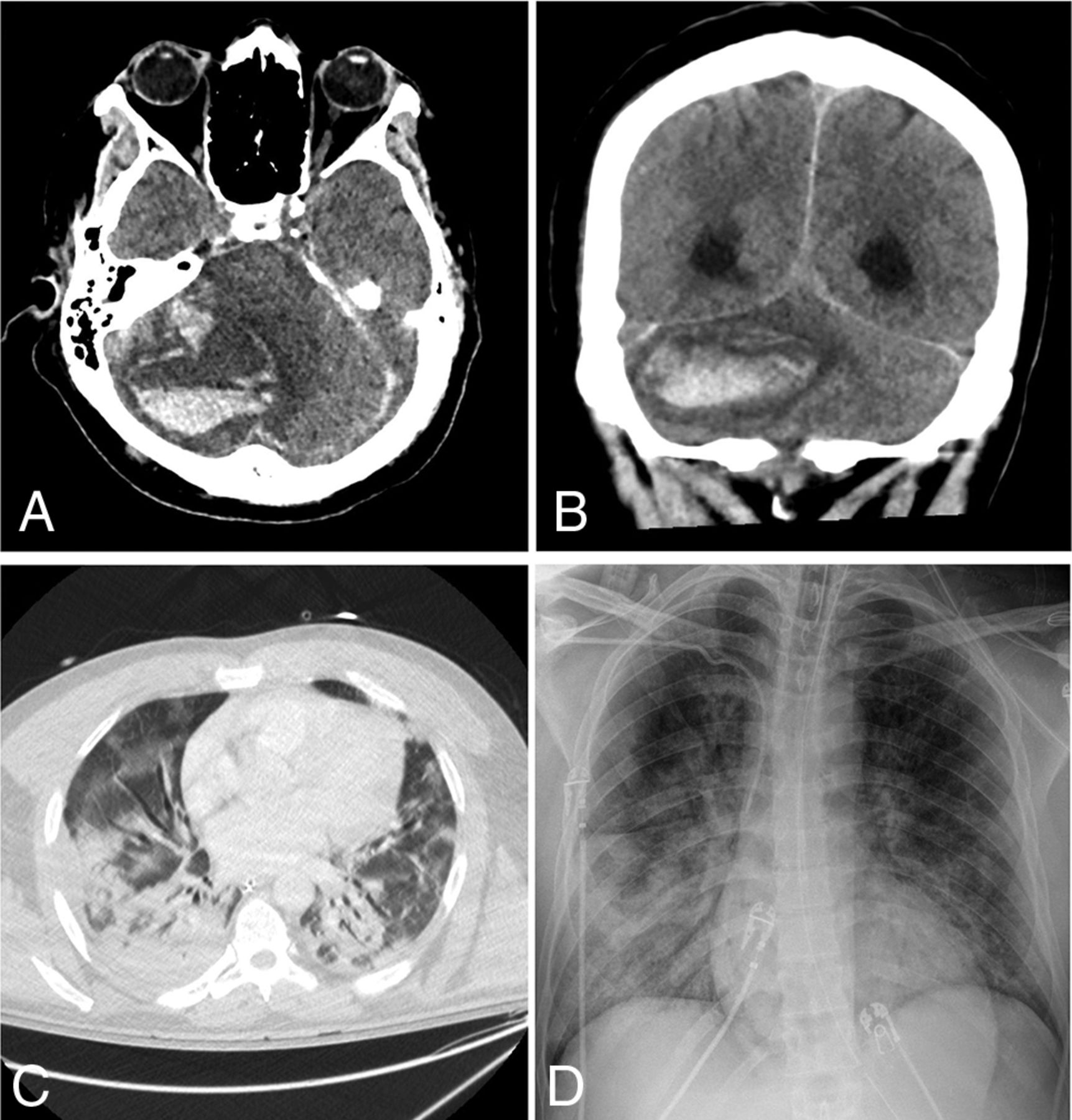
- FIG 1.
A 42-year-old man presented with hypoxemic respiratory failure. Non-contrast-enhanced axial (A) and coronal (B) CT head images demonstrate a new, large, right cerebellar intraparenchymal mixed-attenuation hematoma suggestive of hyperacute or active hemorrhage. C, Chest CT of the patient demonstrates bilateral consolidation and ground-glass opacities involving all lobes, with a CCS score of 18. D, Portable chest radiograph demonstrates bilateral patchy air space opacities with a PXS score of 10.3.
- FIG 2.
An 85-year-old man presented with increasing shortness of breath. Non-contrast-enhanced axial CT of the head demonstrates multiple new foci of hypoattenuation within the bilateral frontal corona radiata (arrows on A) and left centrum semiovale (arrow on B), in keeping with acute infarcts. C, Chest CT of the patient shows diffuse bilateral, right greater-than-left, predominantly consolidation and ground-glass opacities as well as bilateral pleural effusions. The CCS score was calculated as 13. D, Portable chest radiograph demonstrates bilateral patchy opacities with a PXS score of 10.6.
- FIG 3.
A 64-year-old man presented with fever and shortness of breath. Axial FLAIR (A) and diffusion-weighted (B) images demonstrate extensive symmetric confluent T2/FLAIR signal abnormality and restricted diffusion involving the corona radiata bilaterally. C, Chest CT shows bilateral peripheral ground-glass opacities and a small amount of consolidation involving all lobes, with a CCS score of 15. D, Portable chest radiograph demonstrates bilateral lower-zone-predominant peripheral opacities with a PXS score of 8.8.
- FIG 4.
A 67-year-old woman presented with hypoxemic respiratory failure. Axial non-contrast-enhanced head CT image (A) shows no acute findings. B, Chest CT image of the patient demonstrates bilateral peripherally distributed ground-glass opacities involving 4 of the 5 lobes with a CCS score of 8. D, Portable chest radiograph demonstrates bibasilar patchy opacities with a PXS score of 4.4.
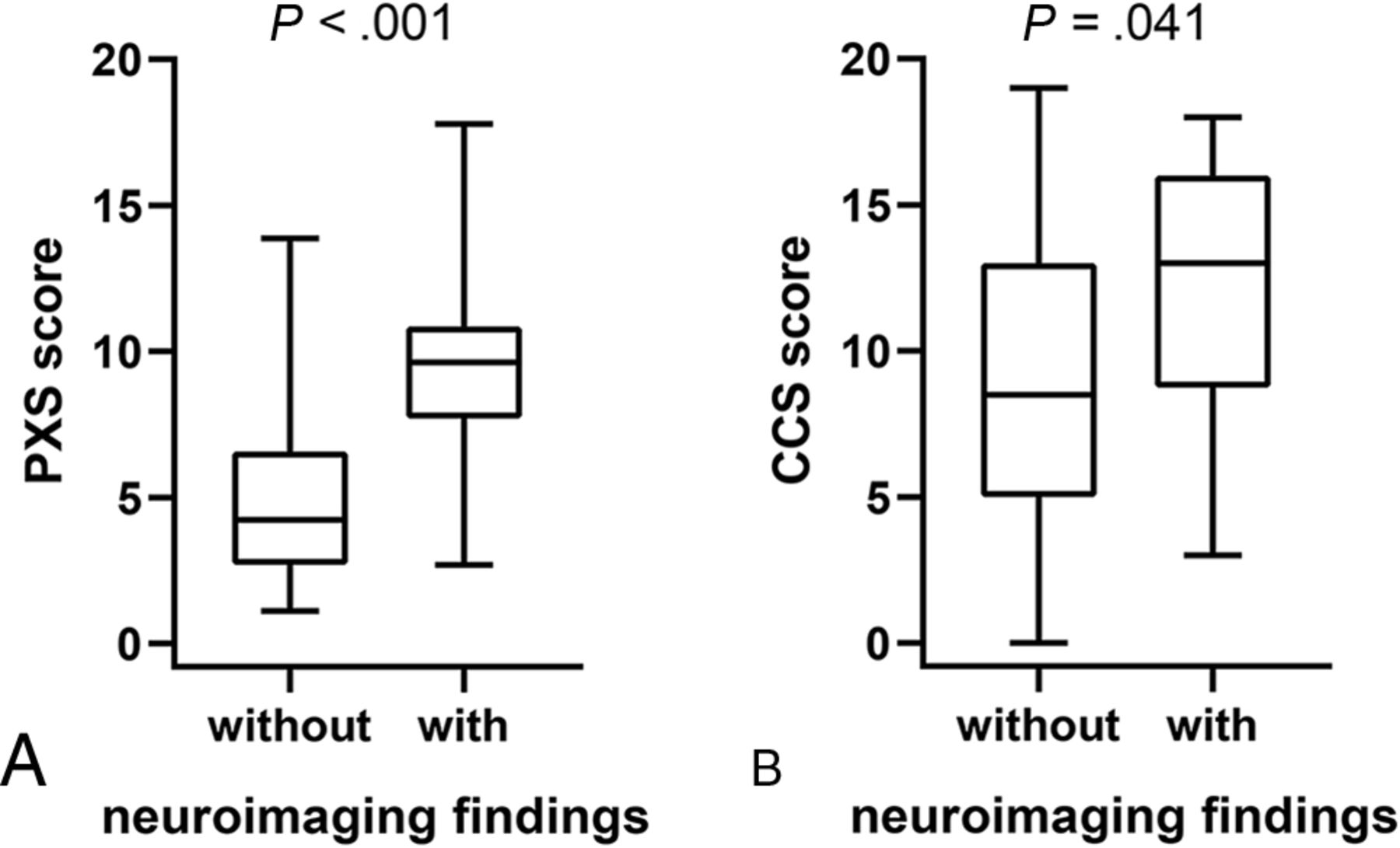
- FIG 5.
A, Comparison of portable chest radiograph severity (PXS) score between patients with and without acute neuroimaging findings. B, Comparison of the CCS score between patients with and without acute neuroimaging findings.
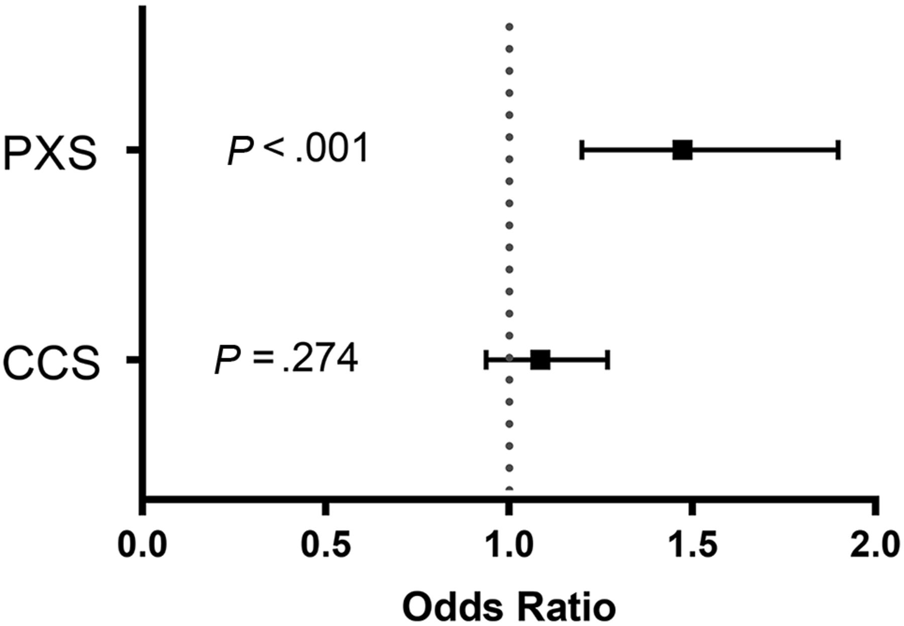
- FIG 6.
Odds ratio of the PXS score and the CCS score in association with the risk of acute neuroimaging findings. A multivariate logistic regression model was used after adjusting for age, sex, the need for intubation, and ICU admission status.
- FIG 7.
ROC curves of the PXS and CCS scores to classify acute neuroimaging findings in patients with COVID-19. A, The area under the ROC curve using the PXS score to classify acute neuroimaging findings was 0.83 (95% CI, 0.72–0.93). The optimal threshold PXS score was 7.55, which corresponded to a sensitivity of 83%, specificity of 81%, positive likelihood ratio of 4.7, and negative likelihood ratio of 0.2. B, The area under the ROC curve using the CCS score to classify acute neuroimaging findings was 0.67 (95% CI, 0.50–0.84). The optimal threshold CCS score was 12.5, which corresponded to a sensitivity of 64%, specificity of 73%, positive likelihood ratio of 2.4, and negative likelihood ratio of 0.5.


Tables
- Table 1:
Summary of patient characteristics, clinical data, and indication for neuroimaging
Demographics With Neuroimaging Findings (n = 24) Without Neuroimaging Findings (n = 69) P Value Age (mean) [SD] (yr) 63 [SD, 16] 66 [SD, 16] .396 Female sex (No.) (%) 8 (33) 23 (33) 1.000 Clinical data Length of stay (mean) [SD] (day) 31 [SD, 12] 23 [SD, 15] .021 ICU admission (No.) (%) 22 (92) 46 (67) .035 Intubation (No.) (%) 21 (88) 36 (52) .005 Death (No.) (%) 5 (21) 12 (17) .945 Indication for neuroimaging .089 Altered mental status 13 (54) 42 (61) Concern for stroke 10 (42) 14 (20) Trauma 0 (0) 10 (15) Seizure 1 (4) 1 (3) Headache 0 (0) 2 (1) With Neuroimaging Findings (n = 24) Without Neuroimaging Findings Acute neuroimaging findings Infarction 7 (29) NA Hemorrhage 7 (29) NA Leukoencephalopathy 6 (25) NA Combination 4 (17) NA Chest CT RSNA category n = 14 n = 66 Typical 9 (64) 34 (52) Indeterminate 1 (7) 18 (27) Atypical 4 (29) 10 (15) Negative 0 (0) 4 (6) Note:—NA indicates not applicable; RSNA, Radiological Society of North America.


Jump to section
Related Articles
Cited By...
- No citing articles found.