Advertisement

Main menu

  • Home
  • Content
    • Current Issue
    • Accepted Manuscripts
    • Article Preview
    • Past Issue Archive
    • Video Articles
    • AJNR Case Collection
    • Case of the Week Archive
    • Case of the Month Archive
    • Classic Case Archive
  • Special Collections
    • AJNR Awards
    • Low-Field MRI
    • Alzheimer Disease
    • ASNR Foundation Special Collection
    • Photon-Counting CT
    • View All
  • Multimedia
    • AJNR Podcasts
    • AJNR SCANtastic
    • Trainee Corner
    • MRI Safety Corner
    • Imaging Protocols
  • For Authors
    • Submit a Manuscript
    • Submit a Video Article
    • Submit an eLetter to the Editor/Response
    • Manuscript Submission Guidelines
    • Statistical Tips
    • Fast Publishing of Accepted Manuscripts
    • Graphical Abstract Preparation
    • Imaging Protocol Submission
    • Author Policies
  • About Us
    • About AJNR
    • Editorial Board
    • Editorial Board Alumni
  • More
    • Become a Reviewer/Academy of Reviewers
    • Subscribers
    • Permissions
    • Alerts
    • Feedback
    • Advertisers
    • ASNR Home

User menu

  • Alerts
  • Log in

Search

  • Advanced search
American Journal of Neuroradiology

American Journal of Neuroradiology

ASHNR American Society of Functional Neuroradiology ASHNR American Society of Pediatric Neuroradiology ASSR
  • Alerts
  • Log in

Advanced Search

  • Home
  • Content
    • Current Issue
    • Accepted Manuscripts
    • Article Preview
    • Past Issue Archive
    • Video Articles
    • AJNR Case Collection
    • Case of the Week Archive
    • Case of the Month Archive
    • Classic Case Archive
  • Special Collections
    • AJNR Awards
    • Low-Field MRI
    • Alzheimer Disease
    • ASNR Foundation Special Collection
    • Photon-Counting CT
    • View All
  • Multimedia
    • AJNR Podcasts
    • AJNR SCANtastic
    • Trainee Corner
    • MRI Safety Corner
    • Imaging Protocols
  • For Authors
    • Submit a Manuscript
    • Submit a Video Article
    • Submit an eLetter to the Editor/Response
    • Manuscript Submission Guidelines
    • Statistical Tips
    • Fast Publishing of Accepted Manuscripts
    • Graphical Abstract Preparation
    • Imaging Protocol Submission
    • Author Policies
  • About Us
    • About AJNR
    • Editorial Board
    • Editorial Board Alumni
  • More
    • Become a Reviewer/Academy of Reviewers
    • Subscribers
    • Permissions
    • Alerts
    • Feedback
    • Advertisers
    • ASNR Home
  • Follow AJNR on Twitter
  • Visit AJNR on Facebook
  • Follow AJNR on Instagram
  • Join AJNR on LinkedIn
  • RSS Feeds

AJNR Awards, New Junior Editors, and more. Read the latest AJNR updates

LetterLetter
Open Access

MR Imaging of the Olfactory Bulbs in Patients with COVID-19 and Anosmia: How to Avoid Misinterpretation

N. Shor, L. Chougar and N. Pyatigorskaya
American Journal of Neuroradiology March 2021, 42 (3) E10-E11; DOI: https://doi.org/10.3174/ajnr.A6921
N. Shor
aService de NeuroradiologieAssistance Publique Hôpitaux de Paris, Hôpital Pitié-SalpêtrièreParis, France
  • Find this author on Google Scholar
  • Find this author on PubMed
  • Search for this author on this site
  • ORCID record for N. Shor
L. Chougar
bService de NeuroradiologieAssistance Publique Hôpitaux de Paris, Hôpital Pitié-SalpêtrièreParis, France
cParis Brain InstituteSorbonne UniversitéParis, France
d“Movement Investigations and Therapeutics” Team
eCentre de NeuroImagerie de RechercheInstitut du CerveauParis, France
  • Find this author on Google Scholar
  • Find this author on PubMed
  • Search for this author on this site
  • ORCID record for L. Chougar
N. Pyatigorskaya
bService de NeuroradiologieAssistance Publique Hôpitaux de Paris, Hôpital Pitié-SalpêtrièreParis, France
cParis Brain InstituteSorbonne UniversitéParis, France
d“Movement Investigations and Therapeutics” Team
eCentre de NeuroImagerie de RechercheInstitut du CerveauParis, France
  • Find this author on Google Scholar
  • Find this author on PubMed
  • Search for this author on this site
  • ORCID record for N. Pyatigorskaya
  • Article
  • Figures & Data
  • Info & Metrics
  • Responses
  • References
  • PDF
Loading

We recently read the article entitled “Brain Imaging of Patients with COVID-19: Findings at an Academic Institution during the Height of the Outbreak in New York City,” a retrospective neuroimaging cohort by Lin et al.1 The authors reported T2-FLAIR postgadolinium olfactory bulb (OB) signal abnormalities in 4 patients positive for coronavirus disease 2019 (COVID-19) with only 1 having documented anosmia. This finding was subsequently interpreted as olfactive neuritis and a correlate of the anosmia.

Anosmia has been identified as one of the first or only recognizable symptoms of the Severe Acute Respiratory Syndrome coronavirus 2 (SARS-CoV-2) infection, accounting for >50% of Western patients.2 It is now known that post-SARS-CoV-1 anosmia could persist for as long as 2 years. It, thus, becomes relevant to identify MR imaging biomarkers of OB involvement, including signal and volume changes, that might be predictive of the olfactory disorder outcome. We, thus, find it important to draw the attention to the OB signal and volume analysis.

The OB signal intensity can vary according to the field strength applied, the MR imaging manufacturer, and the acquisition parameters of T2-FLAIR sequences. Furthermore, it has previously been reported that OBs could appear hyperintense on T2-FLAIR in healthy subjects.3 Lin et al1 reported that they recruited patients who had undergone brain MR imaging from 3 different machines (1.5T and 3T), increasing the risk of signal variation in tiny structures and thus making the results more questionable.

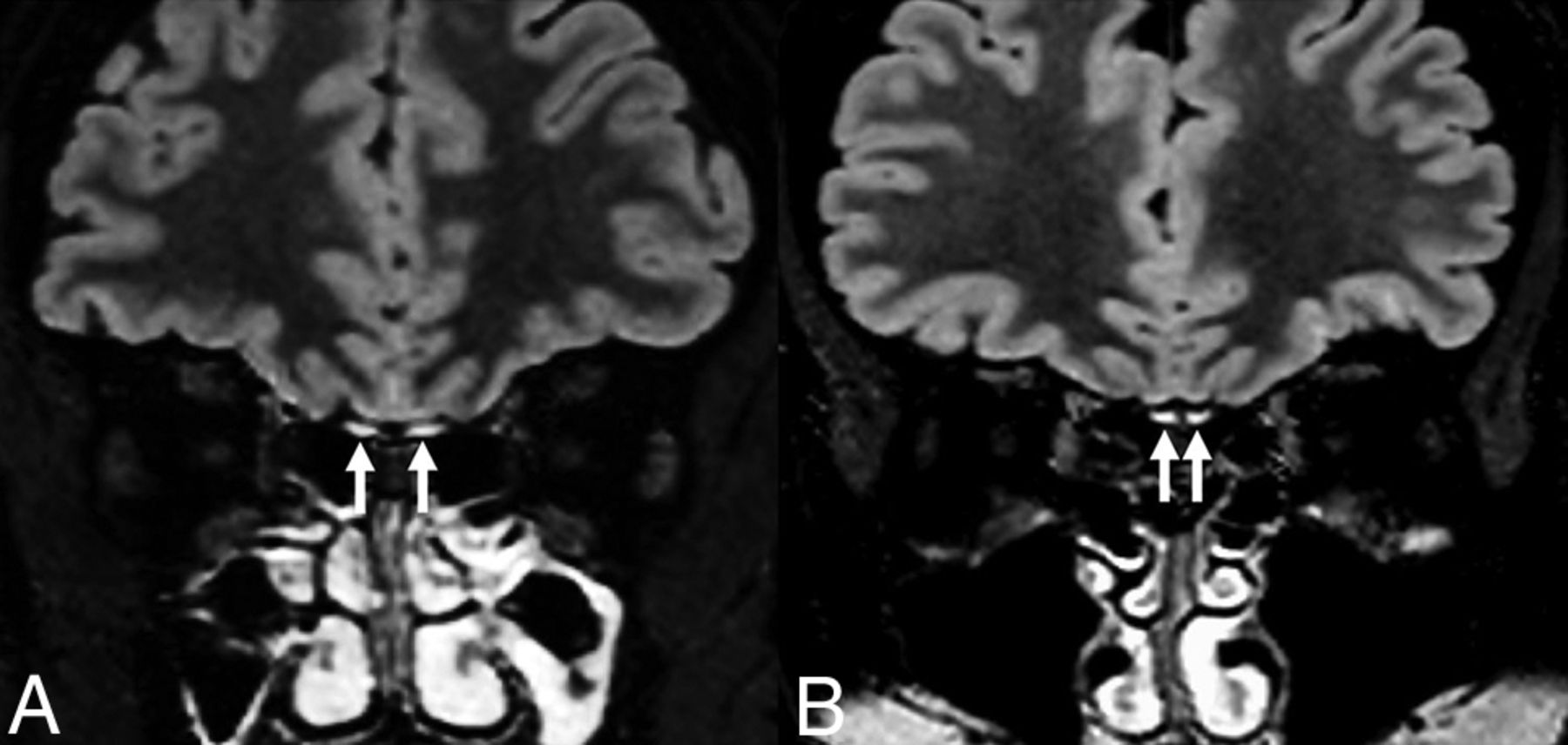
Age- and sex-matched healthy controls scanned on the same MR imaging scanner need to be analyzed to overcome these technical issues and avoid an incorrect OB edema/gadolinium-enhancement description. In our institution, a blind, independent comparison by 2 experienced neuroradiologists of 10 patients with COVID-19–associated anosmia and 10 age- and sex-matched subjects negative for COVID-19 without olfactory dysfunction was performed. It showed that visual analysis of OB high-resolution T2-FLAIR signal could not distinguish the 2 groups because all the subjects presented with the same T2-FLAIR high signal intensity (Fig 1). Thus, the OBs described by Lin et al1 as being abnormally “hyperintense” can probably correspond to a normal signal intensity.

FIG 1.
  • Download figure
  • Open in new tab
  • Download powerpoint
FIG 1.

3T brain MR imaging in a healthy subject (A) and a patient with COVID-19–related anosmia (B). Coronal reformatted 3D FLAIR images show OB hyperintensity (arrows) compared with the cortex in both subjects.

It has been shown that the OB volume decreased in postinfectious anosmia.4 Because the OBs are tiny structures surrounded by CSF, sequences with high resolution such as CISS should be used for volume segmentation. As for the signal on an individual scale, the signal intensity evolution of the OB could be compared with the surrounding structures, such as the cortex or the optic nerves. Subjective appreciation, especially for such a small structure, could lead to misinterpretation.

In summary, OB imaging is challenging, and one should be careful while interpreting its signal and volume, especially in the context of COVID-19.

Footnotes

  • Disclosures: Nadya Pyatigorskaya—UNRELATED: Employment: Assistance publique–Hôpitaux de Paris; Grants/Grants Pending: CRC, Progressive Supranuclear Palsy (PSP)-France*; Payment for Lectures Including Service on Speakers Bureaus: GE Healthcare, Biogen. *Money paid to the institution.

Indicates open access to non-subscribers at www.ajnr.org

References

  1. 1.↵
    1. Lin E,
    2. Lantos JE,
    3. Strauss SB, et al
    . Brain imaging of patients with COVID-19: findings at an academic institution during the height of the outbreak in New York City. AJNR Am J Neuroradiol 2020 Aug 20. [Epub ahead of print] doi:10.3174/ajnr.A6793 pmid:32819899
    Abstract/FREE Full Text
  2. 2.↵
    1. Lechien JR,
    2. Chiesa-Estomba CM,
    3. Hans S, et al
    . Loss of smell and taste in 2013 European patients with mild to moderate COVID-19. Ann Intern Med 2020 May 26. [Epub ahead of print] doi:10.7326/M20-2428 pmid:32449883
    CrossRefPubMed
  3. 3.↵
    1. Chung MS,
    2. Choi WR,
    3. Jeong HY, et al
    . MR imaging–based evaluations of olfactory bulb atrophy in patients with olfactory dysfunction. AJNR Am J Neuroradiol 2018;39:532–37 doi:10.3174/ajnr.A5491 pmid:29269404
    Abstract/FREE Full Text
  4. 4.↵
    1. Yao L,
    2. Yi X,
    3. Pinto JM, et al
    . Olfactory cortex and olfactory bulb volume alterations in patients with post-infectious olfactory loss. Brain Imaging Behav 2018;12:1355–62 doi:10.1007/s11682-017-9807-7 pmid:29234959
    CrossRefPubMed
  • © 2021 by American Journal of Neuroradiology
PreviousNext
Back to top

In this issue

American Journal of Neuroradiology: 42 (3)
American Journal of Neuroradiology
Vol. 42, Issue 3
1 Mar 2021
  • Table of Contents
  • Index by author
  • Complete Issue (PDF)
Advertisement
Print
Download PDF
Email Article

Thank you for your interest in spreading the word on American Journal of Neuroradiology.

NOTE: We only request your email address so that the person you are recommending the page to knows that you wanted them to see it, and that it is not junk mail. We do not capture any email address.

Enter multiple addresses on separate lines or separate them with commas.
MR Imaging of the Olfactory Bulbs in Patients with COVID-19 and Anosmia: How to Avoid Misinterpretation
(Your Name) has sent you a message from American Journal of Neuroradiology
(Your Name) thought you would like to see the American Journal of Neuroradiology web site.
CAPTCHA
This question is for testing whether or not you are a human visitor and to prevent automated spam submissions.
Cite this article
N. Shor, L. Chougar, N. Pyatigorskaya
MR Imaging of the Olfactory Bulbs in Patients with COVID-19 and Anosmia: How to Avoid Misinterpretation
American Journal of Neuroradiology Mar 2021, 42 (3) E10-E11; DOI: 10.3174/ajnr.A6921

Citation Manager Formats

  • BibTeX
  • Bookends
  • EasyBib
  • EndNote (tagged)
  • EndNote 8 (xml)
  • Medlars
  • Mendeley
  • Papers
  • RefWorks Tagged
  • Ref Manager
  • RIS
  • Zotero
0 Responses
Respond to this article
Share
Bookmark this article
MR Imaging of the Olfactory Bulbs in Patients with COVID-19 and Anosmia: How to Avoid Misinterpretation
N. Shor, L. Chougar, N. Pyatigorskaya
American Journal of Neuroradiology Mar 2021, 42 (3) E10-E11; DOI: 10.3174/ajnr.A6921
del.icio.us logo Twitter logo Facebook logo Mendeley logo
  • Tweet Widget
  • Facebook Like
  • Google Plus One
Purchase

Jump to section

  • Article
    • Footnotes
    • References
  • Figures & Data
  • Info & Metrics
  • Responses
  • References
  • PDF

Related Articles

  • No related articles found.
  • PubMed
  • Google Scholar

Cited By...

  • Adapting the UK Biobank brain imaging protocol and analysis pipeline for the C-MORE multi-organ study of COVID-19 survivors
  • Crossref
  • Google Scholar

This article has not yet been cited by articles in journals that are participating in Crossref Cited-by Linking.

More in this TOC Section

  • Letter to the Editor regarding “Automated Volumetric Software in Dementia: Help or Hindrance to the Neuroradiologist?”
  • Reply:
  • Brain AVM’s Nidus: What if We Hadn’t Understood Anything?
Show more LETTERS

Similar Articles

Advertisement

Indexed Content

  • Current Issue
  • Accepted Manuscripts
  • Article Preview
  • Past Issues
  • Editorials
  • Editor's Choice
  • Fellows' Journal Club
  • Letters to the Editor
  • Video Articles

Cases

  • Case Collection
  • Archive - Case of the Week
  • Archive - Case of the Month
  • Archive - Classic Case

More from AJNR

  • Trainee Corner
  • Imaging Protocols
  • MRI Safety Corner
  • Book Reviews

Multimedia

  • AJNR Podcasts
  • AJNR Scantastics

Resources

  • Turnaround Time
  • Submit a Manuscript
  • Submit a Video Article
  • Submit an eLetter to the Editor/Response
  • Manuscript Submission Guidelines
  • Statistical Tips
  • Fast Publishing of Accepted Manuscripts
  • Graphical Abstract Preparation
  • Imaging Protocol Submission
  • Evidence-Based Medicine Level Guide
  • Publishing Checklists
  • Author Policies
  • Become a Reviewer/Academy of Reviewers
  • News and Updates

About Us

  • About AJNR
  • Editorial Board
  • Editorial Board Alumni
  • Alerts
  • Permissions
  • Not an AJNR Subscriber? Join Now
  • Advertise with Us
  • Librarian Resources
  • Feedback
  • Terms and Conditions
  • AJNR Editorial Board Alumni

American Society of Neuroradiology

  • Not an ASNR Member? Join Now

© 2025 by the American Society of Neuroradiology All rights, including for text and data mining, AI training, and similar technologies, are reserved.
Print ISSN: 0195-6108 Online ISSN: 1936-959X

Powered by HighWire