
Article Figures & Data
Figures
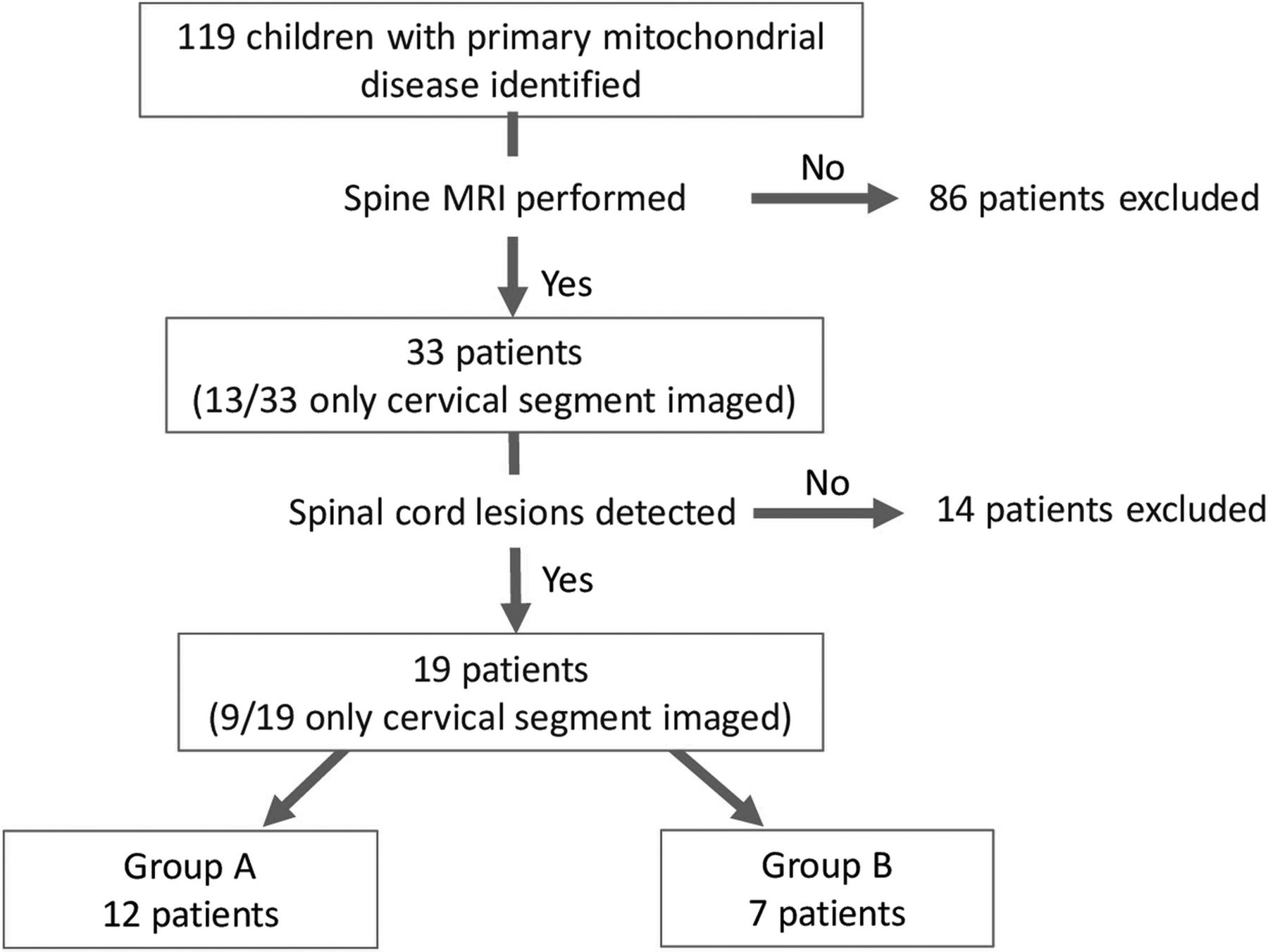
- FIG 1.
Flowchart of patient selection and inclusion criteria for our cohort.
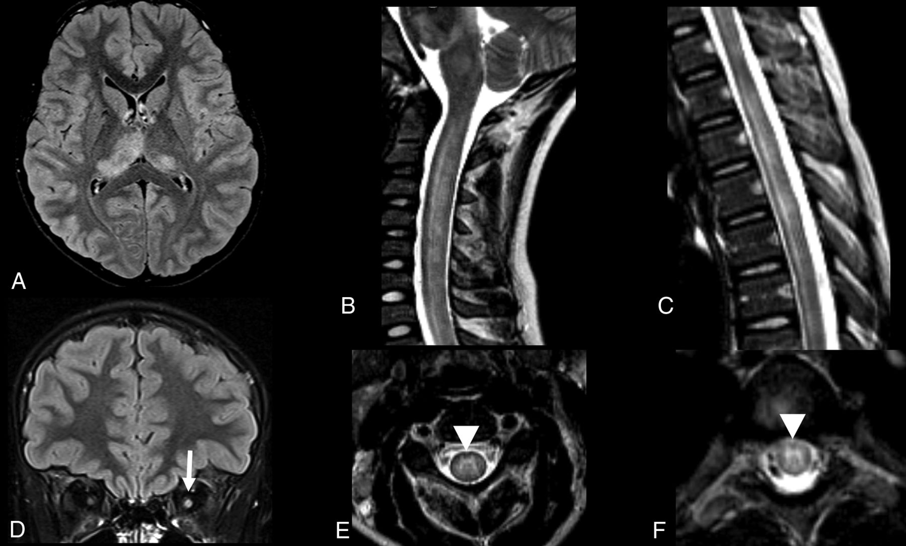
- FIG 2.
Segment distribution of the lesions and different appearances. Three patients with MR imaging spinal cord lesions in different segments of the spinal cord. Imaging in patient 1 (A and D) with an NDUFS1 pathogenic variant (c.365C>T:p.Pro122Leu and c.155 + 1G>A) shows a lesion located in the cervical segment (arrow, A), at the level of C1, with a posterior and central distribution on the axial view (D) and extension to the area postrema. Imaging in patient 2 (B and E) with MT-ND4 pathogenic variant (m.11777C>A) shows a spinal cord lesion with diffuse cross-sectional involvement (E), longitudinally extensive, and involving both cervical and thoracic levels, including the area postrema (arrow, B). Imaging in patient 3 (C and F) with MT-ND5 pathogenic variant (m.13513G>A) shows a thoracolumbar lesion, extending to the conus medullaris, with isolated gray matter involvement and snake eyes appearance.
- FIG 3.
Group A, NMOSD appearance. A 12-year-old male patient with MT-ND4 pathogenic variant (m.11777C>A) with a demyelination-like pattern of the spinal cord lesion and NMOSD-like appearance. Sagittal and axial T2 MR imaging of the cervical spine shows a longitudinal extensive hyperintense lesion in both cervical and thoracic segments with a tumefactive effect and involvement of the area postrema (arrow, A). The lesion has a diffuse cross-sectional involvement of the cord in the axial plane (B).
- FIG 4.
Group A, anti-MOG appearance. A 10-year-old female patient with a family diagnosis of LHON Leber’s hereditary optic neuropathy (heteroplasmic m.14484T>C and homoplasmic m.15256G>A mtDNA variants) and MOG+ demyelination. Brain MR imaging study axial and coronal FLAIR sequences show asymmetric hyperintense lesions in the thalami, more evident on the right side (A), and enlargement and hyperintensity of the intraorbital left optic nerve (arrow, D). Sagittal and axial spinal MR imaging of the cervical (B and E) and thoracic segments (C and F) show longitudinal extensive hyperintense lesions in both segments, with more evident involvement of the central gray matter, giving the “H sign” appearance on the axial planes (arrowheads, E and F).
- FIG 5.
Group A, LBSL appearance. MR imaging of 2 patients with leukoencephalopathy with brain stem and spinal cord involvement and increased lactate (LBSL) appearance. Patient 1 (A–C) is a 6-month-old male patient with an NDUFS1 pathogenic variant. Patient 2 (D–F) is a 1-year-old male patient with a DARS2 pathogenic variant. In both patients, axial T2 sequences show symmetric hyperintense lesions, affecting both corticospinal tracts, in the pons (dotted arrows, A and D). Hyperintense lesions are also observed along the intraparenchymal portions of the trigeminal nerves (arrows, A and D) and medial lemnisci (arrowheads, A and D). Lesions affecting the pyramids (arrowheads, B and E) and inferior cerebellar peduncles (arrows, B and E) are observed in the medulla.On axial T2-weighted cervical spinal MR imaging (C and F), hyperintense lesions are seen in the dorsal columns (arrowheads) and lateral corticospinal tracts (arrows).


