
Article Figures & Data
Figures
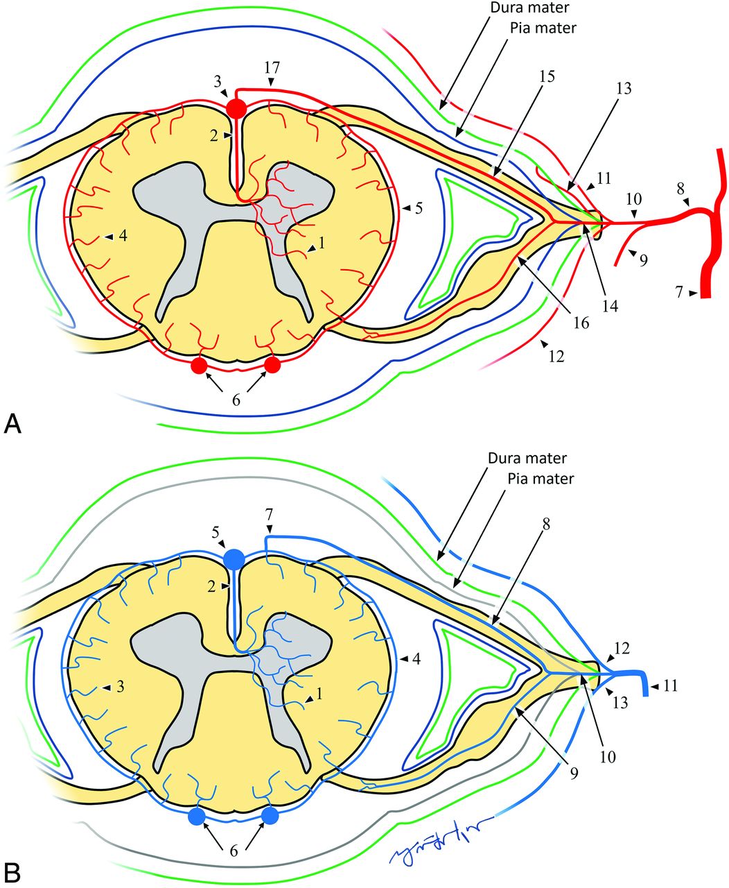
- FIG 1.
A, Normal arterial anatomy: 1, intramedullary branches; 2, sulcal artery; 3, anterior spinal artery; 4, radial perforators; 5, pial arterial plexus; 6, posterior spinal arteries; 7, segmental artery; 8, spinal artery; 9, dorsal branch of the dorsospinal artery; 10, ventral branch of dorsospinal artery; 11, ventral epidural plexus; 12, dorsal epidural plexus; 13, dural artery; 14, radicular artery; 15, ventral radicular artery; 16, dorsal radicular artery; 17, medullary artery. B, Normal venous anatomy: 1, Intramedullary veins; 2, sulcal vein; 3, radial veins; 4, pial venous plexus; 5, anterior spinal vein; 6, posterior spinal veins; 7, medullary vein; 8, ventral radicular vein; 9, dorsal radicular vein; 10, emissary vein; 11, intervertebral vein; 12, branches from ventral epidural venous plexus; 13, branches from dorsal epidural venous plexus.
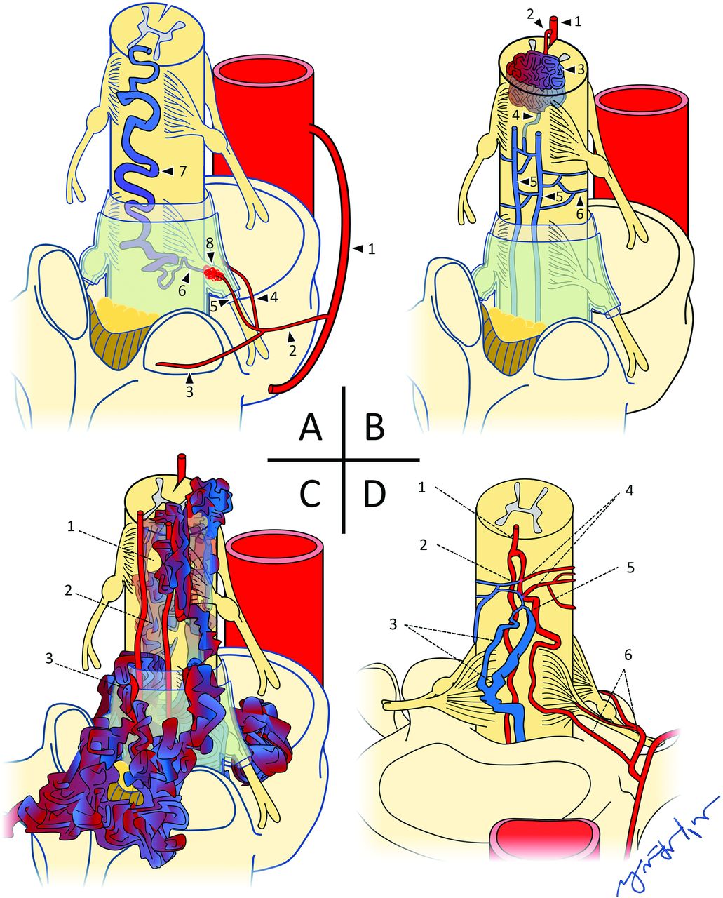
- FIG 2.
A, Type I spinal dural AVF: 1, Intercostal artery; 2, spinal artery; 3, dorsal/muscular branch; 4, radicular artery, ventral branch; 5, radicular artery, dorsal branch; 6, radicular vein; 7, engorged perimedullary vein; 8, dural AVF. B, Type II spinal glomus AVM: 1, anterior spinal artery; 2, feeding arterial branch; 3, intramedullary glomus/nidus; 4, draining branch to pial venous network; 5, posterior spinal veins; 6, pial venous network. C, Type III spinal juvenile/metameric AVM: 1, normal cord tissue within the nidal interstices; 2, intramedullary elements of AVM; 3, extramedullary elements of AVM. D, Type IV IPAVF: 1, anterior spinal artery; 2, fistula; 3, multiple dilated perimedullary veins; 4, multiple contributing arterial feeders; 5, medullary artery; 6, ventral and dorsal radicular arteries.
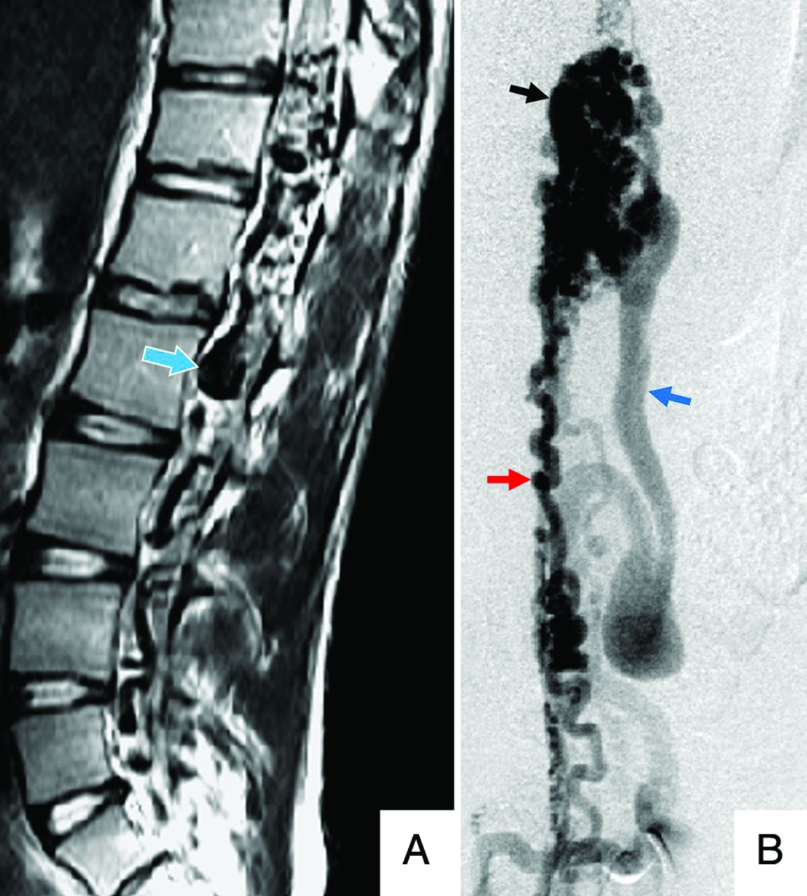
- FIG 3.
A 79-year-old man with a type I spinal dural AVF. A, Sagittal T2 MR imaging of the thoracic spine shows extensive intramedullary edema as signal hyperintensity (white arrow) throughout the cord. The thin peripheral hypointense rim (yellow rectangle) may reflect deoxyhemoglobin within dilated peripheral capillaries. Serpiginous perimedullary flow voids (blue arrow) are most conspicuous along the dorsal aspect of the thoracic cord. B, Frontal view DSA injection of the left L3 segmental artery (white arrow) shows early filling of an ectatic spinal vein (black arrow).
- FIG 4.
A 45-year-old man with a type II spinal glomus AVM. A, Sagittal T2 MR imaging of the cervical spine shows serpiginous intramedullary and perimedullary flow voids (white arrow), with adjacent cord hyperintensity (blue arrow). B, Frontal view DSA injection of the right vertebral artery (red arrow) shows opacification of a feeding arterial branch (black arrow) and nidal elements (dashed outline). C, Lateral view DSA shows early filling of ectatic perimedullary veins (blue arrow).
- FIG 5.
A 14-year-old girl with a type III spinal juvenile/metameric AVM. A, Sagittal T2 MR imaging of the lumbar spine shows numerous ectatic perimedullary and intramedullary flow voids (blue/white arrow). B, Frontal view DSA injection of the right L2 lumbar artery shows opacification of a prominent anterior spinal artery (red arrow), intramedullary and extramedullary nidal elements (black arrow), and early filing of an ectatic perimedullary vein (blue arrow).
- FIG 6.
An 11-year-old boy with a type IV intradural-perimedullary spinal AVF. A, Sagittal T2 MR imaging of the thoracic spine shows enlarged, predominantly ventral, perimedullary flow voids (white arrow) indenting the ventral cord (yellow arrow). B and C, Sequential frontal view DSA images from injection of the left T9 segmental artery show contrast progression through the ASA (red arrow), nidus (black arrow), ectatic left radiculomedullary vein (blue arrow), left common iliac vein (green arrow), and inferior vena cava (white arrow). D, Frontal magnified DSA shows a catheter traversing the opacified ASA (red arrow) and embolization coil (black arrow) and Onyx (Covidien) (yellow arrow) material within the nidus. The draining vein no longer opacifies as a result of shunt obliteration.
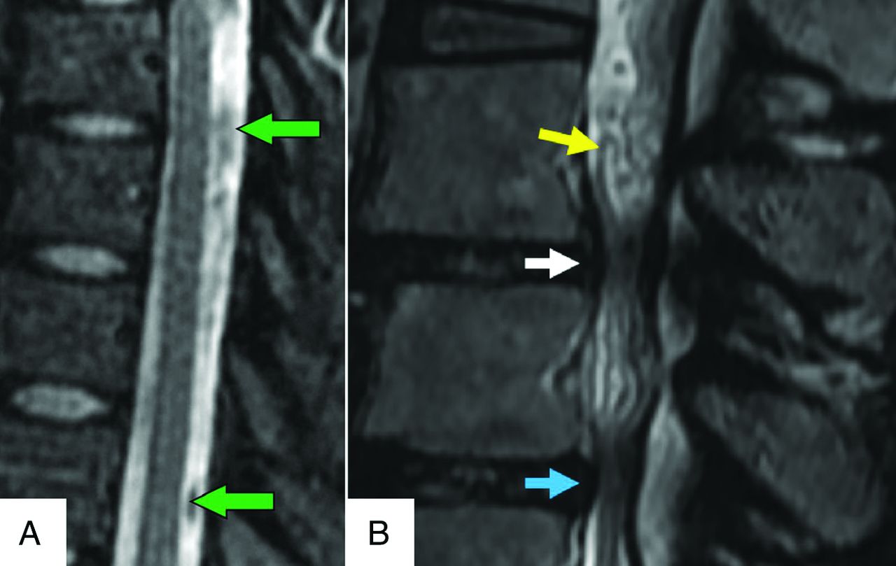
- FIG 7.
A, A 41-year-old woman with normal CSF flow-related signal voids. Sagittal T2 MR imaging of the thoracic spine shows multiple prominent CSF flow voids (green arrows), mimicking the perimedullary vascular flow voids characteristic of spinal vascular shunts. B, A 71-year-old man with spinal stenosis. Sagittal T2 MR imaging of the lumbar spine shows multiple tortuous, redundant-appearing cauda equina nerve roots (yellow arrow) mimicking the perimedullary flow voids of a spinal vascular shunt above the levels of thecal sac stenoses (white and blue arrows).
- FIG 8.
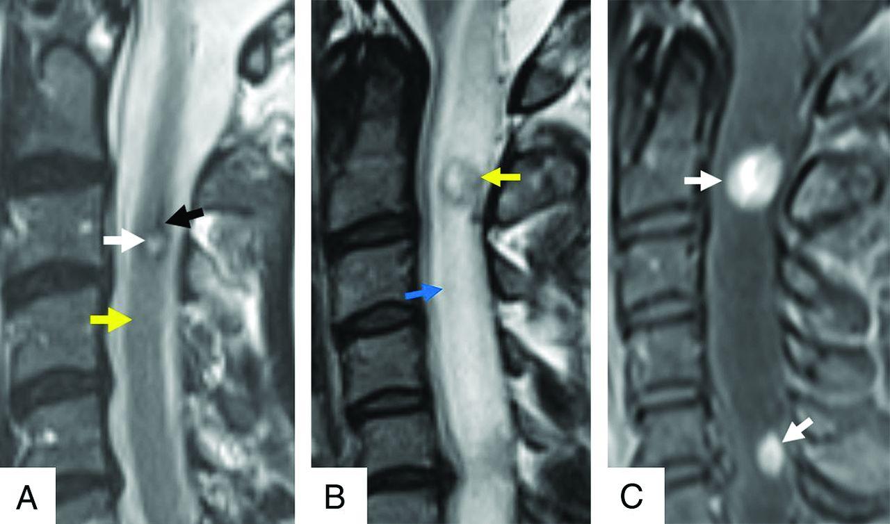
A, A 61-year-old man with a cavernous malformation of the cervical cord. Sagittal T2 MR imaging of the cervical spine shows a small intramedullary mass with central hyperintense signal (white arrow) and a peripheral rim of low signal (black arrow), indicative of blood products in various stages of degradation. There is normal signal in the adjacent cord (yellow arrow). B, A 42-year-old woman with a history of Von Hippel-Lindau disease and cervical cord hemangioblastomas. Sagittal T2 MR imaging shows a small intramedullary mass (yellow arrow) with heterogeneous hyperintense internal signal and surrounding cord edema with cord expansion (blue arrow). C, Sagittal contrast-enhanced T1 with fat saturation depicts intense enhancement (white arrows) within multiple lesions.


Tables
Authors Year Classification Summary Di Chiro et al5 1971 Type I: Single coiled vessel (AVF)Type II: Glomus type (AVM)Type III: Juvenile (AVM) Heros et al6 1986 Type IV: Direct AVF (IPAVF) involving the intrinsic arterial supply of the cord Gueguen et al7 1987 3 Types of classification of IPAVF (type IV) I) Single arterial feeder, small AVF II) Multiple feeders, medium AVF III) Multiple feeders, giant AVF Spetzler et al8 2002 AVF types (and subtypes):ExtraduralIntradural (dorsal or ventral; and single (A) or multiple (B) feeders)AVM types (and subtypes):Extradural-intraduralIntradural (intramedullary, intramedullary-extramedullary, or conus medullaris) Zozulya et al9 2006 Type I: IntramedullaryType II: Intradural or perimedullaryType III: DuralType IV: EpiduralType V: IntravertebralType VI: Combined Takai10 2017 Proposed the addition of Type V: Extradural AVF, with subtypes Va/Vb: with/without intradural venous drainage Shunt Type MRI CTA/MRA DSA I (SDAVF) T2 bright cord edema, ± thin T2 dark rim
Cord expansion
Prominent dorsal perimedullary flow voidsMay localize the involved dural artery along dorsal dural root sleeve
Prominent draining medullary veinDefinitive shunt localization
Spinal arterial stasis in setting of cord edemaII (SGAVM) Eccentric intramedullary flow voids of nidus on T2WI
T2 bright cord edema
Cord expansion
± Prominent perimedullary flow voidsHeterogeneous nidal enhancement
May depict multiplicity of arterial feedersDelineation of arterial feeders
Aneurysms in one-third of cases
± Engorged perimedullary veinsIII (SJAVM) Extensive, ectatic flow voids may involve any tissues of a single metamere
Normal cord tissue within nidal interstices
± Cord compression from large vascular structuresVariable enhancement of the extensively involved vascular structures Numerous ectatic high-flow intra- and extramedullary shunts
Rapid antegrade drainage via intra-, or extramedullary venous structuresIV (IPAVF) Prominent ventral perimedullary flow voids on T2 MR imaging
± Cord compression from perimedullary venous ectasia
± Cord edema/expansion on T2 MR imaging≥1 arterial feeder and draining veins of variable size based on subtype
± Pia arachnoid enhancementProgressively increasing rates of flow with subtypes IVa–c
May depict dilation of small pial surface arteries or venous aneurysms


