
Article Figures & Data
Figures
- FIG 1.
TBSS results show significant differences (P <.05) in DTI measures between the CP and TDC groups. Coronal slices were selected at the level of the CST. From left to right, axial slices were selected at the level of the motor cortex, PLIC, and CerPed, respectively. Mid-sagittal slices were selected at the level of the CC. A, The WM skeleton is shown in green with arrows labeling the CST, somatosensory cortex, parietal lobe, external capsule, PLIC, anterior limb of the internal capsule (ALIC), and corpus callosum. In the coronal view, ROIs for the SCR (red), PLIC (yellow), CerPed (blue), and sub-CerPed (orange) are shown. Significant differences between the CP and TDC groups are shown for FA (B), RD (C), AD (D), and MD (E). The hot colormaps denote whether a DTI measure for the CP group was less than (red-yellow) or greater than (blue-light blue) that in the TDC group. A indicates anterior; FWE, family-wise error; I, inferior; L, left; P, posterior; R, right; S, superior.
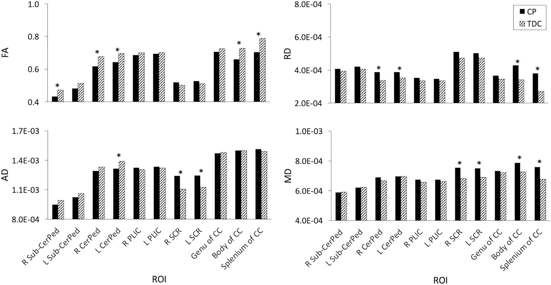
- FIG 2.
Mean differences in DTI measures between the CP and TDC groups within ROIs for the CST and CC. The asterisk indicates significant differences (P <.05). L indicates left; R, right.
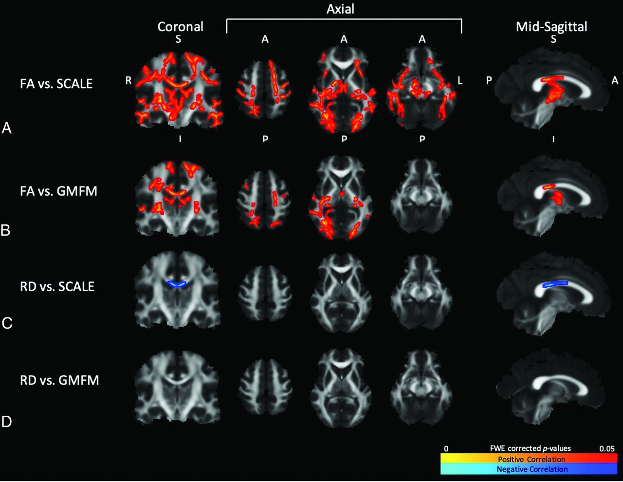
- FIG 3.
TBSS results show significant correlations (P <.05) between DTI measures and clinical measures for the CP group. Coronal slices were selected at the level of the CST. From left to right, axial slices were selected at the level of the motor cortex, PLIC, and CerPed, respectively. Mid-sagittal slices were selected at the level of the CC. Significant correlations are shown for FA vs. SCALE (A), FA vs. GMFM (B), RD vs. SCALE (C), and RD vs. GMFM (D). The hot colormaps denote whether the correlations were positive (red-yellow) or negative (blue-light blue). A indicates anterior; FWE, family-wise error; I, inferior; L, left; P, posterior; R, right; S, superior.


Tables
ROI correlation analyses comparing motor and whole-brain WM regions
Regions Voxel Count Voxels with Significant Correlations FA vs. SCALE (%) FA vs. GMFM (%) RD vs. SCALE (%) RD vs. GMFM (%) CST Sub-CerPed R 375 23 (6.1) 0 (0) 0 (0) 0 (0) Sub-CerPed L 395 0 (0) 0 (0) 0 (0) 0 (0) CerPed R 598 266 (44.5) 0 (0) 0 (0) 0 (0) CerPed L 624 74 (11.9) 0 (0) 0 (0) 0 (0) PLIC R 845 198 (23.4) 74 (8.8) 0 (0) 0 (0) PLIC L 858 20 (2.3) 18 (2.1) 0 (0) 0 (0) SCR R 1294 451 (34.9) 387 (29.9) 0 (0) 0 (0) SCR L 1279 94 (7.3) 25 (2.0) 0 (0) 0 (0) CC Genu 1758 0 (0) 0 (0) 54 (3.1) 0 (0) Body 3138 1177 (37.5) 692 (22.1) 874 (27.9) 0 (0) Splenium 2298 904 (39.3) 615 (26.8) 686 (29.9) 0 (0) Whole brain 126,000 38,251 (30.4) 18,136 (14.4) 2779 (2.2) 0 (0) Note:—L indicates left; R, right.


Jump to section
Related Articles
Cited By...
- Improved Myelination following Camp Leg Power, a Selective Motor Control Intervention for Children with Spastic Bilateral Cerebral Palsy: A Diffusion Tensor MRI Study
- Improved Myelination following Camp Leg Power, a Selective Motor Control Intervention for Children with Spastic Bilateral Cerebral Palsy: A Diffusion Tensor MRI Study