Advertisement

Main menu

  • Home
  • Content
    • Current Issue
    • Accepted Manuscripts
    • Article Preview
    • Past Issue Archive
    • Video Articles
    • AJNR Case Collection
    • Case of the Week Archive
    • Case of the Month Archive
    • Classic Case Archive
  • Special Collections
    • AJNR Awards
    • Low-Field MRI
    • Alzheimer Disease
    • ASNR Foundation Special Collection
    • Photon-Counting CT
    • View All
  • Multimedia
    • AJNR Podcasts
    • AJNR SCANtastic
    • Trainee Corner
    • MRI Safety Corner
    • Imaging Protocols
  • For Authors
    • Submit a Manuscript
    • Submit a Video Article
    • Submit an eLetter to the Editor/Response
    • Manuscript Submission Guidelines
    • Statistical Tips
    • Fast Publishing of Accepted Manuscripts
    • Graphical Abstract Preparation
    • Imaging Protocol Submission
    • Author Policies
  • About Us
    • About AJNR
    • Editorial Board
    • Editorial Board Alumni
  • More
    • Become a Reviewer/Academy of Reviewers
    • Subscribers
    • Permissions
    • Alerts
    • Feedback
    • Advertisers
    • ASNR Home

User menu

  • Alerts
  • Log in

Search

  • Advanced search
American Journal of Neuroradiology

American Journal of Neuroradiology

ASHNR American Society of Functional Neuroradiology ASHNR American Society of Pediatric Neuroradiology ASSR
  • Alerts
  • Log in

Advanced Search

  • Home
  • Content
    • Current Issue
    • Accepted Manuscripts
    • Article Preview
    • Past Issue Archive
    • Video Articles
    • AJNR Case Collection
    • Case of the Week Archive
    • Case of the Month Archive
    • Classic Case Archive
  • Special Collections
    • AJNR Awards
    • Low-Field MRI
    • Alzheimer Disease
    • ASNR Foundation Special Collection
    • Photon-Counting CT
    • View All
  • Multimedia
    • AJNR Podcasts
    • AJNR SCANtastic
    • Trainee Corner
    • MRI Safety Corner
    • Imaging Protocols
  • For Authors
    • Submit a Manuscript
    • Submit a Video Article
    • Submit an eLetter to the Editor/Response
    • Manuscript Submission Guidelines
    • Statistical Tips
    • Fast Publishing of Accepted Manuscripts
    • Graphical Abstract Preparation
    • Imaging Protocol Submission
    • Author Policies
  • About Us
    • About AJNR
    • Editorial Board
    • Editorial Board Alumni
  • More
    • Become a Reviewer/Academy of Reviewers
    • Subscribers
    • Permissions
    • Alerts
    • Feedback
    • Advertisers
    • ASNR Home
  • Follow AJNR on Twitter
  • Visit AJNR on Facebook
  • Follow AJNR on Instagram
  • Join AJNR on LinkedIn
  • RSS Feeds

AJNR Awards, New Junior Editors, and more. Read the latest AJNR updates

Getting new auth cookie, if you see this message a lot, tell someone!
Research ArticleHead and Neck Imaging

A Radiologic Grading System for Assessing the Radiographic Outcome of Treatment in Lymphatic and Lymphatic-Venous Malformations of the Head and Neck

R. De Leacy, D.V. Bageac, S. Manna, B.S. Gershon, D. Kirke, T. Shigematsu, C. Sinclair, D. Chada, P. Som, A. Doshi, K. Nael and A. Berenstein
American Journal of Neuroradiology October 2021, 42 (10) 1859-1864; DOI: https://doi.org/10.3174/ajnr.A7260
R. De Leacy
aFrom the Department of Neurosurgery (R.D.L., D.V.B., S.M., B.S.G., T.S., D.C., A.B.), Icahn School of Medicine at Mount Sinai, New York, New York
  • Find this author on Google Scholar
  • Find this author on PubMed
  • Search for this author on this site
  • ORCID record for R. De Leacy
D.V. Bageac
aFrom the Department of Neurosurgery (R.D.L., D.V.B., S.M., B.S.G., T.S., D.C., A.B.), Icahn School of Medicine at Mount Sinai, New York, New York
  • Find this author on Google Scholar
  • Find this author on PubMed
  • Search for this author on this site
  • ORCID record for D.V. Bageac
S. Manna
aFrom the Department of Neurosurgery (R.D.L., D.V.B., S.M., B.S.G., T.S., D.C., A.B.), Icahn School of Medicine at Mount Sinai, New York, New York
bDepartment of Otolaryngology-Head and Neck Surgery (S.M., D.K., C.S., P.S.), Icahn School of Medicine at Mount Sinai, New York, New York
cDepartment of Radiology (S.M., P.S., A.D., K.N.), Icahn School of Medicine at Mount Sinai, New York
  • Find this author on Google Scholar
  • Find this author on PubMed
  • Search for this author on this site
  • ORCID record for S. Manna
B.S. Gershon
aFrom the Department of Neurosurgery (R.D.L., D.V.B., S.M., B.S.G., T.S., D.C., A.B.), Icahn School of Medicine at Mount Sinai, New York, New York
dCollege of Medicine (B.S.G.), SUNY Downstate, Brooklyn, New York, New York
  • Find this author on Google Scholar
  • Find this author on PubMed
  • Search for this author on this site
  • ORCID record for B.S. Gershon
D. Kirke
bDepartment of Otolaryngology-Head and Neck Surgery (S.M., D.K., C.S., P.S.), Icahn School of Medicine at Mount Sinai, New York, New York
  • Find this author on Google Scholar
  • Find this author on PubMed
  • Search for this author on this site
  • ORCID record for D. Kirke
T. Shigematsu
aFrom the Department of Neurosurgery (R.D.L., D.V.B., S.M., B.S.G., T.S., D.C., A.B.), Icahn School of Medicine at Mount Sinai, New York, New York
  • Find this author on Google Scholar
  • Find this author on PubMed
  • Search for this author on this site
  • ORCID record for T. Shigematsu
C. Sinclair
bDepartment of Otolaryngology-Head and Neck Surgery (S.M., D.K., C.S., P.S.), Icahn School of Medicine at Mount Sinai, New York, New York
  • Find this author on Google Scholar
  • Find this author on PubMed
  • Search for this author on this site
  • ORCID record for C. Sinclair
D. Chada
aFrom the Department of Neurosurgery (R.D.L., D.V.B., S.M., B.S.G., T.S., D.C., A.B.), Icahn School of Medicine at Mount Sinai, New York, New York
  • Find this author on Google Scholar
  • Find this author on PubMed
  • Search for this author on this site
  • ORCID record for D. Chada
P. Som
bDepartment of Otolaryngology-Head and Neck Surgery (S.M., D.K., C.S., P.S.), Icahn School of Medicine at Mount Sinai, New York, New York
cDepartment of Radiology (S.M., P.S., A.D., K.N.), Icahn School of Medicine at Mount Sinai, New York
  • Find this author on Google Scholar
  • Find this author on PubMed
  • Search for this author on this site
  • ORCID record for P. Som
A. Doshi
cDepartment of Radiology (S.M., P.S., A.D., K.N.), Icahn School of Medicine at Mount Sinai, New York
  • Find this author on Google Scholar
  • Find this author on PubMed
  • Search for this author on this site
  • ORCID record for A. Doshi
K. Nael
cDepartment of Radiology (S.M., P.S., A.D., K.N.), Icahn School of Medicine at Mount Sinai, New York
  • Find this author on Google Scholar
  • Find this author on PubMed
  • Search for this author on this site
  • ORCID record for K. Nael
A. Berenstein
aFrom the Department of Neurosurgery (R.D.L., D.V.B., S.M., B.S.G., T.S., D.C., A.B.), Icahn School of Medicine at Mount Sinai, New York, New York
  • Find this author on Google Scholar
  • Find this author on PubMed
  • Search for this author on this site
  • ORCID record for A. Berenstein
  • Article
  • Figures & Data
  • Info & Metrics
  • Responses
  • References
  • PDF
Loading

Article Figures & Data

Figures

  • Tables
  • FIG 1.
    • Download figure
    • Open in new tab
    • Download powerpoint
    FIG 1.

    BDL grade 1 response. A 50-year-old man with dysphagia was found to have a de Serres stage II macrocystic LM of the head. A, Preprocedural T2-weighted axial MR imaging at the level of the hard palate shows a lesion primarily centered within the right parapharyngeal space (white arrow). B, Four months later, following 1 sclerotherapy treatment, T2-weighted axial MR imaging at the same level shows BDL grade 1 complete regression of the lesion on cross-sectional imaging (white arrow).

  • FIG 2.
    • Download figure
    • Open in new tab
    • Download powerpoint
    FIG 2.

    BDL grade 2 response. A 7-year-old girl with a left cheek mass was found to have a de Serres stage II mixed macro-/microcystic LM of the head. A, Preprocedural T2 fat-suppressed axial MR imaging at the level of the maxillary alveolar process shows involvement of the left buccal and parotid spaces (white arrow). B, Seven months later, following 2 sclerotherapy treatments, T2 fat-suppressed axial MR imaging at the same level shows BDL grade 2 near-complete resolution of the LM with trace residuals (white arrow).
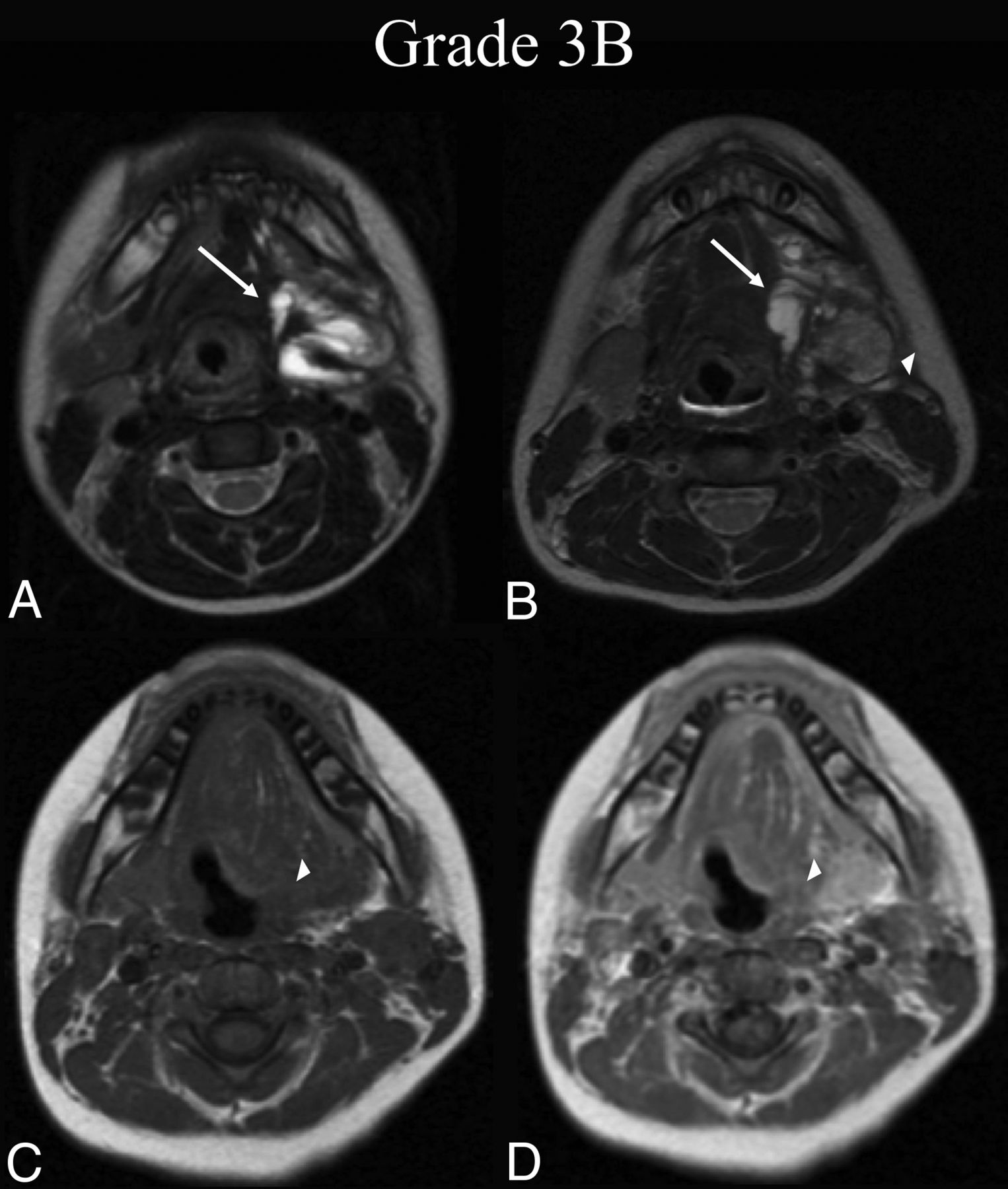
  • FIG 3.
    • Download figure
    • Open in new tab
    • Download powerpoint
    FIG 3.

    BDL grade 3B response. A 6-year-old boy with dysphagia was found to have a de Serres stage II mixed macro-/microsystic LM of the head. A, Preprocedural T2-weighted axial MR imaging at the level of the submandibular glands shows involvement of the left submandibular, sublingual, and parapharyngeal mucosal (not shown) spaces (white arrow). B, Thirty-seven months later, following 1 sclerotherapy treatment, T2-weighted axial MR imaging at the same level shows BDL grade 3B partial regression, with <50% estimated volume of residual malformation (white arrow) and intermediate signal granulation tissue formation within the treatment bed (white arrowhead). C, At the follow-up time point, note precontrast axial T1-weighted MR imaging isointensity of the suspected granuloma with muscle (white arrowhead). D, Postcontrast axial T1 at the same level demonstrates enhancement of the suspected granuloma (white arrowhead), confirming 3B grading.
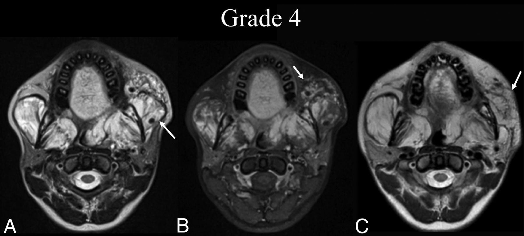
  • FIG 4.
    • Download figure
    • Open in new tab
    • Download powerpoint
    FIG 4.

    BDL grade 4 response. An 18-year-old woman with an extensive bilateral de Serres stage IV mixed macro-/microcystic LVM of the supra- and infrahyoid neck presents for sclerotherapy after receiving multiple treatments at an outside institution. A, Preprocedural T2-weighted axial MR imaging at the level of the alveolar process of the maxilla shows widespread LVM bilaterally (white arrow). B, Preprocedural T1 fat-saturated postcontrast axial MR imaging at the same level demonstrates enhancement of venous structures within the LVM (white arrow). C, Thirty-five months later, following 1 sclerotherapy treatment targeting the left buccal region, repeat T2-weighted axial MR imaging at the same level shows BDL grade 4 partial regression (white arrow) with >50% estimated volume of residual malformation.
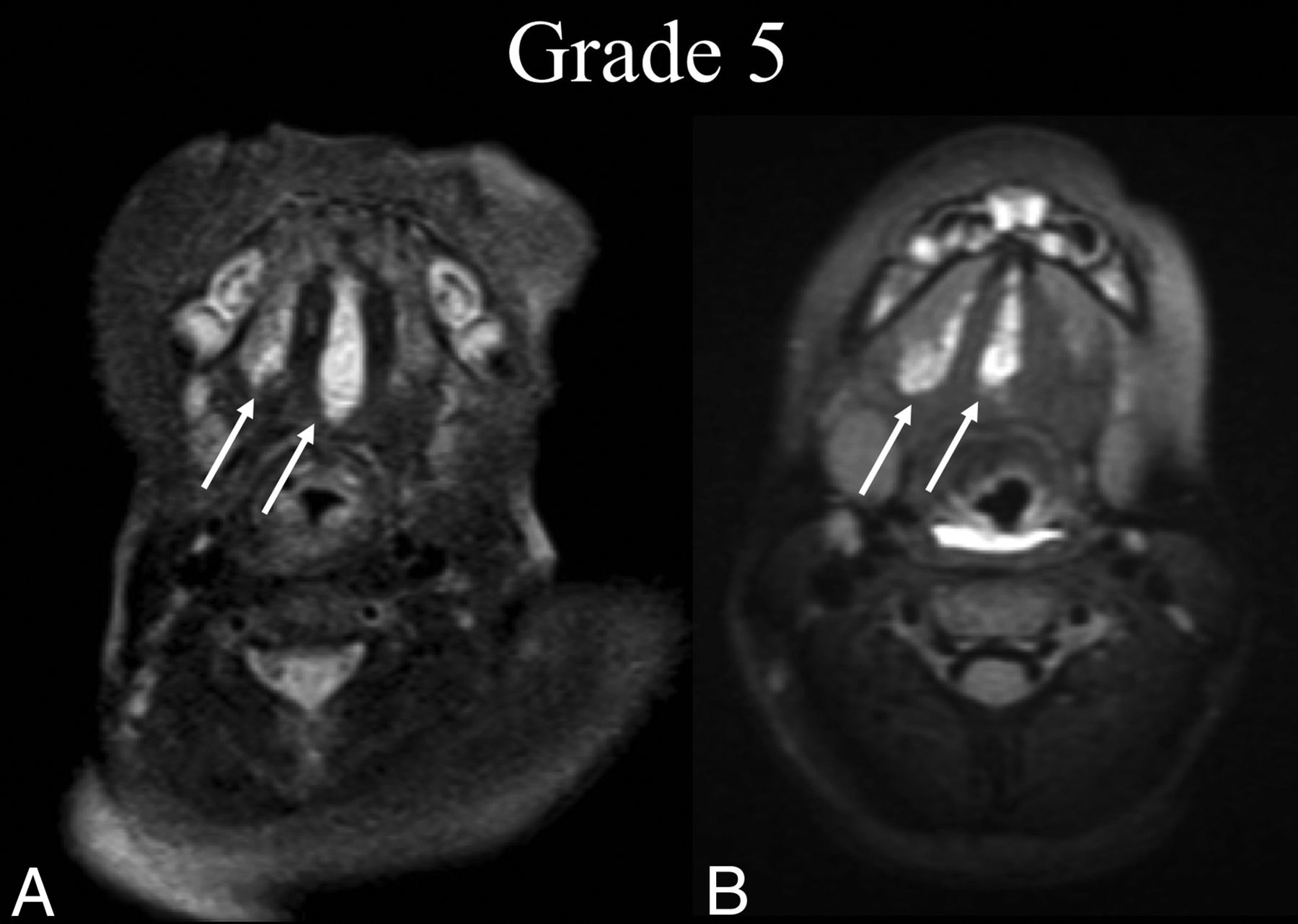
  • FIG 5.
    • Download figure
    • Open in new tab
    • Download powerpoint
    FIG 5.

    BDL grade 5 response. A 2-month-old boy with tongue swelling was found to have a de Serres stage II mixed macro-/microcystic sublingual LM. A, Preprocedural T2 fat-suppressed axial MR imaging at the level of the mandibular symphysis shows involvement of the right and middle aspects of the sublingual space (white arrows). B, Five years later, following 1 sclerotherapy treatment, T2 fat-suppressed axial MR imaging at the same level shows BDL grade 5 recurrence of the LM without a gross interval change from baseline (white arrows).

  • FIG 6.
    • Download figure
    • Open in new tab
    • Download powerpoint
    FIG 6.

    BDL grade 6 response. An 8-year-old boy with right facial prominence was found to have a de Serres stage II mixed macro-/microcystic LM. A, Preprocedural T2 fat-suppressed axial MR imaging at the level of the oropharynx shows trans-spatial involvement of the right neck extending from the right parotid and masticator spaces to the pharyngeal mucosal space (white arrow), sparing the retropharyngeal space. B, One year later, following 2 sclerotherapy treatments, T2 fat-suppressed axial MR imaging at the same level shows BDL grade 6 regression within the treatment bed and extension into the retropharyngeal space, which was previously uninvolved (white arrow).
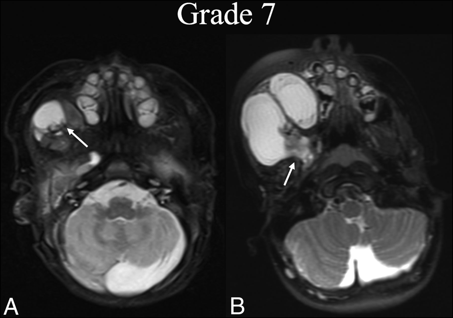
  • FIG 7.
    • Download figure
    • Open in new tab
    • Download powerpoint
    FIG 7.

    BDL grade 7 progression. A neonate boy with right facial prominence was found to have a de Serres stage II macrocystic LM. A, Initial T2 fat-suppressed axial MR imaging at the level of the hard palate shows involvement of the right buccal space (white arrow). B, Three years later, before any intervention, T2 fat-suppressed axial MR imaging at the same level shows BDL grade 7 progression with clear volume increase and extension into the masticator and parapharyngeal (white arrow).

Tables

  • Figures
    • View popup
    Table 1:

    Grading system for assessing the radiographic outcome of treatment in LM-LVM malformations of the head and neck

    GradeDescription
    1Complete regression of the lesion on cross-sectional imaging
    2Near-complete regression with trace residual of the lesion on cross-sectional imaging
    3Partial regression with <50% estimated volume of residual malformation
    4Partial regression with >50% estimated volume of residual malformation
    5Minimal or no gross interval change
    6Regression of malformation in 1 region with progression into a previously uninvolved/untreated area
    7Gross interval progression
    Modifier BGranulation tissue formation in treatment bed
    • View popup
    Table 2:

    Patient age and average treatments

    MedianRange
    Age (yr)6.38(0.33–73.92)
    Imaging interval (mo)32.50(1–131)
    Treatments within imaging interval (mo)1.50(0–13)
    • View popup
    Table 3:

    Patient demographics and LV-LVM characteristics

    No.(%)
    Total56
    Sex
        Male2850.0%
        Female2850.0%
    Classification
        LM3766.1%
        LVM1933.9%
    Architecture
        Macrocystic916.1%
        Microcystic1933.9%
        Mixed2850.0%
    Lesion laterality
        Right1832.1%
        Left1628.6%
        Bilateral2239.3%
    de Serres stage
        I (unilateral infrahyoid)11.8%
        II (unilateral suprahyoid)2748.2%
        III (unilateral suprahyoid and infrahyoid)814.3%
        IV (bilateral suprahyoid)916.1%
        V (bilateral suprahyoid and infrahyoid)1119.6%
    • View popup
    Table 4:

    Frequency of grading outcomes

    Final GradeCountFrequency (%)
    135.4%
    2814.3%
    31730.4%
    4814.3%
    5712.5%
    6610.7%
    758.9%
    B Modifier23.6%
    • View popup
    Table 5:

    BDL interrater reliability

    Interrater Reliability MeasureStatistical ValueCI or P Value
    Krippendorff α statistic0.93(95% CI, 0.89–0.95)
    Intraclass coefficienta0.97(95% CI, 0.96–0.98)
    Spearman ρ (K.N. vs A.D.)0.90P < .001
    Spearman ρ (K.N. vs R.D.L.)0.93P < .001
    Spearman ρ (A.D. vs R.D.L.)0.95P < .001
    • ↵a Intraclass coefficients calculated for both consistency and absolute agreement yielded identical values.

PreviousNext
Back to top

In this issue

American Journal of Neuroradiology: 42 (10)
American Journal of Neuroradiology
Vol. 42, Issue 10
1 Oct 2021
  • Table of Contents
  • Index by author
  • Complete Issue (PDF)
Advertisement
Print
Download PDF
Email Article

Thank you for your interest in spreading the word on American Journal of Neuroradiology.

NOTE: We only request your email address so that the person you are recommending the page to knows that you wanted them to see it, and that it is not junk mail. We do not capture any email address.

Enter multiple addresses on separate lines or separate them with commas.
A Radiologic Grading System for Assessing the Radiographic Outcome of Treatment in Lymphatic and Lymphatic-Venous Malformations of the Head and Neck
(Your Name) has sent you a message from American Journal of Neuroradiology
(Your Name) thought you would like to see the American Journal of Neuroradiology web site.
CAPTCHA
This question is for testing whether or not you are a human visitor and to prevent automated spam submissions.
Cite this article
R. De Leacy, D.V. Bageac, S. Manna, B.S. Gershon, D. Kirke, T. Shigematsu, C. Sinclair, D. Chada, P. Som, A. Doshi, K. Nael, A. Berenstein
A Radiologic Grading System for Assessing the Radiographic Outcome of Treatment in Lymphatic and Lymphatic-Venous Malformations of the Head and Neck
American Journal of Neuroradiology Oct 2021, 42 (10) 1859-1864; DOI: 10.3174/ajnr.A7260

Citation Manager Formats

  • BibTeX
  • Bookends
  • EasyBib
  • EndNote (tagged)
  • EndNote 8 (xml)
  • Medlars
  • Mendeley
  • Papers
  • RefWorks Tagged
  • Ref Manager
  • RIS
  • Zotero
0 Responses
Respond to this article
Share
Bookmark this article
A Radiologic Grading System for Assessing the Radiographic Outcome of Treatment in Lymphatic and Lymphatic-Venous Malformations of the Head and Neck
R. De Leacy, D.V. Bageac, S. Manna, B.S. Gershon, D. Kirke, T. Shigematsu, C. Sinclair, D. Chada, P. Som, A. Doshi, K. Nael, A. Berenstein
American Journal of Neuroradiology Oct 2021, 42 (10) 1859-1864; DOI: 10.3174/ajnr.A7260
del.icio.us logo Twitter logo Facebook logo Mendeley logo
  • Tweet Widget
  • Facebook Like
  • Google Plus One
Purchase

Jump to section

  • Article
    • Abstract
    • ABBREVIATIONS:
    • MATERIALS AND METHODS
    • RESULTS
    • DISCUSSION
    • CONCLUSIONS
    • Footnotes
    • References
  • Figures & Data
  • Info & Metrics
  • Responses
  • References
  • PDF

Related Articles

  • No related articles found.
  • PubMed
  • Google Scholar

Cited By...

  • Percutaneous sclerotherapy for head and neck lymphatic malformations in neonates and infants <=12 months of age
  • Crossref
  • Google Scholar

This article has not yet been cited by articles in journals that are participating in Crossref Cited-by Linking.

More in this TOC Section

  • MRI of the Parasellar Ligaments
  • ASL Sensitivity for Head and Neck Paraganglioma
  • Post SRS Peritumoral Hyperintense Signal of VSs
Show more Head and Neck Imaging

Similar Articles

Advertisement

Indexed Content

  • Current Issue
  • Accepted Manuscripts
  • Article Preview
  • Past Issues
  • Editorials
  • Editor's Choice
  • Fellows' Journal Club
  • Letters to the Editor
  • Video Articles

Cases

  • Case Collection
  • Archive - Case of the Week
  • Archive - Case of the Month
  • Archive - Classic Case

More from AJNR

  • Trainee Corner
  • Imaging Protocols
  • MRI Safety Corner
  • Book Reviews

Multimedia

  • AJNR Podcasts
  • AJNR Scantastics

Resources

  • Turnaround Time
  • Submit a Manuscript
  • Submit a Video Article
  • Submit an eLetter to the Editor/Response
  • Manuscript Submission Guidelines
  • Statistical Tips
  • Fast Publishing of Accepted Manuscripts
  • Graphical Abstract Preparation
  • Imaging Protocol Submission
  • Evidence-Based Medicine Level Guide
  • Publishing Checklists
  • Author Policies
  • Become a Reviewer/Academy of Reviewers
  • News and Updates

About Us

  • About AJNR
  • Editorial Board
  • Editorial Board Alumni
  • Alerts
  • Permissions
  • Not an AJNR Subscriber? Join Now
  • Advertise with Us
  • Librarian Resources
  • Feedback
  • Terms and Conditions
  • AJNR Editorial Board Alumni

American Society of Neuroradiology

  • Not an ASNR Member? Join Now

© 2025 by the American Society of Neuroradiology All rights, including for text and data mining, AI training, and similar technologies, are reserved.
Print ISSN: 0195-6108 Online ISSN: 1936-959X

Powered by HighWire