
Article Figures & Data
Figures
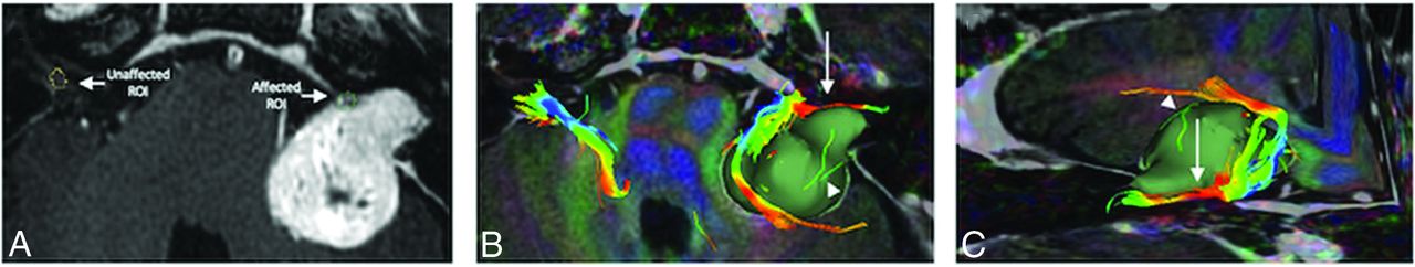
- FIG 1.
T1-weighted anatomic image (A) and 2 fused DTI/T1-weighted images (B and C) with superimposed representation of tumor volume (green) used to locate the ROIs within the distal cisternal/IC segments (A) and to demonstrate the extracted CN VII/VIII tracts (B and C). Note the detailed correspondence of the tumor and its effect on CN VII/VIII (white arrows). The CN VII/VIII can be seen sweeping anterior to the tumor. Also, note scattered, disconnected fibers that are not corresponding to CN configuration in space. These fibers were disregarded anatomically (arrowheads).
- FIG 2.
Microstructural properties of the CN VII/VIII in patients with VS. The averaged mean value of the diffusivity measurements in all patients with VS that demonstrate abnormal WM microstructural changes in their VII/VIII nerves (four asterisks indicate P < .0001 false discovery rate–corrected). DTI-derived values ± standard error of the mean of affected (black bars) and unaffected (gray bars) sides. Compared with the unaffected side, patients had significantly higher FA (A) and lower AD (B), RD (C), and MD (D) on their affected side. Diffusion is in square millimeters/second.
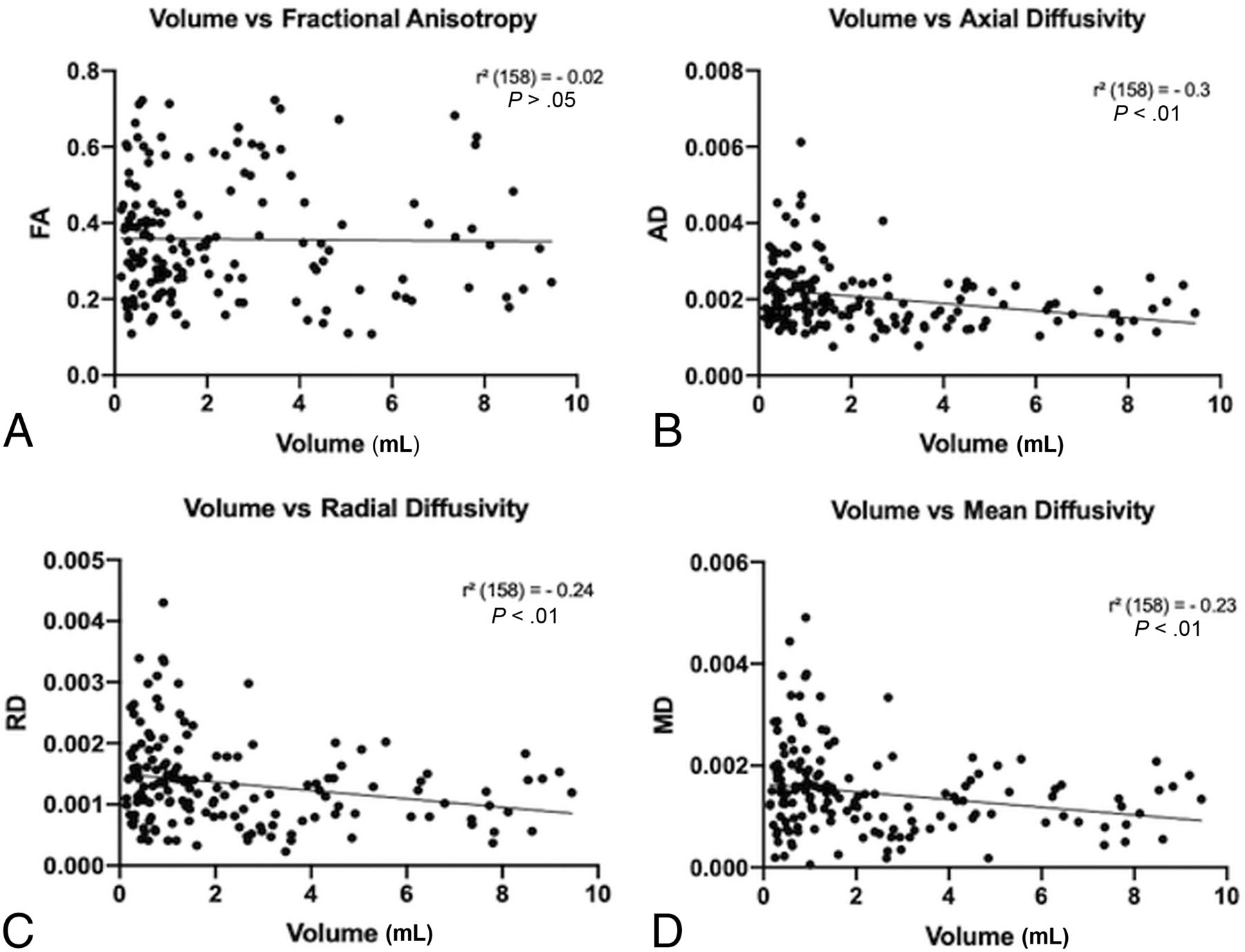
- FIG 3.
Scatterplots of the relationship between tumor volume and microstructural properties in the affected CNs VII/VIII. A weak-but-significant inverse relationship exists between the tumor volume and the specific diffusivities: axial diffusivity (B), radial diffusivity (C), and mean diffusivity (D), but not with FA (A) (significant at P < .01; false discovery rate–corrected). Diffusion is in square millimeters/second.


Tables
The mean, median, and range values for averaged FA and diffusivity
Mean Median Range FA (mm2/s) Affected 0.357 0.332 0.108–0.722 Nonaffected 0.287 0.256 0.114–0.746 AD (mm2/s) Affected 0.002 0.002 0.001–0.006 Nonaffected 0.003 0.003 0.001–0.005 RD (mm2/s) Affected 0.001 0.001 0–0.004 Nonaffected 0.002 0.002 0–0.003 MD (mm2/s) Affected 0.001 0.001 0–0.005 Nonaffected 0.002 0.002 0–0.004


Jump to section
Related Articles
Cited By...
- No citing articles found.