Advertisement

Main menu

  • Home
  • Content
    • Current Issue
    • Accepted Manuscripts
    • Article Preview
    • Past Issue Archive
    • Video Articles
    • AJNR Case Collection
    • Case of the Week Archive
    • Case of the Month Archive
    • Classic Case Archive
  • Special Collections
    • AJNR Awards
    • Low-Field MRI
    • Alzheimer Disease
    • ASNR Foundation Special Collection
    • Photon-Counting CT
    • View All
  • Multimedia
    • AJNR Podcasts
    • AJNR SCANtastic
    • Trainee Corner
    • MRI Safety Corner
    • Imaging Protocols
  • For Authors
    • Submit a Manuscript
    • Submit a Video Article
    • Submit an eLetter to the Editor/Response
    • Manuscript Submission Guidelines
    • Statistical Tips
    • Fast Publishing of Accepted Manuscripts
    • Graphical Abstract Preparation
    • Imaging Protocol Submission
    • Author Policies
  • About Us
    • About AJNR
    • Editorial Board
    • Editorial Board Alumni
  • More
    • Become a Reviewer/Academy of Reviewers
    • Subscribers
    • Permissions
    • Alerts
    • Feedback
    • Advertisers
    • ASNR Home

User menu

  • Alerts
  • Log in

Search

  • Advanced search
American Journal of Neuroradiology

American Journal of Neuroradiology

ASHNR American Society of Functional Neuroradiology ASHNR American Society of Pediatric Neuroradiology ASSR
  • Alerts
  • Log in

Advanced Search

  • Home
  • Content
    • Current Issue
    • Accepted Manuscripts
    • Article Preview
    • Past Issue Archive
    • Video Articles
    • AJNR Case Collection
    • Case of the Week Archive
    • Case of the Month Archive
    • Classic Case Archive
  • Special Collections
    • AJNR Awards
    • Low-Field MRI
    • Alzheimer Disease
    • ASNR Foundation Special Collection
    • Photon-Counting CT
    • View All
  • Multimedia
    • AJNR Podcasts
    • AJNR SCANtastic
    • Trainee Corner
    • MRI Safety Corner
    • Imaging Protocols
  • For Authors
    • Submit a Manuscript
    • Submit a Video Article
    • Submit an eLetter to the Editor/Response
    • Manuscript Submission Guidelines
    • Statistical Tips
    • Fast Publishing of Accepted Manuscripts
    • Graphical Abstract Preparation
    • Imaging Protocol Submission
    • Author Policies
  • About Us
    • About AJNR
    • Editorial Board
    • Editorial Board Alumni
  • More
    • Become a Reviewer/Academy of Reviewers
    • Subscribers
    • Permissions
    • Alerts
    • Feedback
    • Advertisers
    • ASNR Home
  • Follow AJNR on Twitter
  • Visit AJNR on Facebook
  • Follow AJNR on Instagram
  • Join AJNR on LinkedIn
  • RSS Feeds

AJNR Awards, New Junior Editors, and more. Read the latest AJNR updates

Research ArticleExtracranial Vascular
Open Access

Plaque Composition as a Predictor of Plaque Ulceration in Carotid Artery Atherosclerosis: The Plaque At RISK Study

K. Dilba, D.H.K. van Dam-Nolen, A.C. van Dijk, M. Kassem, A.F.W. van der Steen, P.J. Koudstaal, P.J. Nederkoorn, J. Hendrikse, M.E. Kooi, J.J. Wentzel and A. van der Lugt
American Journal of Neuroradiology January 2021, 42 (1) 144-151; DOI: https://doi.org/10.3174/ajnr.A6868
K. Dilba
aFrom the Departments of Radiology and Nuclear Medicine (K.D., D.H.K.v.D.-N., A.C.v.D., A.v.d.L.)
bCardiology (K.D., A.F.W.v.d.S., J.J.W.)
  • Find this author on Google Scholar
  • Find this author on PubMed
  • Search for this author on this site
  • ORCID record for K. Dilba
D.H.K. van Dam-Nolen
aFrom the Departments of Radiology and Nuclear Medicine (K.D., D.H.K.v.D.-N., A.C.v.D., A.v.d.L.)
  • Find this author on Google Scholar
  • Find this author on PubMed
  • Search for this author on this site
  • ORCID record for D.H.K. van Dam-Nolen
A.C. van Dijk
aFrom the Departments of Radiology and Nuclear Medicine (K.D., D.H.K.v.D.-N., A.C.v.D., A.v.d.L.)
cNeurology (A.C.v.D., P.J.K.), Erasmus University Medical Center Rotterdam, Rotterdam, the Netherlands
  • Find this author on Google Scholar
  • Find this author on PubMed
  • Search for this author on this site
  • ORCID record for A.C. van Dijk
M. Kassem
dDepartment of Radiology and Nuclear Medicine (M.K., M.E.K.), CARIM School for Cardiovascular Diseases, Maastricht University Medical Center+, Maastricht, the Netherlands
  • Find this author on Google Scholar
  • Find this author on PubMed
  • Search for this author on this site
  • ORCID record for M. Kassem
A.F.W. van der Steen
bCardiology (K.D., A.F.W.v.d.S., J.J.W.)
  • Find this author on Google Scholar
  • Find this author on PubMed
  • Search for this author on this site
  • ORCID record for A.F.W. van der Steen
P.J. Koudstaal
cNeurology (A.C.v.D., P.J.K.), Erasmus University Medical Center Rotterdam, Rotterdam, the Netherlands
  • Find this author on Google Scholar
  • Find this author on PubMed
  • Search for this author on this site
  • ORCID record for P.J. Koudstaal
P.J. Nederkoorn
eDepartment of Neurology (P.J.N.), University Medical Center Amsterdam, Amsterdam, the Netherlands
  • Find this author on Google Scholar
  • Find this author on PubMed
  • Search for this author on this site
  • ORCID record for P.J. Nederkoorn
J. Hendrikse
fDepartment of Radiology (J.H.), University Medical Center Utrecht, Utrecht, the Netherlands
  • Find this author on Google Scholar
  • Find this author on PubMed
  • Search for this author on this site
  • ORCID record for J. Hendrikse
M.E. Kooi
dDepartment of Radiology and Nuclear Medicine (M.K., M.E.K.), CARIM School for Cardiovascular Diseases, Maastricht University Medical Center+, Maastricht, the Netherlands
  • Find this author on Google Scholar
  • Find this author on PubMed
  • Search for this author on this site
  • ORCID record for M.E. Kooi
J.J. Wentzel
bCardiology (K.D., A.F.W.v.d.S., J.J.W.)
  • Find this author on Google Scholar
  • Find this author on PubMed
  • Search for this author on this site
  • ORCID record for J.J. Wentzel
A. van der Lugt
aFrom the Departments of Radiology and Nuclear Medicine (K.D., D.H.K.v.D.-N., A.C.v.D., A.v.d.L.)
  • Find this author on Google Scholar
  • Find this author on PubMed
  • Search for this author on this site
  • ORCID record for A. van der Lugt
  • Article
  • Figures & Data
  • Info & Metrics
  • Responses
  • References
  • PDF
Loading

Article Figures & Data

Figures

  • Tables
  • FIG 1.
    • Download figure
    • Open in new tab
    • Download powerpoint
    FIG 1.

    Flow diagram explaining the reasons for exclusion of a number of carotid arteries in the final data analysis.
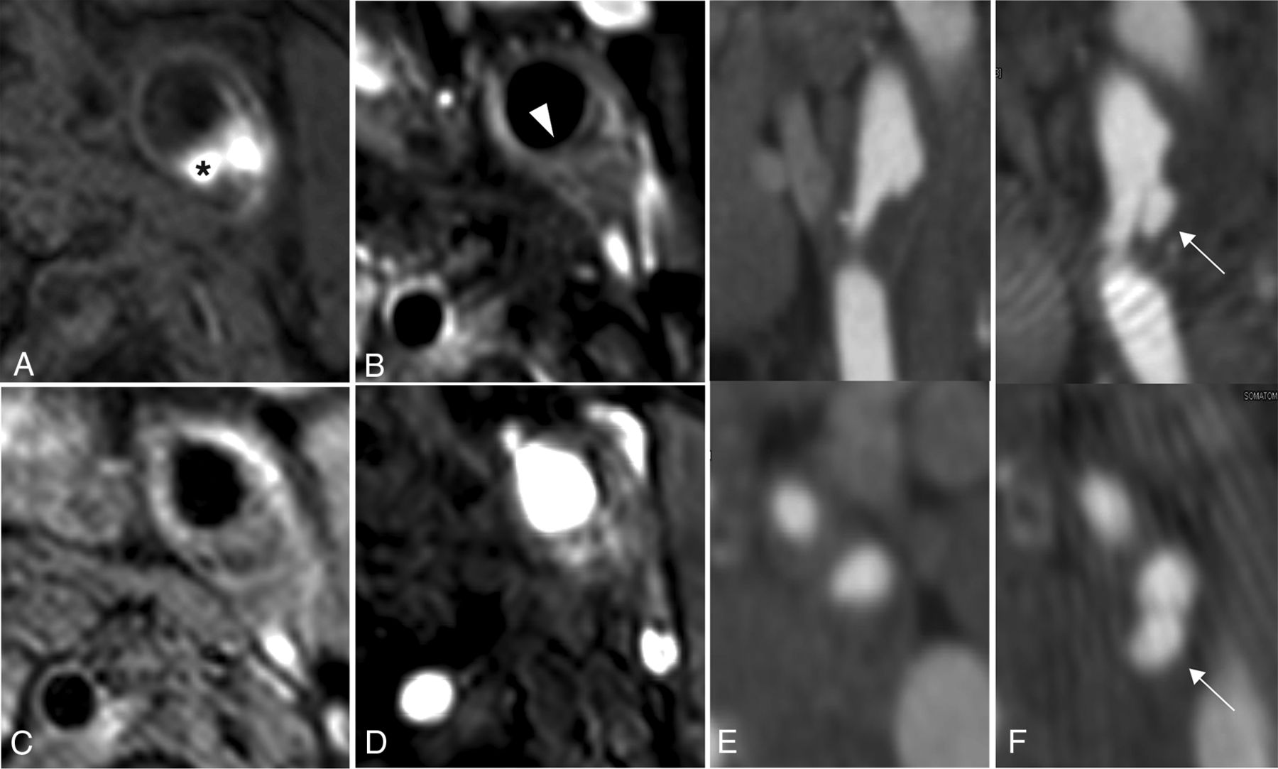
  • FIG 2.
    • Download figure
    • Open in new tab
    • Download powerpoint
    FIG 2.

    Ulceration development during follow-up and plaque composition at baseline of a 56-year-old female patient. MR imaging of carotid plaque proximal to the ulceration: precontrast T1-weighted turbo field echo sequence (A), postcontrast T1-weighted quadruple inversion recovery TSE sequence (B), T2-weighted TSE sequence (C), time-of-flight sequence (D), and MDCTA of irregular plaque at baseline (E) and of ulceration at 2-year follow-up (F). The asterisk marks the intraplaque hemorrhage. The white arrowhead points to thin-or-ruptured fibrous cap. White arrows point to the ulceration.

Tables

  • Figures
    • View popup
    Table 1:

    The comparison of clinical characteristics and scan intervals between plaques with and without an ulcer at baselinea

    All Baseline (n = 180)Ulcer Baseline Absent (n = 131)Ulcer Baseline Present (n = 49)P Value
    Characteristics
     Age (yr)68 (63–73)68 (62–73)69 (64–73).41
     Sex (male)127 (71%)122 (70%)5 (83%).67
     BMI (kg/m2)26 (24–29)25 (24–29)27 (24–29).45
    Classification event0.01
     TIA76 (42%)64 (49%)12 (25%)
     Stroke85 (47%)54 (41%)31 (63%)
     Amaurosis fugax19 (11%)13 (10%)6 (12%)
    Hypertension117 (65%)85 (65%)32 (65%)1.00
    Hypercholesterolemia140 (78%)101 (77%)39 (80%).84
    Diabetes mellitus42 (23%)35 (27%)7 (14%).11
    Current smoker43 (24%)35 (27%)8 (16%).17
    Interval event/MDCTA (days)34 (15–55)36 (16–53)35 (13–59).85
    Interval event/MR imaging (days)47 (30–67)46 (30–67)48 (28–69).87
    Interval MDCTA/MR imaging (days)1 (1–25)1 (1–27)1 (1–17).70
    • Note:—BMI indicates body mass index.

    • ↵a Data are median (IQR) or No. (%).

    • View popup
    Table 2:

    Characteristics of plaques with and without an ulcer at baseline

    Ulcer Absent (n = 131)Median (IQR) or No. (%)Ulcer Present (n = 49) Median (IQR) or No. (%)Binary Logistic Regression OR (95% CI)ROC Analysis AUC (95% CI)
    NASCET (%)8 (0–29)21 (0–37)1.01 (1.00–1.03)0.61 (0.52–0.71)
    ECST (%)55 (45–65)59 (48–70)1.02 (1.00–1.05)
    Minimal diameter (mm)4 (3.3–4.9)3.4 (2.8–4.7)0.06 (0.00–1.00)0.40 (0.30–0.50)
    Total vessel volume (cm3)1.41 (1.16–1.64)1.60 (1.29–2.12)4.9 (2.1–11.4)0.64 (0.54–0.74)
    Lumen volume (cm3)0.56 (0.45–0.72)0.58 (0.46–0.80)2.8 (0.6–12.1)
    Wall volume (cm3)0.85 (0.70–0.98)1.02 (0.77–1.28)12.1 (3.5–42.0)0.67 (0.58–0.77)
    % Wall volume59 (53–66)61 (57–68)1.01 (0.99–1.04)
    LRNC presence78 (60%)38 (78%)2.4 (1.1–5.0)0.59 (0.50–0.68)
    % LRNC volume1 (0–10)15 (1–31)1.7 (1.3–2.2)0.70 (0.60–0.79)
    IPH presence42 (32%)27 (55%)2.6 (1.3–5.1)0.61 (0.52–0.71)
    % IPH volume0 (0–3)5 (0–23)1.7 (1.3–2.2)0.67 (0.56–0.76)
    Calcifications presence MR imaging118 (90%)44 (90%)0.97 (0.33–2.88)
    % Calcifications volume MR imaging5 (2–8)3 (1–7)0.73 (0.50–1.07)
    Calcification presence MDCTA117 (89%)45 (92%)0.74 (0.23–2.38)
    Calcification absolute volume MDCTA (mm3)31.4 (4.3–80.4)15.8 (2.8–51.9)0.99 (0.999–1.002)
    Thin-or-ruptured FC45 (34%)a,b32 (65%)3.4 (1.7–6.7)0.65 (0.56–0.74)
    • Note:—FC indicates fibrous cap.

    • ↵a Four were missing.

    • ↵b Four had bad quality.

    • View popup
    Table 3:

    Characteristics of plaques with and without a new ulceration at 2-year follow-upa

    New Ulceration Absent (n = 67)New Ulceration Present (n = 6)P Value
    NASCET (%)16 (0–34)11 (0–34).95
    ECST (%)57 (47–68)62 (53–71).45
    Minimal diameter (mm)3.8 (2.8–4.6)3.4 (2.9–4.8).80
    Total vessel volume (cm3)1.49 (1.2–1.75)1.61 (1.53–1.87).17
    Lumen volume (cm3)0.54 (0.44–0.77)0.59 (0.55–0.69).43
    Wall volume (cm3)0.86 (0.73–1.00)1.04 (0.97–1.16).029
    % Wall volume60 (52–67)64 (59–67).37
    LRNC presence41 (61%)6 (100%).08
    % LRNC volume2 (0–14)23 (13–31).002
    IPH presence21 (31%)6 (100%).002
    % IPH volume0 (0–5)14 (8–24)<.001
    Calcifications presence MR imaging63 (94%)6 (100%)1.00
    % Calcifications volume MR imaging5 (3–8)6 (2–9).79
    Calcifications presence MDCTA60 (90%)5 (83%).52
    Calcifications, absolute volume MDCTA (mm3)32.0 (7.0–100.3)1.9 (0.8–91.3).15
    Thin-or-ruptured FC28 (42%)6 (100%).009
    • Note:—FC indicates fibrous cap.

    • ↵a Data are median (IQR) or No. (%).

PreviousNext
Back to top

In this issue

American Journal of Neuroradiology: 42 (1)
American Journal of Neuroradiology
Vol. 42, Issue 1
1 Jan 2021
  • Table of Contents
  • Index by author
  • Complete Issue (PDF)
Advertisement
Print
Download PDF
Email Article

Thank you for your interest in spreading the word on American Journal of Neuroradiology.

NOTE: We only request your email address so that the person you are recommending the page to knows that you wanted them to see it, and that it is not junk mail. We do not capture any email address.

Enter multiple addresses on separate lines or separate them with commas.
Plaque Composition as a Predictor of Plaque Ulceration in Carotid Artery Atherosclerosis: The Plaque At RISK Study
(Your Name) has sent you a message from American Journal of Neuroradiology
(Your Name) thought you would like to see the American Journal of Neuroradiology web site.
CAPTCHA
This question is for testing whether or not you are a human visitor and to prevent automated spam submissions.
Cite this article
K. Dilba, D.H.K. van Dam-Nolen, A.C. van Dijk, M. Kassem, A.F.W. van der Steen, P.J. Koudstaal, P.J. Nederkoorn, J. Hendrikse, M.E. Kooi, J.J. Wentzel, A. van der Lugt
Plaque Composition as a Predictor of Plaque Ulceration in Carotid Artery Atherosclerosis: The Plaque At RISK Study
American Journal of Neuroradiology Jan 2021, 42 (1) 144-151; DOI: 10.3174/ajnr.A6868

Citation Manager Formats

  • BibTeX
  • Bookends
  • EasyBib
  • EndNote (tagged)
  • EndNote 8 (xml)
  • Medlars
  • Mendeley
  • Papers
  • RefWorks Tagged
  • Ref Manager
  • RIS
  • Zotero
0 Responses
Respond to this article
Share
Bookmark this article
Plaque Composition as a Predictor of Plaque Ulceration in Carotid Artery Atherosclerosis: The Plaque At RISK Study
K. Dilba, D.H.K. van Dam-Nolen, A.C. van Dijk, M. Kassem, A.F.W. van der Steen, P.J. Koudstaal, P.J. Nederkoorn, J. Hendrikse, M.E. Kooi, J.J. Wentzel, A. van der Lugt
American Journal of Neuroradiology Jan 2021, 42 (1) 144-151; DOI: 10.3174/ajnr.A6868
del.icio.us logo Twitter logo Facebook logo Mendeley logo
  • Tweet Widget
  • Facebook Like
  • Google Plus One
Purchase

Jump to section

  • Article
    • Abstract
    • ABBREVIATIONS:
    • MATERIALS AND METHODS
    • RESULTS
    • DISCUSSION
    • CONCLUSIONS
    • Acknowledgments
    • Footnotes
    • References
  • Figures & Data
  • Info & Metrics
  • Responses
  • References
  • PDF

Related Articles

  • No related articles found.
  • PubMed
  • Google Scholar

Cited By...

  • Identification of biomarkers for ulcerative morphology in carotid atherosclerosis plaques through single-cell RNA sequencing data
  • MR Imaging of Carotid Artery Atherosclerosis: Updated Evidence on High-Risk Plaque Features and Emerging Trends
  • Crossref
  • Google Scholar

This article has not yet been cited by articles in journals that are participating in Crossref Cited-by Linking.

More in this TOC Section

  • Proximal Vertebral Artery Variants and Embryology
  • High-Risk Plaque Features in Carotid MRI
  • Nonstenotic Carotid Plaques and Stroke Review
Show more Extracranial Vascular

Similar Articles

Advertisement

Indexed Content

  • Current Issue
  • Accepted Manuscripts
  • Article Preview
  • Past Issues
  • Editorials
  • Editor's Choice
  • Fellows' Journal Club
  • Letters to the Editor
  • Video Articles

Cases

  • Case Collection
  • Archive - Case of the Week
  • Archive - Case of the Month
  • Archive - Classic Case

More from AJNR

  • Trainee Corner
  • Imaging Protocols
  • MRI Safety Corner
  • Book Reviews

Multimedia

  • AJNR Podcasts
  • AJNR Scantastics

Resources

  • Turnaround Time
  • Submit a Manuscript
  • Submit a Video Article
  • Submit an eLetter to the Editor/Response
  • Manuscript Submission Guidelines
  • Statistical Tips
  • Fast Publishing of Accepted Manuscripts
  • Graphical Abstract Preparation
  • Imaging Protocol Submission
  • Evidence-Based Medicine Level Guide
  • Publishing Checklists
  • Author Policies
  • Become a Reviewer/Academy of Reviewers
  • News and Updates

About Us

  • About AJNR
  • Editorial Board
  • Editorial Board Alumni
  • Alerts
  • Permissions
  • Not an AJNR Subscriber? Join Now
  • Advertise with Us
  • Librarian Resources
  • Feedback
  • Terms and Conditions
  • AJNR Editorial Board Alumni

American Society of Neuroradiology

  • Not an ASNR Member? Join Now

© 2025 by the American Society of Neuroradiology All rights, including for text and data mining, AI training, and similar technologies, are reserved.
Print ISSN: 0195-6108 Online ISSN: 1936-959X

Powered by HighWire