Advertisement

Main menu

  • Home
  • Content
    • Current Issue
    • Accepted Manuscripts
    • Article Preview
    • Past Issue Archive
    • Video Articles
    • AJNR Case Collection
    • Case of the Week Archive
    • Case of the Month Archive
    • Classic Case Archive
  • Special Collections
    • AJNR Awards
    • Low-Field MRI
    • Alzheimer Disease
    • ASNR Foundation Special Collection
    • Photon-Counting CT
    • View All
  • Multimedia
    • AJNR Podcasts
    • AJNR SCANtastic
    • Trainee Corner
    • MRI Safety Corner
    • Imaging Protocols
  • For Authors
    • Submit a Manuscript
    • Submit a Video Article
    • Submit an eLetter to the Editor/Response
    • Manuscript Submission Guidelines
    • Statistical Tips
    • Fast Publishing of Accepted Manuscripts
    • Graphical Abstract Preparation
    • Imaging Protocol Submission
    • Author Policies
  • About Us
    • About AJNR
    • Editorial Board
    • Editorial Board Alumni
  • More
    • Become a Reviewer/Academy of Reviewers
    • Subscribers
    • Permissions
    • Alerts
    • Feedback
    • Advertisers
    • ASNR Home

User menu

  • Alerts
  • Log in

Search

  • Advanced search
American Journal of Neuroradiology

American Journal of Neuroradiology

ASHNR American Society of Functional Neuroradiology ASHNR American Society of Pediatric Neuroradiology ASSR
  • Alerts
  • Log in

Advanced Search

  • Home
  • Content
    • Current Issue
    • Accepted Manuscripts
    • Article Preview
    • Past Issue Archive
    • Video Articles
    • AJNR Case Collection
    • Case of the Week Archive
    • Case of the Month Archive
    • Classic Case Archive
  • Special Collections
    • AJNR Awards
    • Low-Field MRI
    • Alzheimer Disease
    • ASNR Foundation Special Collection
    • Photon-Counting CT
    • View All
  • Multimedia
    • AJNR Podcasts
    • AJNR SCANtastic
    • Trainee Corner
    • MRI Safety Corner
    • Imaging Protocols
  • For Authors
    • Submit a Manuscript
    • Submit a Video Article
    • Submit an eLetter to the Editor/Response
    • Manuscript Submission Guidelines
    • Statistical Tips
    • Fast Publishing of Accepted Manuscripts
    • Graphical Abstract Preparation
    • Imaging Protocol Submission
    • Author Policies
  • About Us
    • About AJNR
    • Editorial Board
    • Editorial Board Alumni
  • More
    • Become a Reviewer/Academy of Reviewers
    • Subscribers
    • Permissions
    • Alerts
    • Feedback
    • Advertisers
    • ASNR Home
  • Follow AJNR on Twitter
  • Visit AJNR on Facebook
  • Follow AJNR on Instagram
  • Join AJNR on LinkedIn
  • RSS Feeds

AJNR Awards, New Junior Editors, and more. Read the latest AJNR updates

Research ArticlePediatric Neuroimaging

Internal Auditory Canal Diverticula among Pediatric Patients: Prevalence and Assessment for Hearing Loss and Anatomic Associations

P.M. Bunch, M.E. Zapadka, C.M. Lack, E.P. Kiell, D.J. Kirse and J.R. Sachs
American Journal of Neuroradiology September 2020, 41 (9) 1712-1717; DOI: https://doi.org/10.3174/ajnr.A6691
P.M. Bunch
aFrom the Departments of Radiology (P.M.B., M.E.Z., C.M.L., J.R.S.)
  • Find this author on Google Scholar
  • Find this author on PubMed
  • Search for this author on this site
  • ORCID record for P.M. Bunch
M.E. Zapadka
aFrom the Departments of Radiology (P.M.B., M.E.Z., C.M.L., J.R.S.)
  • Find this author on Google Scholar
  • Find this author on PubMed
  • Search for this author on this site
  • ORCID record for M.E. Zapadka
C.M. Lack
aFrom the Departments of Radiology (P.M.B., M.E.Z., C.M.L., J.R.S.)
  • Find this author on Google Scholar
  • Find this author on PubMed
  • Search for this author on this site
  • ORCID record for C.M. Lack
E.P. Kiell
bOtolaryngology, Head and Neck Surgery (E.P.K., D.J.K.)
  • Find this author on Google Scholar
  • Find this author on PubMed
  • Search for this author on this site
  • ORCID record for E.P. Kiell
D.J. Kirse
bOtolaryngology, Head and Neck Surgery (E.P.K., D.J.K.)
cPediatrics (D.J.K.), Wake Forest School of Medicine, Winston Salem, North Carolina.
  • Find this author on Google Scholar
  • Find this author on PubMed
  • Search for this author on this site
  • ORCID record for D.J. Kirse
J.R. Sachs
aFrom the Departments of Radiology (P.M.B., M.E.Z., C.M.L., J.R.S.)
  • Find this author on Google Scholar
  • Find this author on PubMed
  • Search for this author on this site
  • ORCID record for J.R. Sachs
  • Article
  • Figures & Data
  • Info & Metrics
  • Responses
  • References
  • PDF
Loading

Article Figures & Data

Figures

  • Tables
  • FIG 1.
    • Download figure
    • Open in new tab
    • Download powerpoint
    FIG 1.

    Axial temporal bone CT image (A) at the level of the inferior aspect of the right internal auditory canal demonstrates the typical appearance of an internal auditory canal diverticulum (arrow). An image obtained at the same level in a different patient without an internal auditory canal diverticulum is provided for comparison (B).

  • FIG 2.
    • Download figure
    • Open in new tab
    • Download powerpoint
    FIG 2.

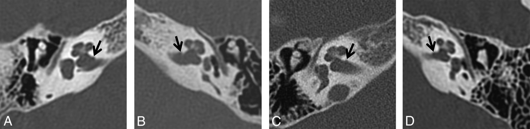
    Visual threshold developed after the initial review and applied during the subsequent review by the 4 neuroradiologist readers. The visual threshold consists of axial temporal bone CT images obtained in 4 different patients and is intended to depict the minimum contour irregularity necessary to be considered an internal auditory canal diverticulum (arrows) for the second review performed in this study.

  • FIG 3.
    • Download figure
    • Open in new tab
    • Download powerpoint
    FIG 3.

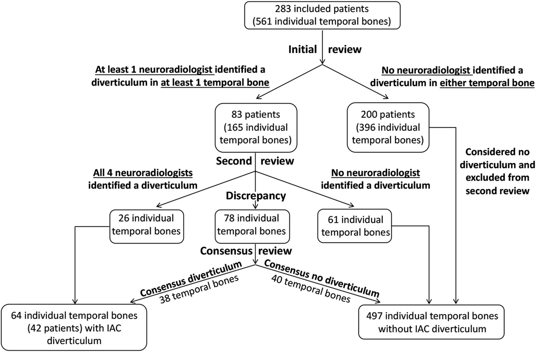
    Flow chart depicting the initial and subsequent image reviews.

  • FIG 4.
    • Download figure
    • Open in new tab
    • Download powerpoint
    FIG 4.

    Axial temporal bone CT images from 4 different patients depict representative examples of subtle IAC contour irregularities (arrows) determined during consensus review of discrepant cases to not reach the visual threshold necessary to be considered an IAC diverticulum.

Tables

  • Figures
    • View popup
    Table 1:

    Characteristics of the study group

    Sex
        Male157 (55%)
        Female126 (45%)
    Age
        Mean (SD) (yr)7.8 (4.8)
        Minimum6 weeks
        Maximum17 years
    Audiogram (temporal bone sides)
        Normal152 (27%)
        Sensorineural loss158 (28%)
        Mixed loss39 (7%)
        Conductive loss112 (20%)
        Unavailable100 (18%)
    • View popup
    Table 2:

    Temporal bone CT findings within the study group

    Individual Temporal Bones (Sides)
    Internal auditory canal diverticulum
        Yes64 (11%)
        No497 (89%)
    Enlarged vestibular aqueduct
        Yes43 (8%)
        No518 (92%)
    Labyrinthine dysplasia
        Yes66 (12%)
        No495 (88%)
    Cochlear cleft
        Yes205 (37%)
        No356 (63%)
    Otospongiosis
        Yes2a (0.4%)
        No559 (99.6%)
    • ↵a Single patient who also had clinical diagnosis of bilateral otospongiosis.

    • View popup
    Table 3:

    Internal auditory canal diverticula association assessment

    Internal Auditory Canal DiverticulumP
    YesNo
    Sexa
        Male28129.13
        Female14112
    Age (mean) (SD) (yr)7.7 (5.0)7.8 (4.8).91
    Audiogramb
        Normal hearing20132.65
        Hearing loss36273
        Conductive1696.86
        Sensorineural16142.48
        Mixed435.79
    Enlarged vestibular aqueductb
        Yes241.21
        No62456
    Labyrinthine dysplasiab
        Yes7591.00
        No57438
    Cochlear cleftb
        Yes30175.07
        No34322
    Otospongiosisb
        Yes20.013
        No62497
    • ↵a Denotes number of patients.

    • ↵b Denotes number of individual temporal bones (sides).

PreviousNext
Back to top

In this issue

American Journal of Neuroradiology: 41 (9)
American Journal of Neuroradiology
Vol. 41, Issue 9
1 Sep 2020
  • Table of Contents
  • Index by author
  • Complete Issue (PDF)
Advertisement
Print
Download PDF
Email Article

Thank you for your interest in spreading the word on American Journal of Neuroradiology.

NOTE: We only request your email address so that the person you are recommending the page to knows that you wanted them to see it, and that it is not junk mail. We do not capture any email address.

Enter multiple addresses on separate lines or separate them with commas.
Internal Auditory Canal Diverticula among Pediatric Patients: Prevalence and Assessment for Hearing Loss and Anatomic Associations
(Your Name) has sent you a message from American Journal of Neuroradiology
(Your Name) thought you would like to see the American Journal of Neuroradiology web site.
CAPTCHA
This question is for testing whether or not you are a human visitor and to prevent automated spam submissions.
Cite this article
P.M. Bunch, M.E. Zapadka, C.M. Lack, E.P. Kiell, D.J. Kirse, J.R. Sachs
Internal Auditory Canal Diverticula among Pediatric Patients: Prevalence and Assessment for Hearing Loss and Anatomic Associations
American Journal of Neuroradiology Sep 2020, 41 (9) 1712-1717; DOI: 10.3174/ajnr.A6691

Citation Manager Formats

  • BibTeX
  • Bookends
  • EasyBib
  • EndNote (tagged)
  • EndNote 8 (xml)
  • Medlars
  • Mendeley
  • Papers
  • RefWorks Tagged
  • Ref Manager
  • RIS
  • Zotero
0 Responses
Respond to this article
Share
Bookmark this article
Internal Auditory Canal Diverticula among Pediatric Patients: Prevalence and Assessment for Hearing Loss and Anatomic Associations
P.M. Bunch, M.E. Zapadka, C.M. Lack, E.P. Kiell, D.J. Kirse, J.R. Sachs
American Journal of Neuroradiology Sep 2020, 41 (9) 1712-1717; DOI: 10.3174/ajnr.A6691
del.icio.us logo Twitter logo Facebook logo Mendeley logo
  • Tweet Widget
  • Facebook Like
  • Google Plus One
Purchase

Jump to section

  • Article
    • Abstract
    • ABBREVIATION:
    • MATERIALS AND METHODS
    • RESULTS
    • DISCUSSION
    • CONCLUSIONS
    • Footnotes
    • References
  • Figures & Data
  • Info & Metrics
  • Responses
  • References
  • PDF

Related Articles

  • No related articles found.
  • PubMed
  • Google Scholar

Cited By...

  • No citing articles found.
  • Crossref
  • Google Scholar

This article has not yet been cited by articles in journals that are participating in Crossref Cited-by Linking.

More in this TOC Section

  • Frontal Paraventricular Cysts
  • Sodium MRI in Pediatric Brain Tumors
  • FRACTURE MR in Congenital Vertebral Anomalies
Show more Pediatric Neuroimaging

Similar Articles

Advertisement

Indexed Content

  • Current Issue
  • Accepted Manuscripts
  • Article Preview
  • Past Issues
  • Editorials
  • Editor's Choice
  • Fellows' Journal Club
  • Letters to the Editor
  • Video Articles

Cases

  • Case Collection
  • Archive - Case of the Week
  • Archive - Case of the Month
  • Archive - Classic Case

More from AJNR

  • Trainee Corner
  • Imaging Protocols
  • MRI Safety Corner
  • Book Reviews

Multimedia

  • AJNR Podcasts
  • AJNR Scantastics

Resources

  • Turnaround Time
  • Submit a Manuscript
  • Submit a Video Article
  • Submit an eLetter to the Editor/Response
  • Manuscript Submission Guidelines
  • Statistical Tips
  • Fast Publishing of Accepted Manuscripts
  • Graphical Abstract Preparation
  • Imaging Protocol Submission
  • Evidence-Based Medicine Level Guide
  • Publishing Checklists
  • Author Policies
  • Become a Reviewer/Academy of Reviewers
  • News and Updates

About Us

  • About AJNR
  • Editorial Board
  • Editorial Board Alumni
  • Alerts
  • Permissions
  • Not an AJNR Subscriber? Join Now
  • Advertise with Us
  • Librarian Resources
  • Feedback
  • Terms and Conditions
  • AJNR Editorial Board Alumni

American Society of Neuroradiology

  • Not an ASNR Member? Join Now

© 2025 by the American Society of Neuroradiology All rights, including for text and data mining, AI training, and similar technologies, are reserved.
Print ISSN: 0195-6108 Online ISSN: 1936-959X

Powered by HighWire