Advertisement

Main menu

  • Home
  • Content
    • Current Issue
    • Accepted Manuscripts
    • Article Preview
    • Past Issue Archive
    • Video Articles
    • AJNR Case Collection
    • Case of the Week Archive
    • Case of the Month Archive
    • Classic Case Archive
  • Special Collections
    • AJNR Awards
    • Low-Field MRI
    • Alzheimer Disease
    • ASNR Foundation Special Collection
    • Photon-Counting CT
    • View All
  • Multimedia
    • AJNR Podcasts
    • AJNR SCANtastic
    • Trainee Corner
    • MRI Safety Corner
    • Imaging Protocols
  • For Authors
    • Submit a Manuscript
    • Submit a Video Article
    • Submit an eLetter to the Editor/Response
    • Manuscript Submission Guidelines
    • Statistical Tips
    • Fast Publishing of Accepted Manuscripts
    • Graphical Abstract Preparation
    • Imaging Protocol Submission
    • Author Policies
  • About Us
    • About AJNR
    • Editorial Board
    • Editorial Board Alumni
  • More
    • Become a Reviewer/Academy of Reviewers
    • Subscribers
    • Permissions
    • Alerts
    • Feedback
    • Advertisers
    • ASNR Home

User menu

  • Alerts
  • Log in

Search

  • Advanced search
American Journal of Neuroradiology

American Journal of Neuroradiology

ASHNR American Society of Functional Neuroradiology ASHNR American Society of Pediatric Neuroradiology ASSR
  • Alerts
  • Log in

Advanced Search

  • Home
  • Content
    • Current Issue
    • Accepted Manuscripts
    • Article Preview
    • Past Issue Archive
    • Video Articles
    • AJNR Case Collection
    • Case of the Week Archive
    • Case of the Month Archive
    • Classic Case Archive
  • Special Collections
    • AJNR Awards
    • Low-Field MRI
    • Alzheimer Disease
    • ASNR Foundation Special Collection
    • Photon-Counting CT
    • View All
  • Multimedia
    • AJNR Podcasts
    • AJNR SCANtastic
    • Trainee Corner
    • MRI Safety Corner
    • Imaging Protocols
  • For Authors
    • Submit a Manuscript
    • Submit a Video Article
    • Submit an eLetter to the Editor/Response
    • Manuscript Submission Guidelines
    • Statistical Tips
    • Fast Publishing of Accepted Manuscripts
    • Graphical Abstract Preparation
    • Imaging Protocol Submission
    • Author Policies
  • About Us
    • About AJNR
    • Editorial Board
    • Editorial Board Alumni
  • More
    • Become a Reviewer/Academy of Reviewers
    • Subscribers
    • Permissions
    • Alerts
    • Feedback
    • Advertisers
    • ASNR Home
  • Follow AJNR on Twitter
  • Visit AJNR on Facebook
  • Follow AJNR on Instagram
  • Join AJNR on LinkedIn
  • RSS Feeds

AJNR Awards, New Junior Editors, and more. Read the latest AJNR updates

  • Getting new auth cookie, if you see this message a lot, tell someone!
  • Getting new auth cookie, if you see this message a lot, tell someone!
Research ArticleAdult Brain

Focal Leptomeningeal Disease with Perivascular Invasion in EGFR-Mutant Non-Small-Cell Lung Cancer

A. Dasgupta, F.Y. Moraes, S. Rawal, P. Diamandis and D.B. Shultz
American Journal of Neuroradiology August 2020, 41 (8) 1430-1433; DOI: https://doi.org/10.3174/ajnr.A6640
A. Dasgupta
aFrom the Department of Radiation Oncology (A.D., F.Y.M., D.B.S.), Division of Neuroimaging, Joint Department of Medical Imaging (S.R.), and Department of Neuropathology (P.D.), Princess Margaret Cancer Centre and University Health Network, Toronto, Ontario, Canada.
  • Find this author on Google Scholar
  • Find this author on PubMed
  • Search for this author on this site
  • ORCID record for A. Dasgupta
F.Y. Moraes
aFrom the Department of Radiation Oncology (A.D., F.Y.M., D.B.S.), Division of Neuroimaging, Joint Department of Medical Imaging (S.R.), and Department of Neuropathology (P.D.), Princess Margaret Cancer Centre and University Health Network, Toronto, Ontario, Canada.
  • Find this author on Google Scholar
  • Find this author on PubMed
  • Search for this author on this site
  • ORCID record for F.Y. Moraes
S. Rawal
aFrom the Department of Radiation Oncology (A.D., F.Y.M., D.B.S.), Division of Neuroimaging, Joint Department of Medical Imaging (S.R.), and Department of Neuropathology (P.D.), Princess Margaret Cancer Centre and University Health Network, Toronto, Ontario, Canada.
  • Find this author on Google Scholar
  • Find this author on PubMed
  • Search for this author on this site
  • ORCID record for S. Rawal
P. Diamandis
aFrom the Department of Radiation Oncology (A.D., F.Y.M., D.B.S.), Division of Neuroimaging, Joint Department of Medical Imaging (S.R.), and Department of Neuropathology (P.D.), Princess Margaret Cancer Centre and University Health Network, Toronto, Ontario, Canada.
  • Find this author on Google Scholar
  • Find this author on PubMed
  • Search for this author on this site
  • ORCID record for P. Diamandis
D.B. Shultz
aFrom the Department of Radiation Oncology (A.D., F.Y.M., D.B.S.), Division of Neuroimaging, Joint Department of Medical Imaging (S.R.), and Department of Neuropathology (P.D.), Princess Margaret Cancer Centre and University Health Network, Toronto, Ontario, Canada.
  • Find this author on Google Scholar
  • Find this author on PubMed
  • Search for this author on this site
  • ORCID record for D.B. Shultz
  • Article
  • Figures & Data
  • Info & Metrics
  • Responses
  • References
  • PDF
Loading

Article Figures & Data

Figures

  • Fig 1.
    • Download figure
    • Open in new tab
    • Download powerpoint
    Fig 1.

    Brain MR imaging for patient 1, a 67-year-old woman with locally advanced EGFRm NSCLC. Sagital (A) and axial (B) projections show enhancement along the parenchyma, with ring-enhancing lesions extending into the right parietal sulci in continuity. The scan was performed for restaging purposes, before any brain-directed treatment.

  • Fig 2.
    • Download figure
    • Open in new tab
    • Download powerpoint
    Fig 2.

    Histopathology. A, Hematoxylin-eosin image shows cohesive epithelioid neoplastic cells closely associated with parenchymal blood vessels (black arrows). Immunohistochemical staining (brown coloration) for CK7 (B) and TTF1 (C) supports a lung origin and highlights the involvement of the perivascular spaces. D, Growth of tumor within the Virchow-Robins spaces (marked brown again with CK7 immunostaining) creates an impression of scattered tumor foci within the unstained brain tissue rather than the typical solitary metastatic mass. E and F, Evidence of leptomeningeal involvement with adenocarcinoma closely associated with and encircling larger thick meningeal-type vessels (black arrows). F, A high-power view of the red box seen in E highlights that most of the increased cellularity is tumor cells.
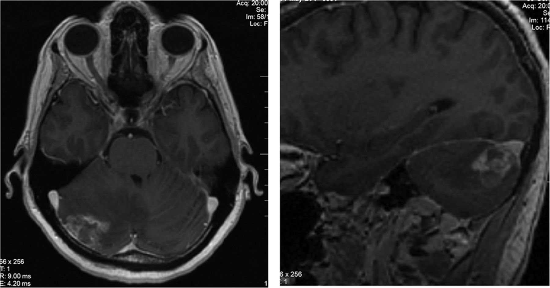
  • Fig 3.
    • Download figure
    • Open in new tab
    • Download powerpoint
    Fig 3.

    Brain MR imaging for patient 2, a 62-year-old man with EGFRm NSCLC. Sagital (A) and axial (B) projections demonstrate an enhancing lesion spreading along the cerebellar folia after several months of treatment with gefitinib.

  • Fig 4.
    • Download figure
    • Open in new tab
    • Download powerpoint
    Fig 4.

    Brain MR imaging for patient 3, a 49-year-old man with EGFRm NSCLC with an enhancing lesion in the left parietal lobe 14 months following WBRT.

  • Fig 5.
    • Download figure
    • Open in new tab
    • Download powerpoint
    Fig 5.

    Brain MR imaging for patient 4, a 77-year-old woman with EGFRm NSCLC. Axial projection demonstrates a right frontal lobe lesion following 18 months of gefitinib treatment.

  • Fig 6.
    • Download figure
    • Open in new tab
    • Download powerpoint
    Fig 6.

    Brain MR imaging for patient 5, a 46-year-old woman with EGFRm NSCLC. Sagital (A) and axial (B) projections demonstrate an enhancing right cerebellar lesion with spread along the cerebellar folia, 1 year following stereotactic radiosurgery and several years of gefitinib treatment.

  • Fig 7.
    • Download figure
    • Open in new tab
    • Download powerpoint
    Fig 7.

    Kaplan-Meier survival plot comparing survival outcomes of FLIP with those of classic leptomeningeal disease in EGFRm NSCLC.

PreviousNext
Back to top

In this issue

American Journal of Neuroradiology: 41 (8)
American Journal of Neuroradiology
Vol. 41, Issue 8
1 Aug 2020
  • Table of Contents
  • Index by author
  • Complete Issue (PDF)
Advertisement
Print
Download PDF
Email Article

Thank you for your interest in spreading the word on American Journal of Neuroradiology.

NOTE: We only request your email address so that the person you are recommending the page to knows that you wanted them to see it, and that it is not junk mail. We do not capture any email address.

Enter multiple addresses on separate lines or separate them with commas.
Focal Leptomeningeal Disease with Perivascular Invasion in EGFR-Mutant Non-Small-Cell Lung Cancer
(Your Name) has sent you a message from American Journal of Neuroradiology
(Your Name) thought you would like to see the American Journal of Neuroradiology web site.
CAPTCHA
This question is for testing whether or not you are a human visitor and to prevent automated spam submissions.
Cite this article
A. Dasgupta, F.Y. Moraes, S. Rawal, P. Diamandis, D.B. Shultz
Focal Leptomeningeal Disease with Perivascular Invasion in EGFR-Mutant Non-Small-Cell Lung Cancer
American Journal of Neuroradiology Aug 2020, 41 (8) 1430-1433; DOI: 10.3174/ajnr.A6640

Citation Manager Formats

  • BibTeX
  • Bookends
  • EasyBib
  • EndNote (tagged)
  • EndNote 8 (xml)
  • Medlars
  • Mendeley
  • Papers
  • RefWorks Tagged
  • Ref Manager
  • RIS
  • Zotero
0 Responses
Respond to this article
Share
Bookmark this article
Focal Leptomeningeal Disease with Perivascular Invasion in EGFR-Mutant Non-Small-Cell Lung Cancer
A. Dasgupta, F.Y. Moraes, S. Rawal, P. Diamandis, D.B. Shultz
American Journal of Neuroradiology Aug 2020, 41 (8) 1430-1433; DOI: 10.3174/ajnr.A6640
del.icio.us logo Twitter logo Facebook logo Mendeley logo
  • Tweet Widget
  • Facebook Like
  • Google Plus One
Purchase

Jump to section

  • Article
    • Abstract
    • ABBREVIATIONS:
    • DISCUSSION
    • Footnotes
    • References
  • Figures & Data
  • Info & Metrics
  • Responses
  • References
  • PDF

Related Articles

  • No related articles found.
  • PubMed
  • Google Scholar

Cited By...

  • No citing articles found.
  • Crossref
  • Google Scholar

This article has not yet been cited by articles in journals that are participating in Crossref Cited-by Linking.

More in this TOC Section

  • Diagnostic Neuroradiology of Monoclonal Antibodies
  • Clinical Outcomes After Chiari I Decompression
  • Segmentation of Brain Metastases with BLAST
Show more Adult Brain

Similar Articles

Advertisement

Indexed Content

  • Current Issue
  • Accepted Manuscripts
  • Article Preview
  • Past Issues
  • Editorials
  • Editor's Choice
  • Fellows' Journal Club
  • Letters to the Editor
  • Video Articles

Cases

  • Case Collection
  • Archive - Case of the Week
  • Archive - Case of the Month
  • Archive - Classic Case

More from AJNR

  • Trainee Corner
  • Imaging Protocols
  • MRI Safety Corner
  • Book Reviews

Multimedia

  • AJNR Podcasts
  • AJNR Scantastics

Resources

  • Turnaround Time
  • Submit a Manuscript
  • Submit a Video Article
  • Submit an eLetter to the Editor/Response
  • Manuscript Submission Guidelines
  • Statistical Tips
  • Fast Publishing of Accepted Manuscripts
  • Graphical Abstract Preparation
  • Imaging Protocol Submission
  • Evidence-Based Medicine Level Guide
  • Publishing Checklists
  • Author Policies
  • Become a Reviewer/Academy of Reviewers
  • News and Updates

About Us

  • About AJNR
  • Editorial Board
  • Editorial Board Alumni
  • Alerts
  • Permissions
  • Not an AJNR Subscriber? Join Now
  • Advertise with Us
  • Librarian Resources
  • Feedback
  • Terms and Conditions
  • AJNR Editorial Board Alumni

American Society of Neuroradiology

  • Not an ASNR Member? Join Now

© 2025 by the American Society of Neuroradiology All rights, including for text and data mining, AI training, and similar technologies, are reserved.
Print ISSN: 0195-6108 Online ISSN: 1936-959X

Powered by HighWire