Advertisement

Main menu

  • Home
  • Content
    • Current Issue
    • Accepted Manuscripts
    • Article Preview
    • Past Issue Archive
    • Video Articles
    • AJNR Case Collection
    • Case of the Week Archive
    • Case of the Month Archive
    • Classic Case Archive
  • Special Collections
    • AJNR Awards
    • Low-Field MRI
    • Alzheimer Disease
    • ASNR Foundation Special Collection
    • Photon-Counting CT
    • View All
  • Multimedia
    • AJNR Podcasts
    • AJNR SCANtastic
    • Trainee Corner
    • MRI Safety Corner
    • Imaging Protocols
  • For Authors
    • Submit a Manuscript
    • Submit a Video Article
    • Submit an eLetter to the Editor/Response
    • Manuscript Submission Guidelines
    • Statistical Tips
    • Fast Publishing of Accepted Manuscripts
    • Graphical Abstract Preparation
    • Imaging Protocol Submission
    • Author Policies
  • About Us
    • About AJNR
    • Editorial Board
    • Editorial Board Alumni
  • More
    • Become a Reviewer/Academy of Reviewers
    • Subscribers
    • Permissions
    • Alerts
    • Feedback
    • Advertisers
    • ASNR Home

User menu

  • Alerts
  • Log in

Search

  • Advanced search
American Journal of Neuroradiology

American Journal of Neuroradiology

ASHNR American Society of Functional Neuroradiology ASHNR American Society of Pediatric Neuroradiology ASSR
  • Alerts
  • Log in

Advanced Search

  • Home
  • Content
    • Current Issue
    • Accepted Manuscripts
    • Article Preview
    • Past Issue Archive
    • Video Articles
    • AJNR Case Collection
    • Case of the Week Archive
    • Case of the Month Archive
    • Classic Case Archive
  • Special Collections
    • AJNR Awards
    • Low-Field MRI
    • Alzheimer Disease
    • ASNR Foundation Special Collection
    • Photon-Counting CT
    • View All
  • Multimedia
    • AJNR Podcasts
    • AJNR SCANtastic
    • Trainee Corner
    • MRI Safety Corner
    • Imaging Protocols
  • For Authors
    • Submit a Manuscript
    • Submit a Video Article
    • Submit an eLetter to the Editor/Response
    • Manuscript Submission Guidelines
    • Statistical Tips
    • Fast Publishing of Accepted Manuscripts
    • Graphical Abstract Preparation
    • Imaging Protocol Submission
    • Author Policies
  • About Us
    • About AJNR
    • Editorial Board
    • Editorial Board Alumni
  • More
    • Become a Reviewer/Academy of Reviewers
    • Subscribers
    • Permissions
    • Alerts
    • Feedback
    • Advertisers
    • ASNR Home
  • Follow AJNR on Twitter
  • Visit AJNR on Facebook
  • Follow AJNR on Instagram
  • Join AJNR on LinkedIn
  • RSS Feeds

AJNR Awards, New Junior Editors, and more. Read the latest AJNR updates

Getting new auth cookie, if you see this message a lot, tell someone!
Research ArticleSpine Imaging and Spine Image-Guided Interventions

Counterpoint: Conventional Fluoroscopy-Guided Selective Cervical Nerve Root Block—A Safe, Effective, and Efficient Modality in the Hands of an Experienced Proceduralist

F.W. Ott, R. Pluhm, K. Ozturk, A.M. McKinney and J.B. Rykken
American Journal of Neuroradiology June 2020, 41 (6) 1112-1119; DOI: https://doi.org/10.3174/ajnr.A6580
F.W. Ott
aFrom the Department of Radiology, University of Minnesota, Minneapolis, Minnesota.
  • Find this author on Google Scholar
  • Find this author on PubMed
  • Search for this author on this site
  • ORCID record for F.W. Ott
R. Pluhm
aFrom the Department of Radiology, University of Minnesota, Minneapolis, Minnesota.
  • Find this author on Google Scholar
  • Find this author on PubMed
  • Search for this author on this site
  • ORCID record for R. Pluhm
K. Ozturk
aFrom the Department of Radiology, University of Minnesota, Minneapolis, Minnesota.
  • Find this author on Google Scholar
  • Find this author on PubMed
  • Search for this author on this site
  • ORCID record for K. Ozturk
A.M. McKinney
aFrom the Department of Radiology, University of Minnesota, Minneapolis, Minnesota.
  • Find this author on Google Scholar
  • Find this author on PubMed
  • Search for this author on this site
  • ORCID record for A.M. McKinney
J.B. Rykken
aFrom the Department of Radiology, University of Minnesota, Minneapolis, Minnesota.
  • Find this author on Google Scholar
  • Find this author on PubMed
  • Search for this author on this site
  • ORCID record for J.B. Rykken
  • Article
  • Figures & Data
  • Info & Metrics
  • Responses
  • References
  • PDF
Loading

Article Figures & Data

Figures

  • Tables
  • Fig 1.
    • Download figure
    • Open in new tab
    • Download powerpoint
    Fig 1.

    The foraminal angle is 50°. White arrow demonstrates the vertebral artery.
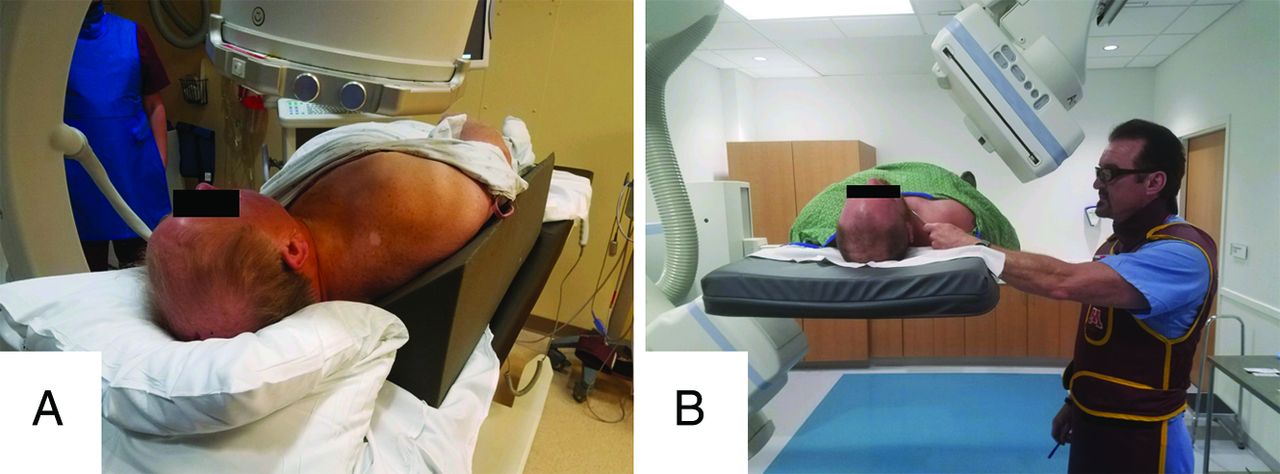
  • Fig 2.
    • Download figure
    • Open in new tab
    • Download powerpoint
    Fig 2.

    A, Patient positioned obliquely on a 45° wedge for right-sided injection. B, The patient positioned supine for right-sided injection with the proceduralist using fluoroscopy to position the clamp before marking the skin. Table and C-arm are raised to eye-level to facilitate proceduralist visualization during the procedure.

  • Fig 3.
    • Download figure
    • Open in new tab
    • Download powerpoint
    Fig 3.

    Contrast syringe connected via 2 short, low-volume tubing to the hub of the 25-ga spinal needle. The patient is positioned for a left-sided injection.

  • Fig 4.
    • Download figure
    • Open in new tab
    • Download powerpoint
    Fig 4.

    Curved clamp tip placed gently on the skin overlying the posterior-inferior aspect of neural foramen for optimal location, taking care to avoid indenting the skin surface.

  • Fig 5.
    • Download figure
    • Open in new tab
    • Download powerpoint
    Fig 5.

    Ideal needle position in the posterior-inferior corner of the neural foramen (A, arrow) and acceptable needle position (B, arrow).
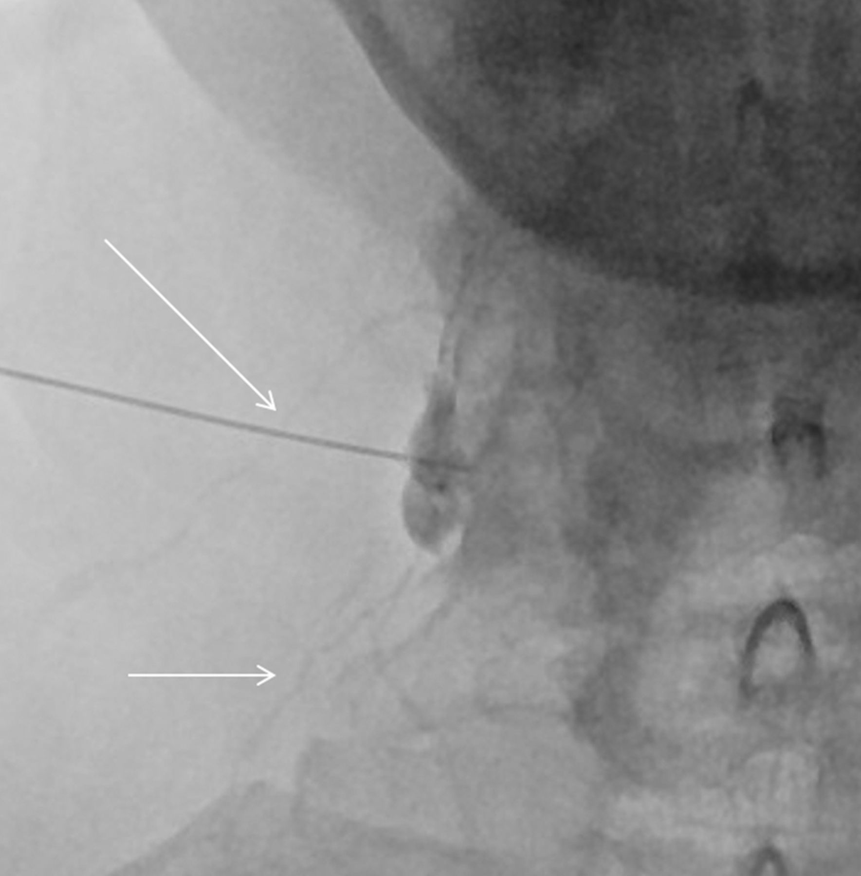
  • Fig 6.
    • Download figure
    • Open in new tab
    • Download powerpoint
    Fig 6.

    Needle tip advanced to the edge of the lateral mass on the anterior-posterior projection (arrow).

  • Fig 7.
    • Download figure
    • Open in new tab
    • Download powerpoint
    Fig 7.

    Contrast in the perineural space along the right C6 nerve root (A, arrow) and clearly outlining the left C7 nerve root (B, arrow). Either result is acceptable.

  • Fig 8.
    • Download figure
    • Open in new tab
    • Download powerpoint
    Fig 8.

    Vascular filling demonstrated during live injection (arrows) requiring needle repositioning.
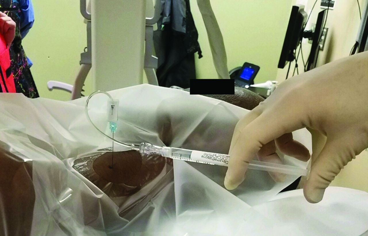
  • Fig 9.
    • Download figure
    • Open in new tab
    • Download powerpoint
    Fig 9.

    Syringe containing 1 mL of dexamethasone, 10 mg/mL; 1 mL of 1% lidocaine; and a small amount of air connected to the remaining tubing segment closest to the hub. The syringe should be held so that the air floats anti-dependently during the injection, not horizontally as shown in this picture, which was taken before injection.

  • Fig 10.
    • Download figure
    • Open in new tab
    • Download powerpoint
    Fig 10.

    Flow chart demonstrates the effectiveness of CF-SCNRB with regard to postprocedural pain assessment at 15 minutes and 2 weeks.

Tables

  • Figures
    • View popup
    Table 1:

    Patient demographics

    Study PopulationMaleFemale
    Average age (yr)51.550.2
    No.123131
    Percentage48.4%51.6%
    • View popup
    Table 2:

    Levels of SCNRBs

    Level of InjectionNo. of Patients
    C49
    C525
    C6115
    C776
    C829
    • View popup
    Table 3:

    Pre- and postprocedural pain level assessmenta

    No. of Patients
    Primary outcomes
     >50% pain relief at 15 min185 (75.2%)
     <50% pain relief at 15 min61 (24.8%)
     >50% pain reduction from baseline at 2 wk165 (67.1%)
     <50% pain reduction from baseline at 2 wk81 (32.9%)
     No preprocedural pain (patient-reported numbness or paresthesia)8 (3.1%)
     Alleviation of preprocedural numbness or paresthesia at 2 wk7 of 8 (87.5%)
    Minor adverse reactions or events
     Vasovagal reaction3 (1.2%)
     Transient pseudo-Horner syndrome (sympathetic blockade)1 (0.4%)
     New onset of diffuse headache2 (0.8%)
     Increase in usual pain in a familiar distribution8 (3.1%)
    • ↵a Transient arm numbness postinjection was not included as an untoward effect because it is expected when exposing cervical nerve roots to a lidocaine containing mixture.

    • View popup
    Table 4:

    Radiation exposure based on timea

    Approximate Corresponding Radiation Incurred
    CT-guided average effective dose0.36 ± 0.41 mSv
    CF average fluoroscopy time of 24.3 sec0.17 mSv
    • ↵a Average CT-guided effective dose was calculated from dose-length product values reported by Lazarus et al11 over 228 SCNRB cases using a conversion factor of 5.4 uSv/mGy × cm for cervical spine examinations developed by Huda et al.21 CF radiation dose is referenced and extrapolated from a direct comparison by Schmid et al27 using an Alderson Rando phantom.

PreviousNext
Back to top

In this issue

American Journal of Neuroradiology: 41 (6)
American Journal of Neuroradiology
Vol. 41, Issue 6
1 Jun 2020
  • Table of Contents
  • Index by author
  • Complete Issue (PDF)
Advertisement
Print
Download PDF
Email Article

Thank you for your interest in spreading the word on American Journal of Neuroradiology.

NOTE: We only request your email address so that the person you are recommending the page to knows that you wanted them to see it, and that it is not junk mail. We do not capture any email address.

Enter multiple addresses on separate lines or separate them with commas.
Counterpoint: Conventional Fluoroscopy-Guided Selective Cervical Nerve Root Block—A Safe, Effective, and Efficient Modality in the Hands of an Experienced Proceduralist
(Your Name) has sent you a message from American Journal of Neuroradiology
(Your Name) thought you would like to see the American Journal of Neuroradiology web site.
CAPTCHA
This question is for testing whether or not you are a human visitor and to prevent automated spam submissions.
Cite this article
F.W. Ott, R. Pluhm, K. Ozturk, A.M. McKinney, J.B. Rykken
Counterpoint: Conventional Fluoroscopy-Guided Selective Cervical Nerve Root Block—A Safe, Effective, and Efficient Modality in the Hands of an Experienced Proceduralist
American Journal of Neuroradiology Jun 2020, 41 (6) 1112-1119; DOI: 10.3174/ajnr.A6580

Citation Manager Formats

  • BibTeX
  • Bookends
  • EasyBib
  • EndNote (tagged)
  • EndNote 8 (xml)
  • Medlars
  • Mendeley
  • Papers
  • RefWorks Tagged
  • Ref Manager
  • RIS
  • Zotero
0 Responses
Respond to this article
Share
Bookmark this article
Counterpoint: Conventional Fluoroscopy-Guided Selective Cervical Nerve Root Block—A Safe, Effective, and Efficient Modality in the Hands of an Experienced Proceduralist
F.W. Ott, R. Pluhm, K. Ozturk, A.M. McKinney, J.B. Rykken
American Journal of Neuroradiology Jun 2020, 41 (6) 1112-1119; DOI: 10.3174/ajnr.A6580
del.icio.us logo Twitter logo Facebook logo Mendeley logo
  • Tweet Widget
  • Facebook Like
  • Google Plus One
Purchase

Jump to section

  • Article
    • Abstract
    • ABBREVIATIONS:
    • MATERIALS AND METHODS
    • RESULTS
    • DISCUSSION
    • CONCLUSIONS
    • References
  • Figures & Data
  • Info & Metrics
  • Responses
  • References
  • PDF

Related Articles

  • No related articles found.
  • PubMed
  • Google Scholar

Cited By...

  • No citing articles found.
  • Crossref
  • Google Scholar

This article has not yet been cited by articles in journals that are participating in Crossref Cited-by Linking.

More in this TOC Section

  • Optimization of Photon Counting CT Myelography
  • Characteristics of SIH Type I Culprit Lesions
  • Management Outcomes For VO Spine Biopsy
Show more Spine Imaging and Spine Image-Guided Interventions

Similar Articles

Advertisement

Indexed Content

  • Current Issue
  • Accepted Manuscripts
  • Article Preview
  • Past Issues
  • Editorials
  • Editor's Choice
  • Fellows' Journal Club
  • Letters to the Editor
  • Video Articles

Cases

  • Case Collection
  • Archive - Case of the Week
  • Archive - Case of the Month
  • Archive - Classic Case

More from AJNR

  • Trainee Corner
  • Imaging Protocols
  • MRI Safety Corner
  • Book Reviews

Multimedia

  • AJNR Podcasts
  • AJNR Scantastics

Resources

  • Turnaround Time
  • Submit a Manuscript
  • Submit a Video Article
  • Submit an eLetter to the Editor/Response
  • Manuscript Submission Guidelines
  • Statistical Tips
  • Fast Publishing of Accepted Manuscripts
  • Graphical Abstract Preparation
  • Imaging Protocol Submission
  • Evidence-Based Medicine Level Guide
  • Publishing Checklists
  • Author Policies
  • Become a Reviewer/Academy of Reviewers
  • News and Updates

About Us

  • About AJNR
  • Editorial Board
  • Editorial Board Alumni
  • Alerts
  • Permissions
  • Not an AJNR Subscriber? Join Now
  • Advertise with Us
  • Librarian Resources
  • Feedback
  • Terms and Conditions
  • AJNR Editorial Board Alumni

American Society of Neuroradiology

  • Not an ASNR Member? Join Now

© 2025 by the American Society of Neuroradiology All rights, including for text and data mining, AI training, and similar technologies, are reserved.
Print ISSN: 0195-6108 Online ISSN: 1936-959X

Powered by HighWire