
Article Figures & Data
Figures
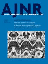
- Fig 1.
Perirolandic sign in 4 different patients with POLG-related disorders (A, An 8-month-old female), (B and C, A 3 year-old-male), (D, An 1-year-old male) and (E and F, A 9-month-old female). Signal changes around the central sulcus were variable with varying degrees of conspicuity. A, T2WI. Signal changes are subtle and focal, evident only in the left precentral gyrus (open arrows). B, T2WI. Signal changes are subtle and focal, evident in the left pre- and postcentral gyrus (open arrows), but more conspicuous in the DWI (open arrows, C). D, DWI. Linear signal changes involving mainly the cortex surrounding the right central sulcus (open arrows). E and F, DWI and ADC map, respectively. Marked signal changes in both right pre- and postcentral gyri.
- Fig 2.
MR imaging thalamic signal changes in 3 different patients with POLG-related disorders (A, A 9-month-old female), (B, A 3-year-old female), and (C, A 3-year-old male). Thalamic signal changes were also variable with varying degrees of conspicuity. A, DWI. Signal changes were subtle and focal with restricted diffusion in the right thalamus (open arrow). B, FLAIR. Signal changes involved both thalami, more conspicuous on the left side (open arrows). C, T2WI. Signal changes were bilateral and symmetric involving both thalami (open arrows).
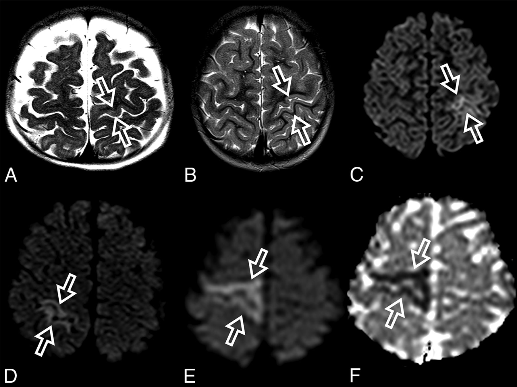
- Fig 3.
ADC map images of a 7-month-year old male with POLG-related disorder, demonstrating a diffuse pattern of leukoencephalopathy with restricted diffusion of the periventricular white matter of the bilateral temporal lobes in A, the occipital lobes in B, and also of the bilateral fornix in C and corpus callosum in D.
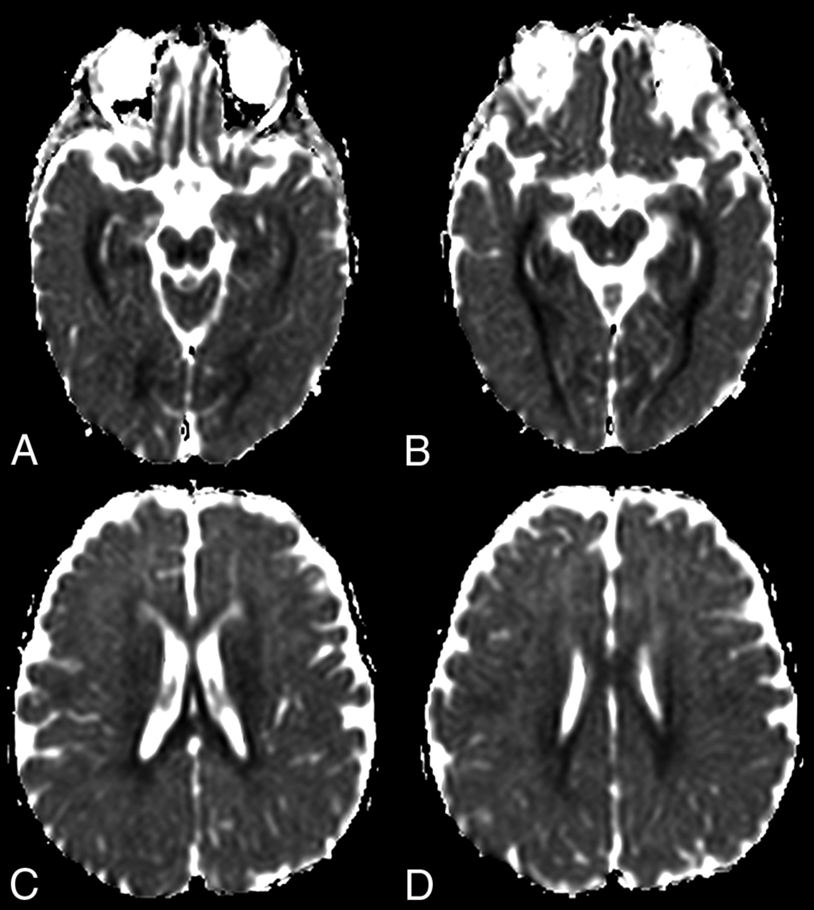
- Fig 4.
Bilateral occipital volume loss, gliosis, and encephalomalacia in a 16-year-old female patient with POLG-related disorder (open arrows).
- Fig 5.
Increased ASL perfusion in 3 different patients with POLG-related disorders (A and B), (C and D), and (E and F). Increased perfusion regions are seen in the right perirolandic region in A (open arrow) and in the left occipital lobe in B (open arrow). Bilateral parietal and occipital increased perfusion is seen in C (open arrows) and in the bilateral cerebellar hemispheres (open arrows) in D. Increased ASL perfusion is seen in the left thalamus in E (solid arrow) and in the left parietal and occipital lobes in E and F (open arrows).


Jump to section
Related Articles
Cited By...
- No citing articles found.