
Article Figures & Data
Figures
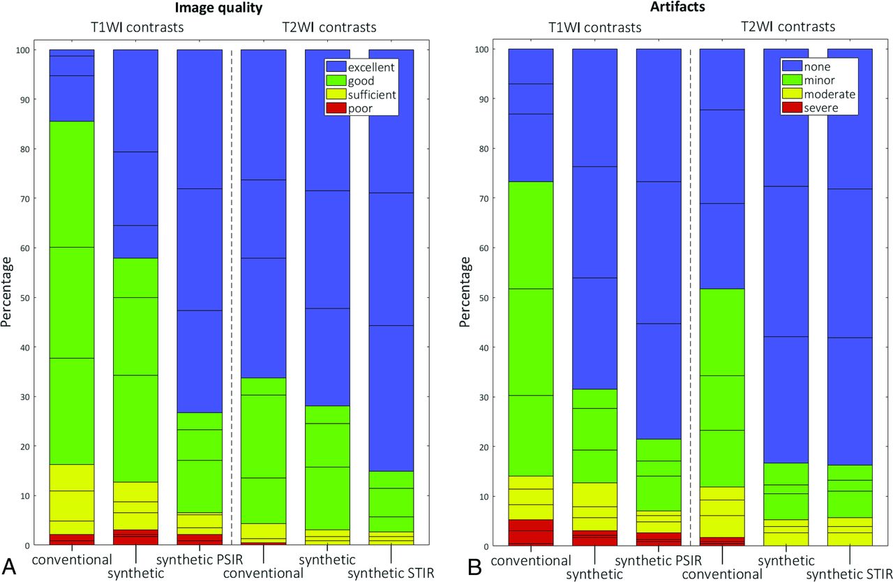
- Fig 1.
Qualitative comparison of T1WI and T2WI quality (A) and image artifacts (B). Horizontal lines within colored portions indicate scoring per rater. PSIR indicates phase-sensitive inversion recovery.
- Fig 2.
Upper row, Conventional T1WI (A) and T2WI (B and C). Lower row, Synthetic T1WI (D) and T2WIs (E and F). Focal WM lesions (small arrows) are clearly visible on both T1WIs. Cystic periventricular leukomalacia (arrows) is seen around the ventricles in both T2WIs.
- Fig 3.
Bland-Altman plots comparing different volumetric metrics calculated with Synthetic MR imaging (labeled SyMRI) and MANTiS. The red line indicates the average relative difference, and the blue dashed lines indicate the average ±1.96.
- Fig 4.
Synthetic CSF segmentation maps: SyMRI algorithm without optimization for the immature neonatal brain and CSF haze in the WM (blue) (A); improved SyMRI (Version 11.1) at the same level (B). Example of CSF overlap to calculate the Dice similarity coefficients (C). The synthetic CSF segment is red, and the MANTiS segment is blue. Regions where the segments overlap are purple. Voxels of the SyMRI CSF segment containing <20% CSF are not shown.


Tables
Category Assessment Scale Overall image quality Poor, sufficient, good, excellent Artifacts Severe, moderate, minor, none Visualization of anatomic structures Legible, illegible Posterior cross-roads (T2/T2 STIR only) Central sulcus Lentiform nucleus Ventrolateral thalamus Dorsal pons Diagnostic performance Present, absent Focal WM lesions Cystic degeneration Sensitivity Specificity Accuracy P Value WM lesions (n = 23/76) Conventional 91% 86% 89% 1.00 Synthetic 89% 77% 86% Cysts (n = 7/76) Conventional 96% 57% 92% .29 Synthetic 100% 64% 95%


Jump to section
Related Articles
Cited By...
- Synthetic MRI and MR Fingerprinting-Derived Relaxometry of Antenatal Human Brainstem Myelination: A Postmortem-Based Quantitative Imaging Study
- Accelerated Nonenhanced 3D T1-MPRAGE Using Wave-Controlled Aliasing in Parallel Imaging for Infant Brain Imaging
- Accelerated Synthetic MRI with Deep Learning-Based Reconstruction for Pediatric Neuroimaging
- Impact of Prematurity on the Tissue Properties of the Neonatal Brain Stem: A Quantitative MR Approach
- Pediatric Head CT: Automated Quantitative Analysis with Quantile Regression