Advertisement

Main menu

  • Home
  • Content
    • Current Issue
    • Accepted Manuscripts
    • Article Preview
    • Past Issue Archive
    • Video Articles
    • AJNR Case Collection
    • Case of the Week Archive
    • Case of the Month Archive
    • Classic Case Archive
  • Special Collections
    • AJNR Awards
    • Low-Field MRI
    • Alzheimer Disease
    • ASNR Foundation Special Collection
    • Photon-Counting CT
    • View All
  • Multimedia
    • AJNR Podcasts
    • AJNR SCANtastic
    • Trainee Corner
    • MRI Safety Corner
    • Imaging Protocols
  • For Authors
    • Submit a Manuscript
    • Submit a Video Article
    • Submit an eLetter to the Editor/Response
    • Manuscript Submission Guidelines
    • Statistical Tips
    • Fast Publishing of Accepted Manuscripts
    • Graphical Abstract Preparation
    • Imaging Protocol Submission
    • Author Policies
  • About Us
    • About AJNR
    • Editorial Board
    • Editorial Board Alumni
  • More
    • Become a Reviewer/Academy of Reviewers
    • Subscribers
    • Permissions
    • Alerts
    • Feedback
    • Advertisers
    • ASNR Home

User menu

  • Alerts
  • Log in

Search

  • Advanced search
American Journal of Neuroradiology

American Journal of Neuroradiology

ASHNR American Society of Functional Neuroradiology ASHNR American Society of Pediatric Neuroradiology ASSR
  • Alerts
  • Log in

Advanced Search

  • Home
  • Content
    • Current Issue
    • Accepted Manuscripts
    • Article Preview
    • Past Issue Archive
    • Video Articles
    • AJNR Case Collection
    • Case of the Week Archive
    • Case of the Month Archive
    • Classic Case Archive
  • Special Collections
    • AJNR Awards
    • Low-Field MRI
    • Alzheimer Disease
    • ASNR Foundation Special Collection
    • Photon-Counting CT
    • View All
  • Multimedia
    • AJNR Podcasts
    • AJNR SCANtastic
    • Trainee Corner
    • MRI Safety Corner
    • Imaging Protocols
  • For Authors
    • Submit a Manuscript
    • Submit a Video Article
    • Submit an eLetter to the Editor/Response
    • Manuscript Submission Guidelines
    • Statistical Tips
    • Fast Publishing of Accepted Manuscripts
    • Graphical Abstract Preparation
    • Imaging Protocol Submission
    • Author Policies
  • About Us
    • About AJNR
    • Editorial Board
    • Editorial Board Alumni
  • More
    • Become a Reviewer/Academy of Reviewers
    • Subscribers
    • Permissions
    • Alerts
    • Feedback
    • Advertisers
    • ASNR Home
  • Follow AJNR on Twitter
  • Visit AJNR on Facebook
  • Follow AJNR on Instagram
  • Join AJNR on LinkedIn
  • RSS Feeds

AJNR Awards, New Junior Editors, and more. Read the latest AJNR updates

Research ArticlePediatric Neuroimaging

Tumor Response Assessment in Diffuse Intrinsic Pontine Glioma: Comparison of Semiautomated Volumetric, Semiautomated Linear, and Manual Linear Tumor Measurement Strategies

L.A. Gilligan, M.D. DeWire-Schottmiller, M. Fouladi, P. DeBlank and J.L. Leach
American Journal of Neuroradiology May 2020, 41 (5) 866-873; DOI: https://doi.org/10.3174/ajnr.A6555
L.A. Gilligan
aFrom the Departments of Radiology (L.A.G., J.L.L.)
cDepartment of Graduate Medical Education (L.A.G., M.D.D.-S.), Mount Carmel Health System, Columbus, Ohio
  • Find this author on Google Scholar
  • Find this author on PubMed
  • Search for this author on this site
  • ORCID record for L.A. Gilligan
M.D. DeWire-Schottmiller
band Cancer and Blood Diseases Institute (M.D.D.-S., M.F.), Cincinnati Children’s Hospital Medical Center, Cincinnati, Ohio
cDepartment of Graduate Medical Education (L.A.G., M.D.D.-S.), Mount Carmel Health System, Columbus, Ohio
  • Find this author on Google Scholar
  • Find this author on PubMed
  • Search for this author on this site
  • ORCID record for M.D. DeWire-Schottmiller
M. Fouladi
band Cancer and Blood Diseases Institute (M.D.D.-S., M.F.), Cincinnati Children’s Hospital Medical Center, Cincinnati, Ohio
d and Departments of Pediatrics (M.F., P.D.)
  • Find this author on Google Scholar
  • Find this author on PubMed
  • Search for this author on this site
  • ORCID record for M. Fouladi
P. DeBlank
d and Departments of Pediatrics (M.F., P.D.)
  • Find this author on Google Scholar
  • Find this author on PubMed
  • Search for this author on this site
  • ORCID record for P. DeBlank
J.L. Leach
aFrom the Departments of Radiology (L.A.G., J.L.L.)
e Radiology (J.L.L.), University of Cincinnati College of Medicine, Cincinnati, Ohio.
  • Find this author on Google Scholar
  • Find this author on PubMed
  • Search for this author on this site
  • ORCID record for J.L. Leach
  • Article
  • Figures & Data
  • Info & Metrics
  • Responses
  • References
  • PDF
Loading

Article Figures & Data

Figures

  • Tables
  • Fig 1.
    • Download figure
    • Open in new tab
    • Download powerpoint
    Fig 1.

    Sample case demonstrating measurement methods. A, Manual clinical transaxial (2D) measurements. Largest dimension identified on axial images and perpendicular short axis dimension performed and reported in the clinical radiology report and used for derivation of tumor response. B, Semiautomated 2D measurements. Tumor margins are traced (red outline) on each image, and automated 2D measurements (largest long axis dimension and perpendicular short axis dimension, blue lines) are automatically derived, along with tumor volume in the mint Lesion software package. C, Semiautomated 2D measurements and volumes performed during the treatment course. Imaging performed after cycles 2, 4, 6, and 8.
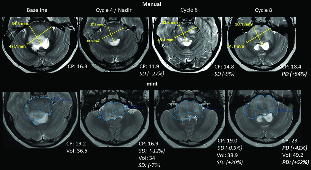
  • Fig 2.
    • Download figure
    • Open in new tab
    • Download powerpoint
    Fig 2.

    Sample case demonstrating clinical 2D measurements (clinical CP) and semiautomated 2D and volumetric measurements (semiautomated CP) during the treatment course. Note that in this case, although there were differences in orientation of the measurements with the semiautomated process, response classification was the same compared with manual clinical CP measurements.
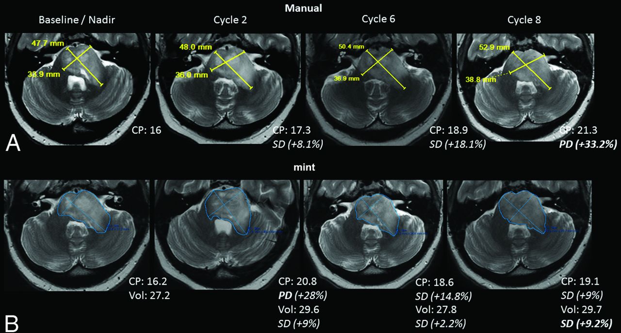
  • Fig 3.
    • Download figure
    • Open in new tab
    • Download powerpoint
    Fig 3.

    Sample case demonstrating clinical 2D measurements (clinical CP) and semiautomated 2D and volumetric measurements (semiautomated CP) during the treatment course. In this case, per protocol, imaging progression based on clinical CP (A) was called after cycle 8 (33.2% increase in CP). With semiautomated CP (B), progressive disease would have been called (based solely on imaging) after cycle 2 (28% increase). This is due to a different section choice as a maximum transaxial dimension and slightly different measurement orientation (B, cycle 2). Subsequently, however, on the basis of a protocol comparing with smallest CP during treatment (baseline), stable disease would have been called. Note that although the CP increased 28% (PD) after cycle 2, the tumor volume only increased 9% (SD). Such discrepancies were common when comparing treatment-response strategies.
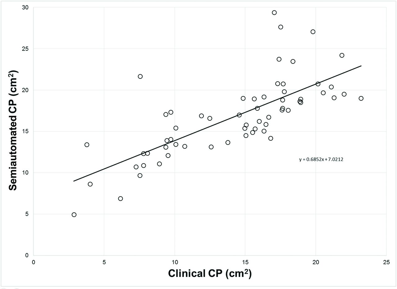
  • Fig 4.
    • Download figure
    • Open in new tab
    • Download powerpoint
    Fig 4.

    Clinically derived tumor CP versus semiautomated software–derived tumor CP for all time points with a linear trendline (r = 0.74, P < .0001).

  • Fig 5.
    • Download figure
    • Open in new tab
    • Download powerpoint
    Fig 5.

    Bland-Altman plot demonstrating bias between clinical and semiautomated tumor CP for all time points. The solid line indicates a mean bias between techniques. Dashed lines indicate ±2 SDs of the mean (95% limits of agreement). Overall, clinical CP measured less than semiautomated CP (mean bias, −2.5). Outliers (>1.96 SDs) were predominantly noted in larger tumors.

  • Fig 6.
    • Download figure
    • Open in new tab
    • Download powerpoint
    Fig 6.

    Correlation of percentage change from prior examination in clinical CP versus semiautomated CP (r = 0.36, P = .011).
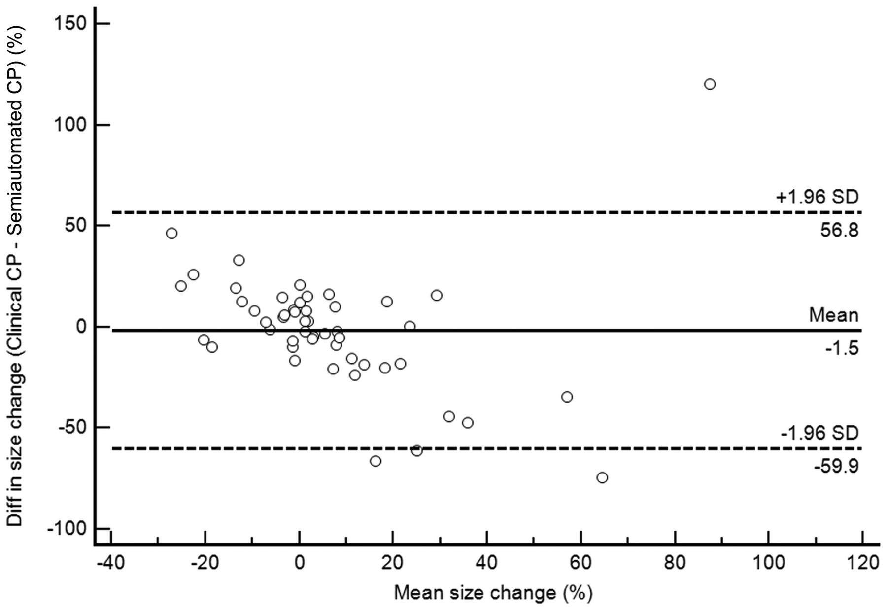
  • Fig 7.
    • Download figure
    • Open in new tab
    • Download powerpoint
    Fig 7.

    Bland-Altman plot demonstrating bias between the percentage change in clinical and semiautomated tumor CP from a prior examination. The solid line indicates mean bias between techniques. Dashed lines indicate ±2 SDs of the mean (95% limits of agreement). Overall, percentage change in clinical CP was smaller than the percentage change in semiautomated CP (mean bias, −1.5%, 95% limits of agreement, −59.9, +56.8) between time points.

Tables

  • Figures
    • View popup
    Table 1:

    Response assessment classifications of 50 MR imaging time points for 3 tumor-management strategiesa

    Clinical CPSemiautomated CPVolume (25/50)Volume (40/65)
    PRSDPDPRSDPDPRSDPDPRSDPD
    Clinical CP
     PR000000000
     SD22514223161328
     PD027027036
     Concordance64%60%76%
    Semiautomated CP
     PR200110
     SD02070252
     PD05160912
     Concordance76%76%
    Volume
     PR110
     SD0250
     PD0914
     Concordance80%
    • ↵a Two different tumor-response criteria were applied to the volume data (25/50 uses the same criteria as used for CP-response determinations, 40/65 denotes PR as >65% decrease from baseline and PD as >40% increase from the prior lowest tumor size). Unless otherwise noted, values represent the number of MR imaging time points.

    • View popup
    Table 2:

    Descriptive statistics of the percentage change in tumor size from baseline or nadir to follow-up examination (assigned per clinical protocol) for each response assessment classification per tumor measurement strategy

    TRCMean (%)Standard Deviation (%)Minimum (%)Maximum (%)
    CPcCPsaVol1Vol2CPcCPsaVol1Vol2CPcCPsaVol1Vol2CPcCPsaVol1Vol2
    PD57837579647881592718942227224271271
    SD61416813283220-27-68-735125918339
    • Note:—CPc indicates clinical cross-product; CPsa, semiautomated cross-product; Vol1, volume (25/50% tumor response criteria); Vol2, volume (40/65% tumor response criteria).

PreviousNext
Back to top

In this issue

American Journal of Neuroradiology: 41 (5)
American Journal of Neuroradiology
Vol. 41, Issue 5
1 May 2020
  • Table of Contents
  • Index by author
  • Complete Issue (PDF)
Advertisement
Print
Download PDF
Email Article

Thank you for your interest in spreading the word on American Journal of Neuroradiology.

NOTE: We only request your email address so that the person you are recommending the page to knows that you wanted them to see it, and that it is not junk mail. We do not capture any email address.

Enter multiple addresses on separate lines or separate them with commas.
Tumor Response Assessment in Diffuse Intrinsic Pontine Glioma: Comparison of Semiautomated Volumetric, Semiautomated Linear, and Manual Linear Tumor Measurement Strategies
(Your Name) has sent you a message from American Journal of Neuroradiology
(Your Name) thought you would like to see the American Journal of Neuroradiology web site.
CAPTCHA
This question is for testing whether or not you are a human visitor and to prevent automated spam submissions.
Cite this article
L.A. Gilligan, M.D. DeWire-Schottmiller, M. Fouladi, P. DeBlank, J.L. Leach
Tumor Response Assessment in Diffuse Intrinsic Pontine Glioma: Comparison of Semiautomated Volumetric, Semiautomated Linear, and Manual Linear Tumor Measurement Strategies
American Journal of Neuroradiology May 2020, 41 (5) 866-873; DOI: 10.3174/ajnr.A6555

Citation Manager Formats

  • BibTeX
  • Bookends
  • EasyBib
  • EndNote (tagged)
  • EndNote 8 (xml)
  • Medlars
  • Mendeley
  • Papers
  • RefWorks Tagged
  • Ref Manager
  • RIS
  • Zotero
0 Responses
Respond to this article
Share
Bookmark this article
Tumor Response Assessment in Diffuse Intrinsic Pontine Glioma: Comparison of Semiautomated Volumetric, Semiautomated Linear, and Manual Linear Tumor Measurement Strategies
L.A. Gilligan, M.D. DeWire-Schottmiller, M. Fouladi, P. DeBlank, J.L. Leach
American Journal of Neuroradiology May 2020, 41 (5) 866-873; DOI: 10.3174/ajnr.A6555
del.icio.us logo Twitter logo Facebook logo Mendeley logo
  • Tweet Widget
  • Facebook Like
  • Google Plus One
Purchase

Jump to section

  • Article
    • Abstract
    • ABBREVIATIONS:
    • MATERIALS AND METHODS
    • RESULTS
    • DISCUSSION
    • CONCLUSIONS
    • Footnotes
    • References
  • Figures & Data
  • Info & Metrics
  • Responses
  • References
  • PDF

Related Articles

  • No related articles found.
  • PubMed
  • Google Scholar

Cited By...

  • Automated Pediatric Brain Tumor Imaging Assessment Tool from CBTN: Enhancing Suprasellar Region Inclusion and Managing Limited Data with Deep Learning
  • Development and Practical Implementation of a Deep Learning-Based Pipeline for Automated Pre- and Postoperative Glioma Segmentation
  • Crossref
  • Google Scholar

This article has not yet been cited by articles in journals that are participating in Crossref Cited-by Linking.

More in this TOC Section

Pediatric Neuroimaging

  • Frontal Paraventricular Cysts
  • Sodium MRI in Pediatric Brain Tumors
  • FRACTURE MR in Congenital Vertebral Anomalies
Show more Pediatric Neuroimaging

Functional

  • Kurtosis and Epileptogenic Tubers: A Pilot Study
  • Glutaric Aciduria Type 1: DK vs. Conventional MRI
  • Multiparametric MRI in PEDS Pontine Glioma
Show more Functional

Similar Articles

Advertisement

Indexed Content

  • Current Issue
  • Accepted Manuscripts
  • Article Preview
  • Past Issues
  • Editorials
  • Editor's Choice
  • Fellows' Journal Club
  • Letters to the Editor
  • Video Articles

Cases

  • Case Collection
  • Archive - Case of the Week
  • Archive - Case of the Month
  • Archive - Classic Case

More from AJNR

  • Trainee Corner
  • Imaging Protocols
  • MRI Safety Corner
  • Book Reviews

Multimedia

  • AJNR Podcasts
  • AJNR Scantastics

Resources

  • Turnaround Time
  • Submit a Manuscript
  • Submit a Video Article
  • Submit an eLetter to the Editor/Response
  • Manuscript Submission Guidelines
  • Statistical Tips
  • Fast Publishing of Accepted Manuscripts
  • Graphical Abstract Preparation
  • Imaging Protocol Submission
  • Evidence-Based Medicine Level Guide
  • Publishing Checklists
  • Author Policies
  • Become a Reviewer/Academy of Reviewers
  • News and Updates

About Us

  • About AJNR
  • Editorial Board
  • Editorial Board Alumni
  • Alerts
  • Permissions
  • Not an AJNR Subscriber? Join Now
  • Advertise with Us
  • Librarian Resources
  • Feedback
  • Terms and Conditions
  • AJNR Editorial Board Alumni

American Society of Neuroradiology

  • Not an ASNR Member? Join Now

© 2025 by the American Society of Neuroradiology All rights, including for text and data mining, AI training, and similar technologies, are reserved.
Print ISSN: 0195-6108 Online ISSN: 1936-959X

Powered by HighWire