
Article Figures & Data
Figures
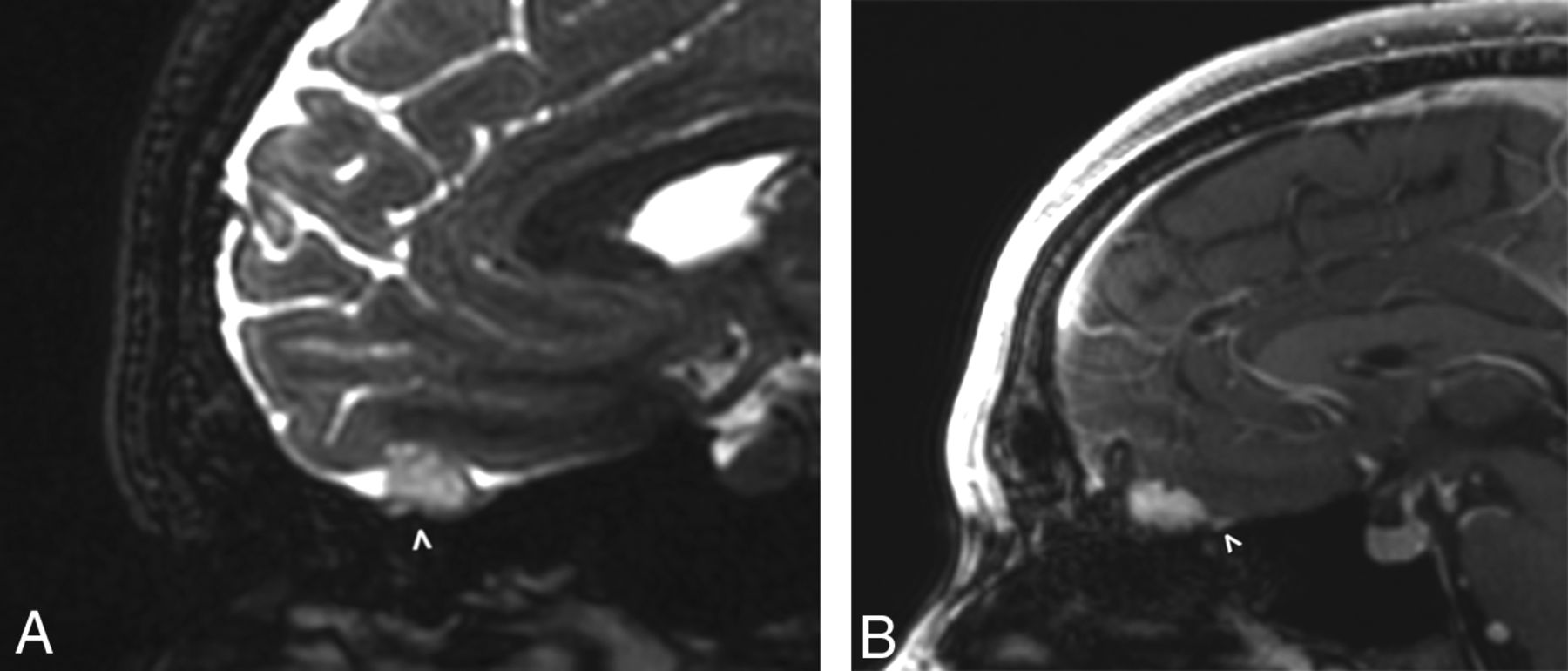
- Fig 1.
A 27-year-old man with mild hyperprolactinemia (case 1). Sagittal FIESTA (A) demonstrates a T2 hyperintense apparent extra-axial mass (arrowhead) within the left anterior cranial fossa. On the sagittal postcontrast T1-weighted image (B), the mass has uniform enhancement with a subtle adjacent dural tail (arrowhead). On imaging, this was believed to be a meningioma, but at resection, it was determined to be a pilocytic astrocytoma.
- Fig 2.
A 46-year-old woman with left inferior frontal grade III diffuse anaplastic oligodendroglioma previously treated with resection and chemoradiation (case 3). Axial and coronal FLAIR images obtained 8 years after the initial treatment (A) demonstrate a left temporal resection cavity (asterisk) and unchanged FLAIR signal in the left frontal lobe (arrow). There is subtle nodular FLAIR signal abnormality (arrowheads) of the left olfactory bulb. Axial and coronal FLAIR images obtained 16 years after the initial treatment (B) show minimal change around the resection cavity (asterisk) but progressive enlargement of the hyperintense left olfactory bulb (arrowheads).
- Fig 3.
A 53-year-old man with a right temporal anaplastic oligodendroglioma previously treated with subtotal resection and chemoradiation (case 4). Axial FLAIR image (A) obtained 7 years after the initial treatment demonstrates masslike FLAIR signal abnormality within the right olfactory cortex as well as the right olfactory tract and bulb (arrowhead), compatible with oligodendroglioma involvement. Coronal FLAIR image (B) obtained at the same time illustrates an asymmetric masslike FLAIR signal abnormality within the right olfactory tract and bulb (arrowhead), while the contralateral olfactory bulb remains normal in caliber.
- Fig 4.
A 10-year-old boy with right anterior temporal grade III anaplastic astrocytoma detected 8 years prior (case 5). Coronal FLAIR image (A) demonstrates a nonenhancing masslike signal abnormality within the right olfactory tract (arrowhead), while an axial FLAIR image (B) obtained at the same time demonstrates a nonenhancing masslike signal abnormality within the left internal auditory canal (arrowhead) as well as in the prepontine cistern, along the presumed location of the left abducens nerve/CN VI (arrow). Combined, findings are compatible with leptomeningeal disease spread involving multiple cranial nerves.
- Fig 5.
A 15-year-old boy presenting for follow-up of diffuse midline glioma initially diagnosed 2 years earlier and treated with chemoradiation (case 6). An axial FLAIR image (A) demonstrates nonenhancing T2/FLAIR signal abnormality of the bilateral olfactory nerves (arrowheads) and left dorsal pons (arrow), as well as cortical thickening and signal abnormality in the left anterior temporal lobe. Coronal FLAIR image (B) illustrates thickening and FLAIR signal abnormality involving the bilateral olfactory nerves (arrowheads) as well as multiple cortical areas in the bilateral frontal lobes (arrows). Findings are compatible with multifocal glioma involvement.
- Fig 6.
A 37-year-old man with a history of right frontal operculum GBM resected 4 years prior (case 10). Coronal FLAIR image demonstrates masslike FLAIR signal abnormality in the left olfactory nerve and surrounding left inferior frontal lobe (arrowhead), pathologically proved to reflect disease recurrence on resection.
- Fig 7.
A 36-year-old man with a history of IDH1 wild-type GBM centered in the corpus callosum, treated with radiation and TMZ (case 11). Coronal postcontrast T1-weighted echo-spoiled gradient echo image (A) obtained 1 year from initial treatment demonstrates abnormal enlargement and patchy enhancement of the left olfactory nerve (arrowhead). On a follow-up coronal postcontrast T1-weighted spin-echo image obtained 3 months later (B), the enhancing mass has enlarged (arrowhead) and appears to involve the leptomeningeal space, compatible with progressive recurrence.
- Fig 8.
A 31-year-old man originally treated for right frontal lobe grade III anaplastic astrocytoma 6 years prior and now presenting with tumor transformation to an IDH1-mutant, 1p19q-codeleted GBM (case 12). Coronal and axial FLAIR images on re-presentation demonstrate a bulky nonenhancing masslike signal abnormality involving the right olfactory cortex (arrowheads) and olfactory nerve (arrow), compatible with GBM involvement.


Jump to section
Related Articles
Cited By...
- No citing articles found.