
Article Figures & Data
Figures
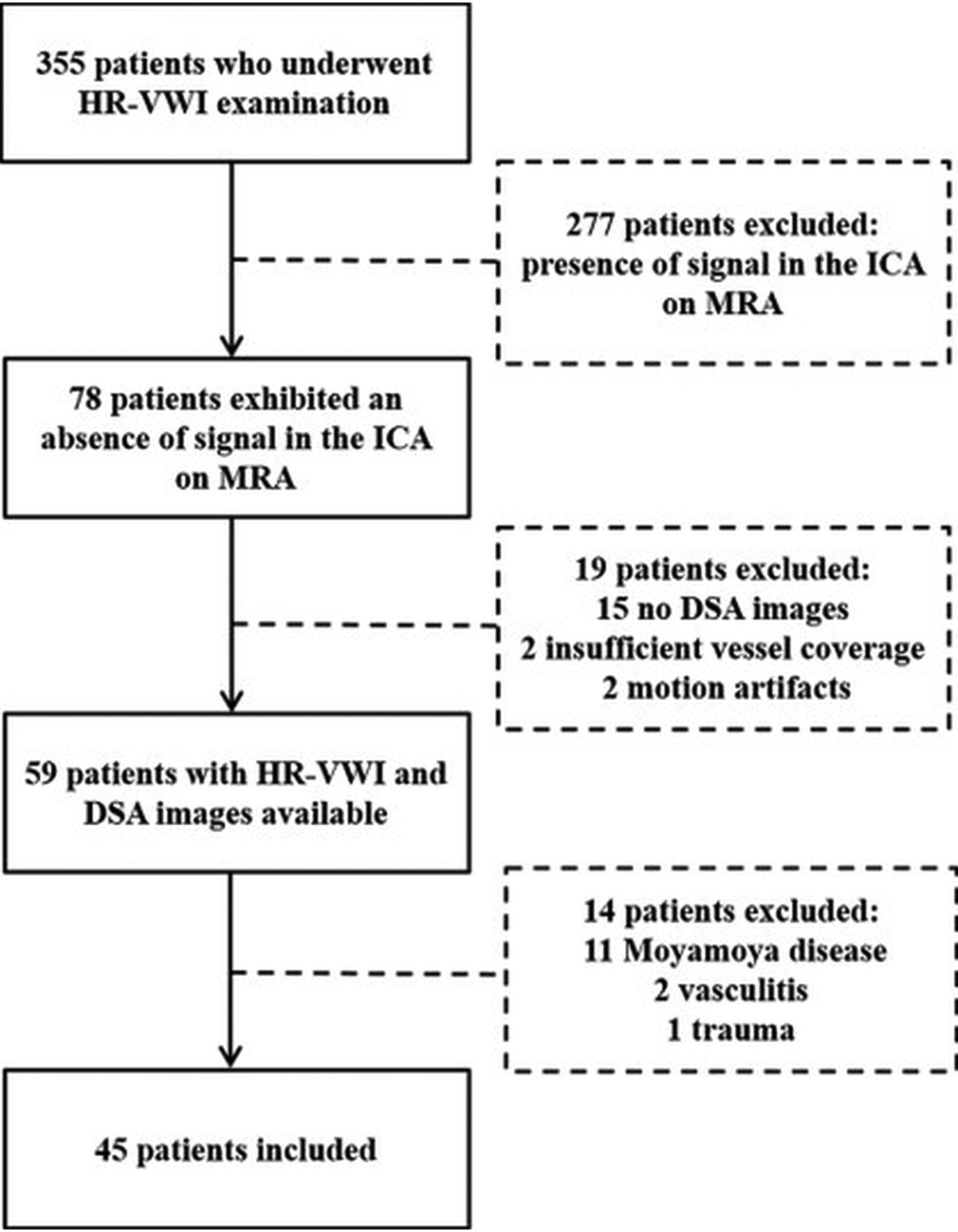
- Fig 1.
Flow chart of patient selection.
- Fig 2.
An example of tandem occlusion of the ICA. A, MRA shows an absence of signal in the left ICA. B, DSA presents contrast cutoff (arrow) at the level of the carotid bulb with distal ICA reconstitution (arrowhead). C, HR-VWI shows occlusion from the carotid bulb to the clinoid segment of the left ICA (arrowhead), with reconstitution at the supraclinoid segment, which was considered unsuitable for recanalization. LICA indicates left ICA.
- Fig 3.
An example of apparent right internal carotid tandem occlusion on MRA (A) and preoperative DSA (B, arrow). C, HR-VWI shows a patent ICA with a narrow residual cavity (arrowhead), defined as near-occlusion with full collapse, which was considered suitable for recanalization. D, Postoperative DSA demonstrates successful recanalization with TICI 3. RICA indicates right ICA.
- Fig 4.
The extent of occlusion of 45 patients and their suitability for recanalization evaluated by DSA and VWI. The black part represents occlusion or near-occlusion. OA indicates ophthalmic artery.


Tables
HR-VWI DSA Total IICA Occlusion EICA Occlusion Tandem Occlusion Near-Occlusion IICA occlusion 4 (8.9%) 0 (0.0%) 2 (4.4%) 0 (0.0%) 6 (13.3%) EICA occlusion 0 (0.0%) 1 (2.2%) 6 (13.3%) 0 (0.0%) 7 (15.6%) Tandem occlusiona 0 (0.0%) 0 (0.0%) 18 (40.0%) 0 (0.0%) 18 (40.0%) Near-occlusion 0 (0.0%) 0 (0.0%) 8 (17.8%) 6 (13.3%) 14 (31.1%) Total 4 (8.9%) 1 (2.2%) 34 (75.6%) 6 (13.3%) 45 (100%) ↵a Tandem occlusion, concomitant extracranial ICA ipsilateral to the intracranial ICA occlusion. Fisher exact test: P < .001.
- Table 2:
Performance of HR-VWI versus DSA in assessing tandem occlusion and suitability for recanalization of ICAa
DSA HR-VWI Present Absent Suitable Unsuitable HR-VWI present 18 (40.0%) 0 (0.0%) 4 (8.9%) 14 (31.1%) HR-VWI absent 16 (35.6%) 11 (24.4%) 27 (60.0%) 0 (0.0%) DSA (suitability for recanalization) Suitable 9 (20.0%) 11 (24.4%) 20 (44.4%) 0 (0.0%) Unsuitable 25 (55.6%) 0 (0.0%) 11 (24.4%) 14 (31.1%) ↵a “Present” means tandem occlusion–positive; “Absent” means tandem occlusion–negative. The table shows the proportional relationship between the presence of tandem occlusion and the suitablity for recanalization. The percentage was calculated on the basis of a total number of patients of 45.


