
Article Figures & Data
Figures
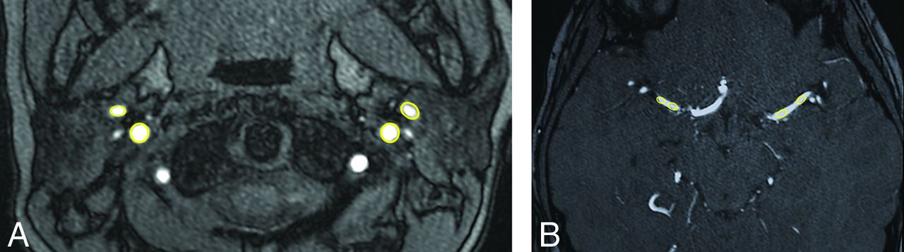
- Fig 1.
Measurement of arterial signal intensity from TOF-MRA using ROI analysis. A, Prestenotic ROI. B, Poststenotic ROI.
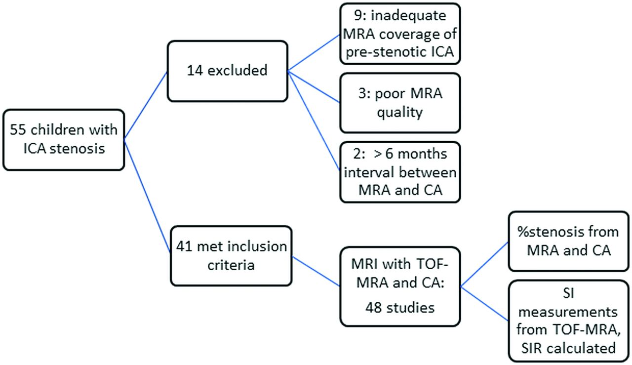
- Fig 2.
Flow chart showing patient selection for our study. SIR indicates signal intensity ratios.
- Fig 3.
A–C, A 15-month-old boy with intracranial vasculopathy and bilateral intracranial arterial stenosis. A, Maximum-intensity-projection reformat from TOF-MRA shows bilateral terminal ICA stenosis, measured on MRA as right: 100%; left: 86%. SI ratios were measured as right: 1.0; left: 1.3. B, The child presented with extensive MCA territory infarct. C, MR imaging performed 5 years later. Axial FLAIR image shows no new infarcts in the left cerebral hemisphere. D–F. A 15-year-old boy with right-sided intracranial arteriopathy. D, Maximum-intensity-projection reformat from TOF-MRA shows 60% stenosis of the supraclinoid right ICA, with additional mild narrowing of the proximal A1 anterior cerebral artery and a normal appearance of the left ICA. The SI ratios were measured as right: 0.60; left: 1.3. E, The patient presented with acute infarction of the right basal ganglia. F, MRA performed 22 months later shows near-complete resolution of the supraclinoid ICA stenosis, consistent with transient cerebral arteriopathy.
- Fig 4.
Receiver operator characteristic curve for post-/prestenotic SI ratios to diagnose severe (>70%) intracranial arterial stenosis. The area under the curve was 0.86, with a determined cutoff of 1.00 (see text).


Tables
Sites Right 14 Left 12 Bilateral 22 Associated with infarct 36/70 (51.4%) Specific location Cervical and petrous 4 (6%) Cavernous 3 (4%) Postclinoid 20 (29%) Postcommunicating and terminal 21 (30%) Postterminal (A1 ACA or M1 MCA) 22 (31%) Note:—ACA indicates anterior cerebral artery.
- Table 2:
Underlying diagnosis in our cohort of children with stroke and intracranial arterial stenosis (n = 41)
Clinical Diagnosis No. Moyamoya disease 15 Neurofibromatosis type 1 with Moyamoya 6 Trisomy-21 with Moyamoya 3 Dissection 2 Primary CNS vasculitis 3 Varicella vasculitis 2 TCA 3 Sickle cell disease with Moyamoya 3 Other Systemic large vessel vasculitis 1 Hemolytic uremic syndrome 1 Thalassemia with Moyamoya 1 Hurler syndrome 1 Note:—TCA indicates transient cerebral arteriopathy.
- Table 3:
ICA stenosis severity, comparison of MRA with catheter angiography evaluations (n = 96 arteries: 70 stenotic and 26 nonstenotic)
% Stenosis Arteries on MRA (No.) Arteries on CA (No.) ≤50 31 29 51–69 16 21 70–99 4 32 100 46 14


