
Article Figures & Data
Figures
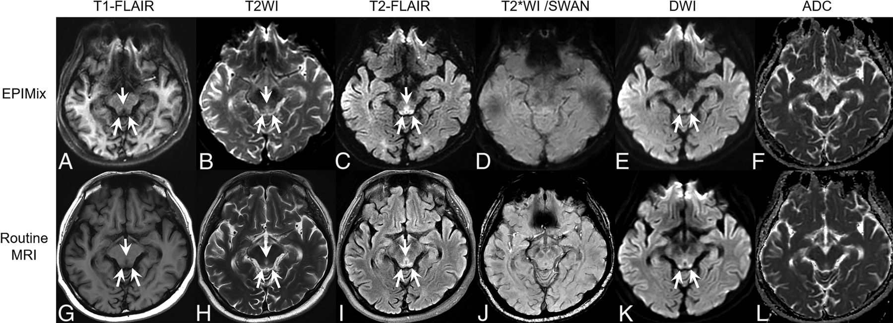
- Fig 1.
A 59-year-old woman with intracranial hemorrhage. A right thalamic hemorrhage with intraventricular extension to the right lateral ventricle shows mainly hyperintensity on T1-FLAIR (A and G), hyperintensity on T2WI (B and H) and T2-FLAIR (C and I), blooming artifacts on T2*WI (D) and SWAN (J), and diffusion restriction on DWI (E and K) (arrows in each sequence). These findings are well-visualized on both EPIMix MR imaging (A–F) and routine MR imaging (G–L).
- Fig 2.
A 48-year-old woman with brain abscesses. There are 2 irregularly shaped masslike lesions with perilesional edema in the right temporal lobe. The internal content shows hypointensity on T1-FLAIR (A and G) and hyperintensity on T2WI (B and H) and T2-FLAIR (C and I) (arrows). The internal content shows diffusion restriction on DWI (E and K) (arrows). There are blooming artifacts on T2*WI (D) and SWAN (J) (arrows). These findings are well-visualized on both EPIMix MR imaging (A–F) and routine MR imaging (G–L).
- Fig 3.
A 52-year-old man with Wernicke encephalopathy. Signal changes in the tectal plate of the midbrain and periaqueductal gray matter showing hypointensity on T1-FLAIR (A and G), hyperintensity on T2WI (B and H) and T2-FLAIR (C and I), and hyperintensity on DWI (E and K) are well-visualized on both EPIMix MR imaging (A–F) and routine MR imaging (G–L) (arrows in each sequence).


Tables
Acquisition parameters of EPIMix and routine protocols
Imaging Parameter EPIMix MR Imaging Routine Brain MR Imaging T1-FLAIR T2WI T2-FLAIR DWI T2*WI T1-FLAIR T2WI T2-FLAIR DWI SWAN FOV (cm) 24 24 24 24 24 22 22 22 22 22 Section thickness (mm) 5 5 5 5 5 5 5 5 5 1.2 TR (ms) 1300 2447 5818 2447 2542 2400 5175 9000 5417 32.8 TI (ms) 570.2 2751 850 2465 TE (ms) 19.3 109 115 109 30.5 25.8 119.4 102.7 73.1 22.9 ETL (ms) 5 26 32 1 3 Frequency matrix 180 180 180 180 180 360 512 288 128 288 Phase matrix 180 180 180 180 180 280 512 288 192 260 Flip angle 90° 90° 90° 90° 90° 90° 90° 90° 90° 15° Bandwidth (kHz) 250 250 250 250 250 50 50 41.67 250 41.67 Parallel imaging acceleration factor ARC 3 ARC 3 ASSET 2.5 ASSET 2.5 Net scan time (min:sec) 1:12 1:58 2:29 2:25 1:19 2:33 Total scan time (min:sec) 1:28 1:58 2:56 2:49 1:37 2:39 Note:—ARC indicates autocalibrating reconstruction for Cartesian imaging; ASSET, array spatial sensitivity encoding technique; ETL, echo-train length.


