
Article Figures & Data
Figures
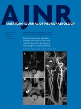
- FIG 1.
Axial SWI of a premature male infant born at 30 weeks’ gestation diagnosed with grade III GM-IVH shows significant siderosis in the ependymal surfaces at the level of the lateral ventricles (A, arrowheads), third ventricle (B, white arrowheads), cerebral aqueduct (B, black arrowhead), and fourth ventricle (C, arrowheads). MR imaging was performed 14 weeks after birth. Note significant SS around the mesencephalon and pons.
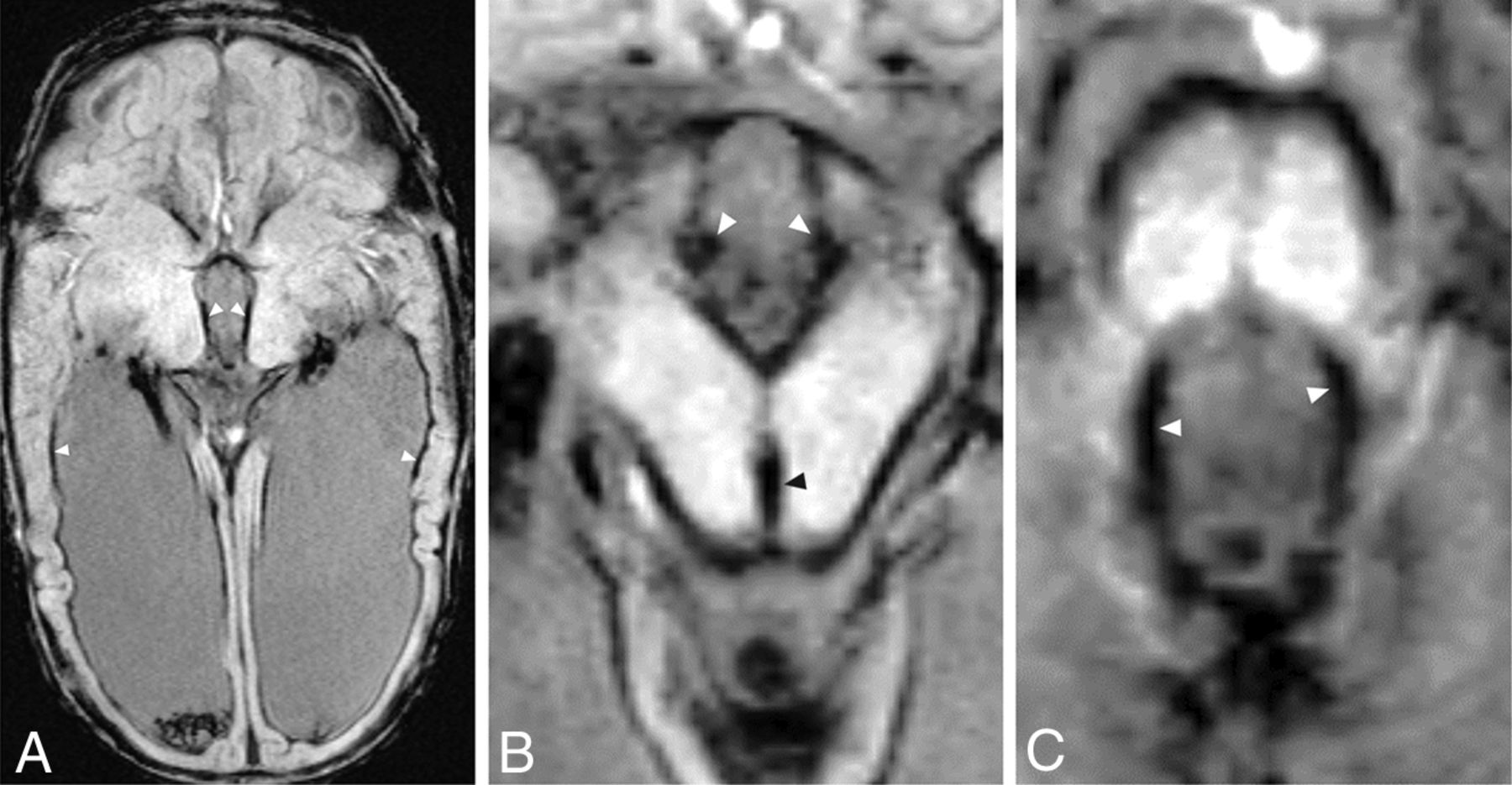
- FIG 2.
Axial SWI depicting SS in the same patient as in Fig 1 at the level of the cervical spinal cord (A), medulla oblongata (B), pons (C), and mesencephalon (D). Arrowheads indicate the point of maximal SS depth within each structure. Note the circumferential distribution of SS around each of these brain stem structures and significant vermian SS.
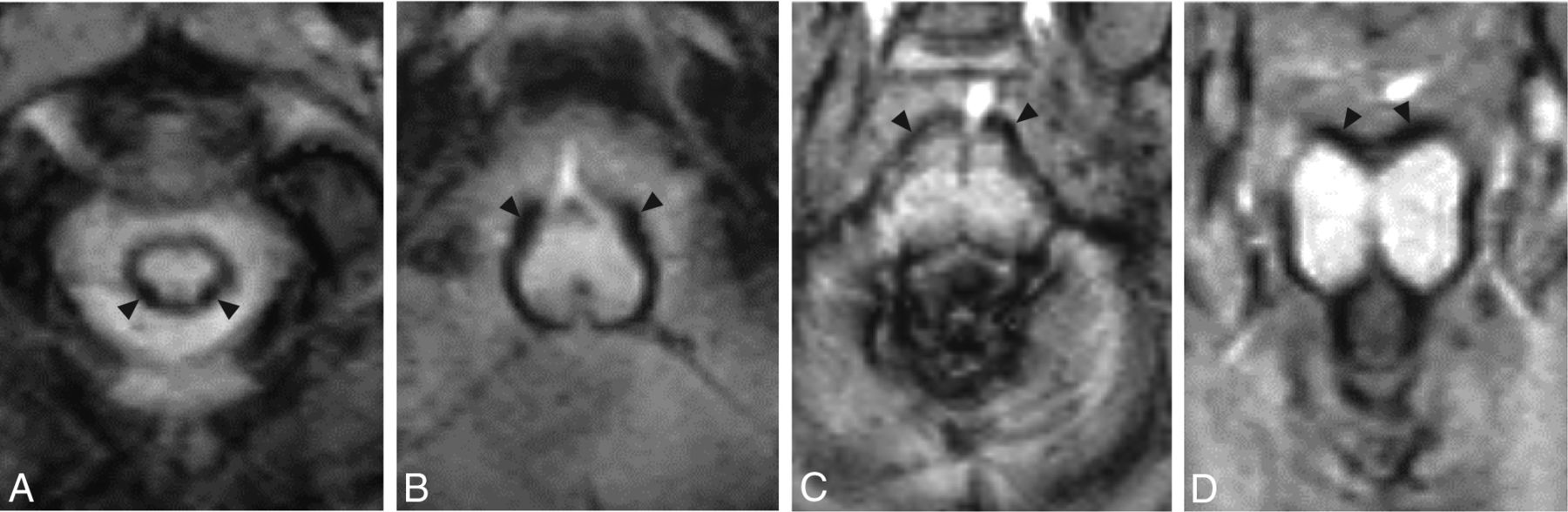
- FIG 3.
Sample axial SWI views at the level of the medulla oblongata in infants diagnosed with GM-IVH grades I (A), II (B), III (C), and IV (D). Note that severity of SS, measured by SS depth and distribution, increases from lower grade to higher grade GM-IVH. Also note the multiple punctate hemorrhages seen in the cerebellum in grades III and IV GM-IVH.
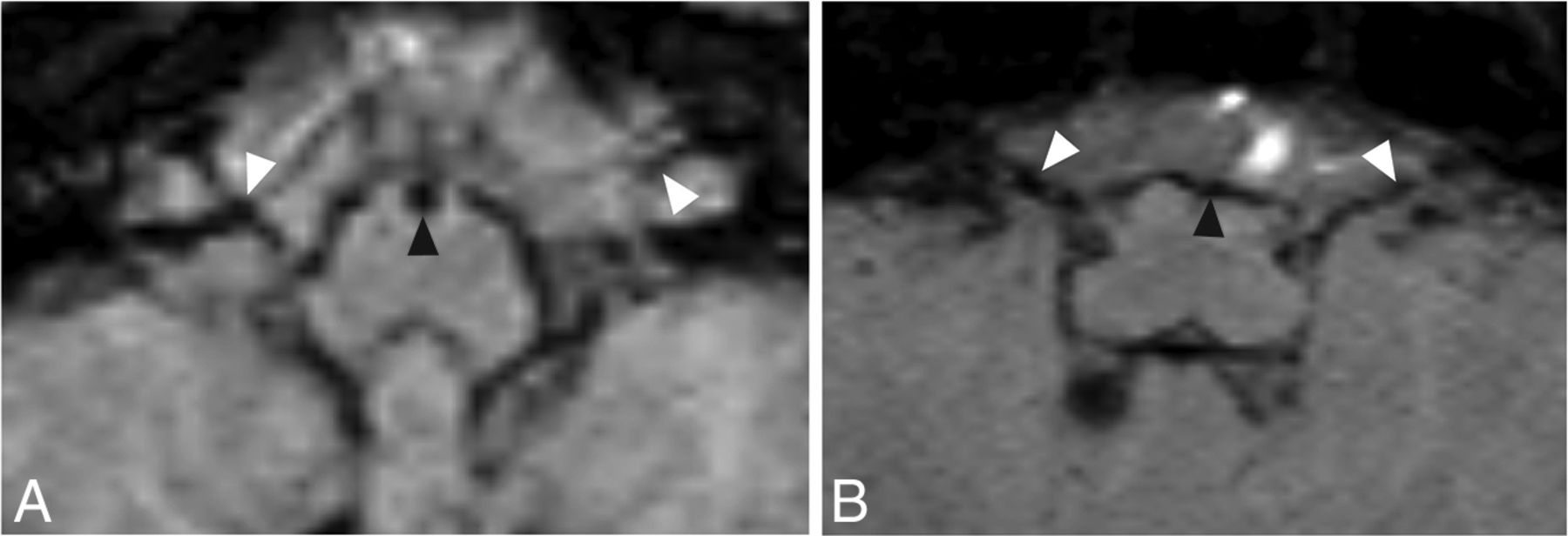
- FIG 4.
Axial MR images from 2 patients at the level of CNs VII and VIII seen on T2-weighted imaging (A and D), T1-weighted imaging (B and E), and SWI (C and F). The arrowhead indicates the positions of the cisternal segment of CNs VII and VIII. Leptomeningeal thickening and significant iron accumulation can be seen in CNs VII and VIII bilaterally. Images were obtained from a boy born at 28 weeks’ gestation with grade III GM-IVH (A–C) and a boy born at 24 weeks’ gestation with grade III GM-IVH (D–F).
- FIG 5.
Initial axial SWI shows significant SS as early as 12 days after birth in a male patient with grade IV GM-IVH in the medulla oblongata (A, black arrowhead) and the CN VII/VIII complex (A, white arrowheads). Follow-up axial SWI of this patient reveals significant residual SS in the medulla oblongata (B, black arrowhead) and CN VII/VIII complex (B, white arrowheads) at 235 days after birth. Note that significant residual SS can also be seen in the CN VII/VIII complex at both time points.


Tables
GM-IVH Grade I II III IV P Brain stem SS (%) 45.5 95.5 95.5 95.5 <.001 Brain stem SS distribution 0 (140.6) 168.8 (140.6) 213.8 (106.9) 247.5 (163.1) <.001 Spinal cord SSD (mm) 0 (0–0.8) 0.65 (0–1.1) 0.8 (0–1.5) 0.75 (0–1.5) <.001 Medulla oblongata SSD (mm) 0 (0–0.8) 0.75 (0–1.2) 1 (0–1.7) 1 (0–2) <.001 Pons SSD (mm) 0 (0–1.2) 0.6 (0–1.1) 0.8 (0–1.7) 0.85 (0–1.8) <.001 Mesencephalon SSD (mm) 0 (0–0.9) 0.6 (0–1.1) 0.8 (0–1.6) 0.8 (0–2.3) <.001 Lateral ventricle SSD (mm) 0.5 (0–1) 0.9 (0–3.3) 1 (0–2) 0.95 (0.4–2.5) <.001 Third ventricle SSD (mm) 0 (0–0.2) 0 (0–1.6) 0 (0–2.2) 0.6 (0–1.2) <.001 Fourth ventricle SSD (mm) 0 (0–0.8) 0.1 (0–0.9) 0.4 (0–1.9) 0.65 (0–2.2) <.001 CN VII/VIII siderosis (%) 9.1 54.5 72.7 54.5 <.001 Total ventricular HV on HUS (mm3) 64.9 (79.8) 491.1 (358.7) 2168.7 (2469) 4319.4 (9731.5) <.001 Total cerebellar HV (mm3) 0 (0–13.6) 0 (0–6333) 0 (0–530) 0.5 (0–1123) .12 Cerebellar SS present (%) 31.8 77.3 72.7 59.1 .009 Cerebellar SS depth (mm) 0 (0–0.25) 0.3 (0–2) 0.35 (0–2.1) 0.43 (0–2.2) .001 Cerebellar hematoma present (%) 27.3 45.5 27.3 50 .26 Note:—HV indicates hematoma volume; SSD, superficial siderosis depth.
↵a Numeric variables are presented as median (minimum-maximum).
GM-IVH Grade Total Ventricular HV (mm3) Cerebellar HV (mm3) r P r P r P Spinal cord SSD (mm) 0.55 <.001 0.55 <.001 0.29 .07 Medulla oblongata SSD (mm) 0.60 <.001 0.63 <.001 0.30 .005 Pons SSD (mm) 0.52 <.001 0.61 <.001 0.20 .06 Mesencephalon SSD (mm) 0.59 <.001 0.62 <.001 0.34 .001 Brain stem SS distribution 0.54 <.001 0.62 <.001 0.25 .018 Lateral ventricle SSD (mm) 0.48 <.001 0.51 <.001 0.32 .02 Third ventricle SSD (mm) 0.46 <.001 0.66 <.001 0.28 .009 Fourth ventricle SSD (mm) 0.44 <.001 0.59 <.001 0.21 .049 Choroid lateral ventricle SSD (mm) 0.67 <.001 0.72 <.001 0.26 .013 Choroid third ventricle SSD (mm) 0.61 <.001 0.73 <.001 0.32 .003 Choroid fourth ventricle SSD (mm) 0.40 <.001 0.47 <.001 0.23 .03 Cerebellar SSD (mm) 0.37 <.001 0.50 <.001 0.46 <.001 Total ventricular HV on HUS (mm3) 0.85 <.001 n/a n/a 0.22 .04 Cerebellar HV (mm3) 0.17 .11 0.22 .04 n/a n/a Note:—n/a indicates not applicable; HV, hematoma volume; SSD, superficial siderosis depth; HUS, head ultrasound.
↵a GMH-IVH hemorrhage was detected by HUS and classified by using the Papile classification.
Brain Stem SS Absent (n = 15) Brain Stem SS Present (n = 73) P Gestational age at birth (wk) 28.6 ± 2.9 27.3 ± 2.9 .11 Sex (% male) 53.3 60.3 .62 Birth weight (g) 1299.5 ± 830 1071.7 ± 407.4 .11 GM-IVH grade I (0) III (2) <.001 Total ventricular HV on HUS (mm3) 91.6 (96.3) 935.1 (2978.8) <.001 Cerebellar hematoma (%) 20 35.6 .24 Cerebellar HV (mm3) 0 (0) 0 (7.9) .12 CV hematoma + (%) 0 19.2 .12 CV HV (mm3) 0 (0) 0 (0) .07 Cerebellar SS + (%) 0 42.5 .001 Lateral ventricle SSD (mm) 0 (0–1) 0.9 (0–3.3) <.001 Third ventricle SSD (mm) 0 (0–0) 0 (0–2.2) <.001 Fourth ventricle SSD (mm) 0 (0–0) 0.4 (0–2.2) <.001 CV SS + (%) 0 72.6 <.001 CN VII/VIII SS + (%) 0 57.5 <.001 CV SS depth (mm) 0 (0–0) 0.4 (0–2.8) <.001 Cerebellar SS depth (mm) 0 (0–0) 0 (0–2.5) .002 Note:—CV indicates cerebellar vermis; GM-IVH, Germinal matrix intraventricular hemorrhage; HV, hematoma volume; SS, superficial siderosis; CN, cranial nerve; +, positive, pathology found; SSD, superficial siderosis depth.
a Numeric variables are presented as mean ± SD or median (minimum-maximum) where appropriate.


