Advertisement

Main menu

  • Home
  • Content
    • Current Issue
    • Accepted Manuscripts
    • Article Preview
    • Past Issue Archive
    • Video Articles
    • AJNR Case Collection
    • Case of the Week Archive
    • Case of the Month Archive
    • Classic Case Archive
  • Special Collections
    • AJNR Awards
    • Low-Field MRI
    • Alzheimer Disease
    • ASNR Foundation Special Collection
    • Photon-Counting CT
    • View All
  • Multimedia
    • AJNR Podcasts
    • AJNR SCANtastic
    • Trainee Corner
    • MRI Safety Corner
    • Imaging Protocols
  • For Authors
    • Submit a Manuscript
    • Submit a Video Article
    • Submit an eLetter to the Editor/Response
    • Manuscript Submission Guidelines
    • Statistical Tips
    • Fast Publishing of Accepted Manuscripts
    • Graphical Abstract Preparation
    • Imaging Protocol Submission
    • Author Policies
  • About Us
    • About AJNR
    • Editorial Board
    • Editorial Board Alumni
  • More
    • Become a Reviewer/Academy of Reviewers
    • Subscribers
    • Permissions
    • Alerts
    • Feedback
    • Advertisers
    • ASNR Home

User menu

  • Alerts
  • Log in

Search

  • Advanced search
American Journal of Neuroradiology

American Journal of Neuroradiology

ASHNR American Society of Functional Neuroradiology ASHNR American Society of Pediatric Neuroradiology ASSR
  • Alerts
  • Log in

Advanced Search

  • Home
  • Content
    • Current Issue
    • Accepted Manuscripts
    • Article Preview
    • Past Issue Archive
    • Video Articles
    • AJNR Case Collection
    • Case of the Week Archive
    • Case of the Month Archive
    • Classic Case Archive
  • Special Collections
    • AJNR Awards
    • Low-Field MRI
    • Alzheimer Disease
    • ASNR Foundation Special Collection
    • Photon-Counting CT
    • View All
  • Multimedia
    • AJNR Podcasts
    • AJNR SCANtastic
    • Trainee Corner
    • MRI Safety Corner
    • Imaging Protocols
  • For Authors
    • Submit a Manuscript
    • Submit a Video Article
    • Submit an eLetter to the Editor/Response
    • Manuscript Submission Guidelines
    • Statistical Tips
    • Fast Publishing of Accepted Manuscripts
    • Graphical Abstract Preparation
    • Imaging Protocol Submission
    • Author Policies
  • About Us
    • About AJNR
    • Editorial Board
    • Editorial Board Alumni
  • More
    • Become a Reviewer/Academy of Reviewers
    • Subscribers
    • Permissions
    • Alerts
    • Feedback
    • Advertisers
    • ASNR Home
  • Follow AJNR on Twitter
  • Visit AJNR on Facebook
  • Follow AJNR on Instagram
  • Join AJNR on LinkedIn
  • RSS Feeds

AJNR Awards, New Junior Editors, and more. Read the latest AJNR updates

Research ArticleClinical Report

Artery of Davidoff and Schechter Supply in Dural Arteriovenous Fistulas

K.D. Bhatia, H. Kortman, T. Wälchli, I. Radovanovic, V.M. Pereira and T. Krings
American Journal of Neuroradiology February 2020, 41 (2) 300-304; DOI: https://doi.org/10.3174/ajnr.A6380
K.D. Bhatia
aFrom the Division of Neuroradiology (K.D.B., H.K., V.M.P., T.K.)
  • Find this author on Google Scholar
  • Find this author on PubMed
  • Search for this author on this site
  • ORCID record for K.D. Bhatia
H. Kortman
aFrom the Division of Neuroradiology (K.D.B., H.K., V.M.P., T.K.)
  • Find this author on Google Scholar
  • Find this author on PubMed
  • Search for this author on this site
  • ORCID record for H. Kortman
T. Wälchli
bJoint Department of Medical Imaging, and Division of Neurosurgery (T.W., I.R., V.M.P., T.K.), Department of Surgery, Toronto Western Hospital, Toronto, Ontario, Canada
cDepartment of Fundamental Neurobiology (T.W., I.R.), Krembil Research Institute, Toronto Western Hospital, University Health Network, University of Toronto, Toronto, Ontario, Canada
dGroup of CNS Angiogenesis and Neurovascular Link, and Physician-Scientist Program (T.W.), Institute for Regenerative Medicine, Neuroscience Center Zurich, Zurich, Switzerland
eDepartment of Health Sciences and Technology (T.W.), Swiss Federal Institute of Technology, Zurich, Zurich, Switzerland
fDivision of Neurosurgery Program (T.W.), University Hospital Zurich, Zurich, Switzerland.
  • Find this author on Google Scholar
  • Find this author on PubMed
  • Search for this author on this site
  • ORCID record for T. Wälchli
I. Radovanovic
bJoint Department of Medical Imaging, and Division of Neurosurgery (T.W., I.R., V.M.P., T.K.), Department of Surgery, Toronto Western Hospital, Toronto, Ontario, Canada
cDepartment of Fundamental Neurobiology (T.W., I.R.), Krembil Research Institute, Toronto Western Hospital, University Health Network, University of Toronto, Toronto, Ontario, Canada
  • Find this author on Google Scholar
  • Find this author on PubMed
  • Search for this author on this site
  • ORCID record for I. Radovanovic
V.M. Pereira
aFrom the Division of Neuroradiology (K.D.B., H.K., V.M.P., T.K.)
bJoint Department of Medical Imaging, and Division of Neurosurgery (T.W., I.R., V.M.P., T.K.), Department of Surgery, Toronto Western Hospital, Toronto, Ontario, Canada
  • Find this author on Google Scholar
  • Find this author on PubMed
  • Search for this author on this site
  • ORCID record for V.M. Pereira
T. Krings
aFrom the Division of Neuroradiology (K.D.B., H.K., V.M.P., T.K.)
bJoint Department of Medical Imaging, and Division of Neurosurgery (T.W., I.R., V.M.P., T.K.), Department of Surgery, Toronto Western Hospital, Toronto, Ontario, Canada
  • Find this author on Google Scholar
  • Find this author on PubMed
  • Search for this author on this site
  • ORCID record for T. Krings
  • Article
  • Figures & Data
  • Supplemental
  • Info & Metrics
  • Responses
  • References
  • PDF
Loading

Article Figures & Data

Figures

  • Tables
  • FIG 1.
    • Download figure
    • Open in new tab
    • Download powerpoint
    FIG 1.

    A, Cerebral DSA (patient 3) via a left vertebral artery injection demonstrating arterial supply to a superior sagittal sinus dural AVF by enlarged bilateral arteries of Davidoff and Schechter. Anterior-posterior projection in the arterial phase shows a bilateral enlarged ADS arising from the superior aspect of the P1–2 junctions of the posterior cerebral arteries and converging (black arrows) into an enlarged falcine artery at the falcotentorial junction, with early venous filling of the superior sagittal sinus. Note the presence of cortical venous reflux. B, Lateral projection shows a bilateral enlarged ADS (black arrows) extending posteriorly along the free edges of the tentorium cerebelli and converging adjacent to the falcotentorial junction.

  • FIG 2.
    • Download figure
    • Open in new tab
    • Download powerpoint
    FIG 2.

    A, Cerebral DSA and endovascular treatment (patient 1) of a falcotentorial dural AVF supplied by the bilateral arteries of Davidoff and Schechter. Anterior-posterior projection of a right vertebral artery injection in the arterial phase shows an enlarged left ADS (black arrow) with early filling of the vein of Galen. Note the mildly enlarged right ADS arising from the right P1–2 junction (white arrow) and the infratentorial venous reflux. B, Axial MIP reconstruction of a 3D rotational angiogram performed via right vertebral artery injection shows the bilateral origins of the ADS from the P1–2 junctions of the posterior cerebral arteries (white arrows). C, Anterior-posterior projection of a microcatheter injection within the right ADS shows early venous filling along the straight and left transverse sinuses. D, Lateral projection of a microcatheter injection within the right ADS shows the course of the vessel along the free edge of the tentorium to the falcotentorial junction, with early venous filling into the vein of Galen. E, Nonsubtracted anterior-posterior projection during right vertebral artery injection after a second-stage transvenous embolization shows no residual early venous filling, with an Onyx cast filling the vein of Galen. F, Spot lateral radiograph of the skull shows the Onyx cast following transarterial and transvenous embolizations.
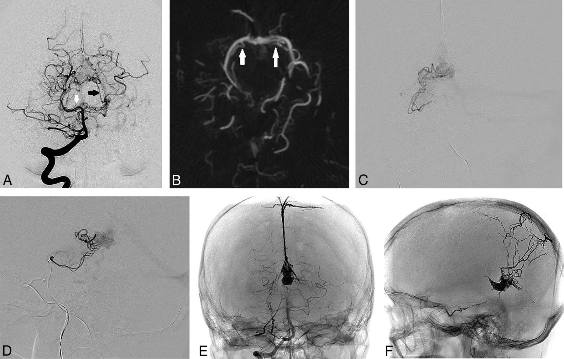
  • FIG 3.
    • Download figure
    • Open in new tab
    • Download powerpoint
    FIG 3.

    A, Cerebral DSA and endovascular treatment (patient 2) of a falcotentorial dural AVF supplied by the bilateral arteries of Davidoff and Schechter. Lateral projection of a left vertebral artery injection shows supply to the dural AVF by the left posterior meningeal artery (white arrow) and faint supply from the ADS (black arrow). B, Lateral projection of a left vertebral artery injection after embolization using Onyx from the posterior meningeal artery shows the residual ADS supply (black arrow) to the inferior surface of the vein of Galen more clearly, with reduced competitive flow. C, Anterior-posterior projection of a microcatheter injection via the left ADS distally shows the remnant fistulous flow to the junction of the vein of Galen and straight sinus at the falcotentorial junction. D, Lateral projection of a microcatheter injection via the left ADS distally shows the residual supply to the dural AVF with deep venous reflux. E, Anterior-posterior projection of a left vertebral artery injection in the arterial phase shows the residual supply to the dural AVF by the enlarged left ADS (black arrow). F, Anterior-posterior projection of a right vertebral artery injection after embolization of the left ADS distal supply using coils and Onyx shows no residual early venous filling with cure of the AVF.

Tables

  • Figures
    • View popup
    Table:

    Risk-reduction strategies for endovascular treatment of falcotentorial dural AVFs

    CategoryRisk-Reduction StrategyPatients Treated
    Anatomic assessment; to identify supply from ADSMagnified high-frame-rate angiography1–6
    3D rotational angiography with multiplanar reconstructions1–6
    Transarterial embolization; strategies to avoid reflux across ADSInitial embolization from non-ADS arterial supplies (to reduce competitive flow)1–6
    Close monitoring for linear reflux anteriorly from the vein of Galen (along the expected course of ADS)1–6
    Embolization directly via ADS; strategies to treat via ADS while avoiding refluxDirect cannulation of ADS with embolization if distal access achieved (pressure-cooker technique to minimize reflux)2
    Aborting embolization attempts from ADS if distal access not achieved (insufficient safety margin)1
    Strategies for residual ADS supplyStaged embolization over multiple sessions as required1, 3, 5
    Transvenous approach (eg, reverse pressure-cooker technique)1
PreviousNext
Back to top

In this issue

American Journal of Neuroradiology: 41 (2)
American Journal of Neuroradiology
Vol. 41, Issue 2
1 Feb 2020
  • Table of Contents
  • Index by author
  • Complete Issue (PDF)
Advertisement
Print
Download PDF
Email Article

Thank you for your interest in spreading the word on American Journal of Neuroradiology.

NOTE: We only request your email address so that the person you are recommending the page to knows that you wanted them to see it, and that it is not junk mail. We do not capture any email address.

Enter multiple addresses on separate lines or separate them with commas.
Artery of Davidoff and Schechter Supply in Dural Arteriovenous Fistulas
(Your Name) has sent you a message from American Journal of Neuroradiology
(Your Name) thought you would like to see the American Journal of Neuroradiology web site.
CAPTCHA
This question is for testing whether or not you are a human visitor and to prevent automated spam submissions.
Cite this article
K.D. Bhatia, H. Kortman, T. Wälchli, I. Radovanovic, V.M. Pereira, T. Krings
Artery of Davidoff and Schechter Supply in Dural Arteriovenous Fistulas
American Journal of Neuroradiology Feb 2020, 41 (2) 300-304; DOI: 10.3174/ajnr.A6380

Citation Manager Formats

  • BibTeX
  • Bookends
  • EasyBib
  • EndNote (tagged)
  • EndNote 8 (xml)
  • Medlars
  • Mendeley
  • Papers
  • RefWorks Tagged
  • Ref Manager
  • RIS
  • Zotero
0 Responses
Respond to this article
Share
Bookmark this article
Artery of Davidoff and Schechter Supply in Dural Arteriovenous Fistulas
K.D. Bhatia, H. Kortman, T. Wälchli, I. Radovanovic, V.M. Pereira, T. Krings
American Journal of Neuroradiology Feb 2020, 41 (2) 300-304; DOI: 10.3174/ajnr.A6380
del.icio.us logo Twitter logo Facebook logo Mendeley logo
  • Tweet Widget
  • Facebook Like
  • Google Plus One
Purchase

Jump to section

  • Article
    • Abstract
    • ABBREVIATIONS:
    • MATERIALS AND METHODS
    • RESULTS
    • DISCUSSION
    • CONCLUSIONS
    • Footnotes
    • References
  • Figures & Data
  • Supplemental
  • Info & Metrics
  • Responses
  • References
  • PDF

Related Articles

  • Erratum
  • PubMed
  • Google Scholar

Cited By...

  • Endovascular Treatment for Tentorial Dural Arteriovenous Fistulas: A Retrospective Single-Center Study
  • Endovascular Management of Intracranial Dural AVFs: Transvenous Approach
  • Endovascular Management of Intracranial Dural Arteriovenous Fistulas: Transarterial Approach
  • Endovascular Management of Intracranial Dural AVFs: Principles
  • Clinical, angiographic, and treatment characteristics of cranial dural arteriovenous fistulas with pial arterial supply
  • Crossref
  • Google Scholar

This article has not yet been cited by articles in journals that are participating in Crossref Cited-by Linking.

More in this TOC Section

Clinical Report

  • Clinical and Imaging Features of Primary Intradural Extramedullary Ewing Sarcoma
  • A Practical Guide to Microtia Scoring: Step-by-Step
  • Revisiting a Rare Anomaly Described 25 Years Ago in the AJNR: A Journey from Pediatric Hemifacial Microsomia and Middle Cranial Fossa Aplasia to CSF-Lymphatic Fistula and Spontaneous Intracranial Hypotension as an Adult
Show more Clinical Report

Neurointervention

  • Clinical and Imaging Features of Primary Intradural Extramedullary Ewing Sarcoma
  • A Practical Guide to Microtia Scoring: Step-by-Step
  • Revisiting a Rare Anomaly Described 25 Years Ago in the AJNR: A Journey from Pediatric Hemifacial Microsomia and Middle Cranial Fossa Aplasia to CSF-Lymphatic Fistula and Spontaneous Intracranial Hypotension as an Adult
Show more Neurointervention

Similar Articles

Advertisement

Indexed Content

  • Current Issue
  • Accepted Manuscripts
  • Article Preview
  • Past Issues
  • Editorials
  • Editor's Choice
  • Fellows' Journal Club
  • Letters to the Editor
  • Video Articles

Cases

  • Case Collection
  • Archive - Case of the Week
  • Archive - Case of the Month
  • Archive - Classic Case

More from AJNR

  • Trainee Corner
  • Imaging Protocols
  • MRI Safety Corner
  • Book Reviews

Multimedia

  • AJNR Podcasts
  • AJNR Scantastics

Resources

  • Turnaround Time
  • Submit a Manuscript
  • Submit a Video Article
  • Submit an eLetter to the Editor/Response
  • Manuscript Submission Guidelines
  • Statistical Tips
  • Fast Publishing of Accepted Manuscripts
  • Graphical Abstract Preparation
  • Imaging Protocol Submission
  • Evidence-Based Medicine Level Guide
  • Publishing Checklists
  • Author Policies
  • Become a Reviewer/Academy of Reviewers
  • News and Updates

About Us

  • About AJNR
  • Editorial Board
  • Editorial Board Alumni
  • Alerts
  • Permissions
  • Not an AJNR Subscriber? Join Now
  • Advertise with Us
  • Librarian Resources
  • Feedback
  • Terms and Conditions
  • AJNR Editorial Board Alumni

American Society of Neuroradiology

  • Not an ASNR Member? Join Now

© 2025 by the American Society of Neuroradiology All rights, including for text and data mining, AI training, and similar technologies, are reserved.
Print ISSN: 0195-6108 Online ISSN: 1936-959X

Powered by HighWire