
Article Figures & Data
Figures
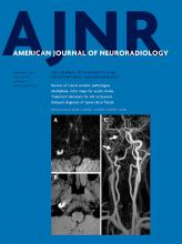
- FIG 1.
Right-sided M1 segment MCA occlusion (arrow). ColorViz summation maps (upper row) show predominantly green vessels in the affected territory (arrowheads), indicating a 1-phase delay; The vessel extent is identical to that of the unaffected hemisphere. The findings are consistent with good pial artery filling and collateral flow. Corresponding conventional mCTA images are displayed in the lower row. The patient received intravenous alteplase, and follow-up MR imaging 24 hours after symptom onset (upper and lower right) shows infarction of the caudate head and lentiform nucleus only, with sparing of the cortex.
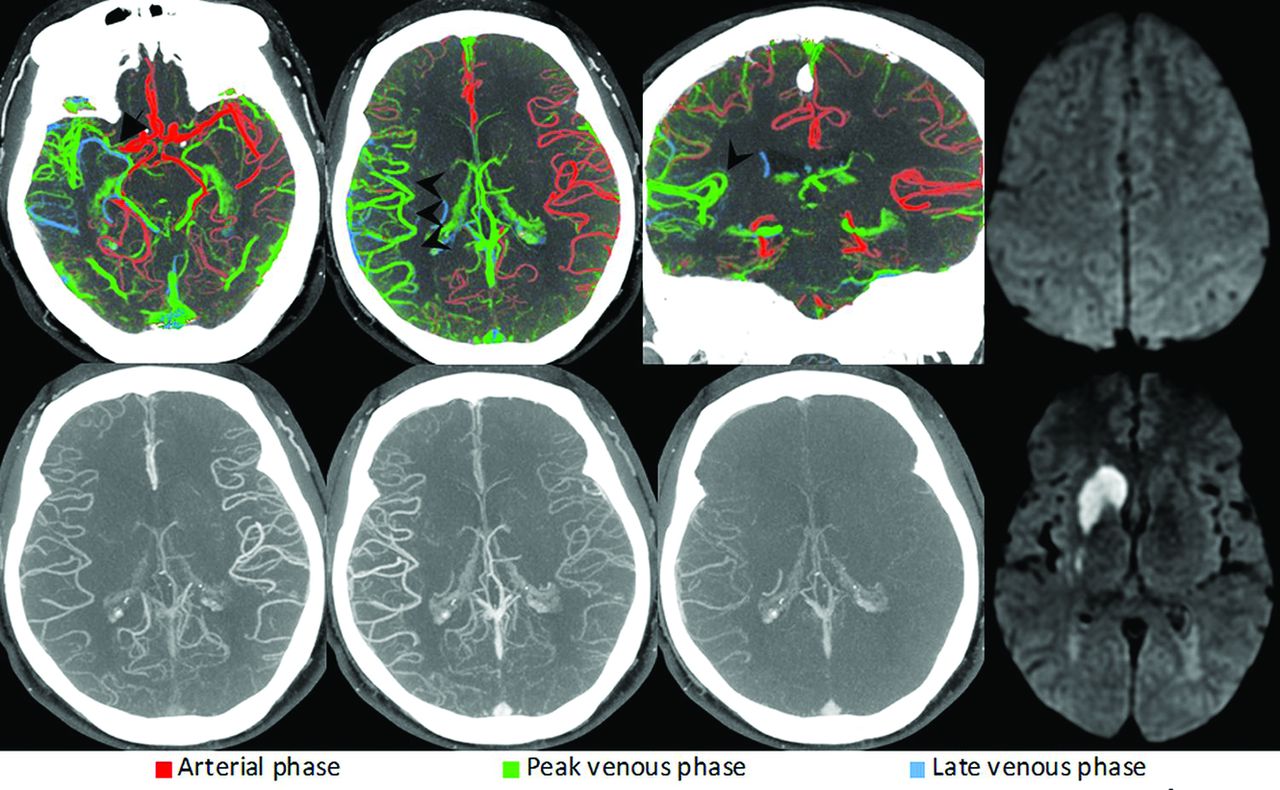
- FIG 2.
Left-sided M1 segment MCA occlusion (arrow). ColorViz summation maps (upper row) show predominantly green vessels (arrowheads) in the affected territory, indicating a 1-phase delay. However, the vessel extent is reduced compared with the unaffected hemisphere. Hence, the findings are consistent with intermediate pial artery filling and collateral flow. Corresponding conventional mCTA images are displayed in the lower row. The patient was treated with intravenous alteplase, and follow-up CT after 24 hours (upper and lower right) reveals an incomplete infarction pattern. While the insula, M1, and M2 regions are infarcted, the M3, M4, M5, and M6 regions are spared. Infarction is also present in the left PCA territory, indicating a fetal left PCA origin.
- FIG 3.
Left-sided ICA carotid L-occlusion (arrow). ColorViz summation maps (upper row) show a severely reduced vessel extent compared with the unaffected hemisphere (arrowheads). The few visible opacified vessels are predominantly blue, suggesting a 2-phase delay. This is consistent with poor pial artery filling and collateral flow. The patient was treated with antithrombotic therapy, and follow-up MR imaging after 24 hours (upper and lower right) shows complete infarction of the left MCA territory.
- FIG 4.
Left-sided M4 segment MCA occlusion. ColorViz summation images (upper row) show a focal area with blue and green vessels in the left frontal lobe (black arrows), ie, an area with delayed pial artery filling and washout, suggestive of a distal vessel occlusion. The thrombus itself cannot be visualized; the occlusion was detected on the basis of arterial flow information only. Corresponding conventional mCTA images in the lower row also show a slight delay in left frontal pial artery filling and washout; however, the occlusion is not as easily appreciable as in the color-coded summation maps. The patient received intravenous alteplase. Follow-up MR imaging after 24 hours (upper right) shows an acute left frontal lobe infarction, thereby confirming the suspected site of intracranial occlusion.
- FIG 5.
Right-sided A2 segment ACA occlusion (arrows). ColorViz summation images (upper row) show green and blue vessels distal to the occlusion (arrowheads), indicating impaired arterial filling within the right ACA territory. Corresponding conventional mCTA images are shown in the lower row. The patient received intravenous alteplase. Follow-up CT after 24 hours (upper and lower right) shows infarction in the right ACA territory.
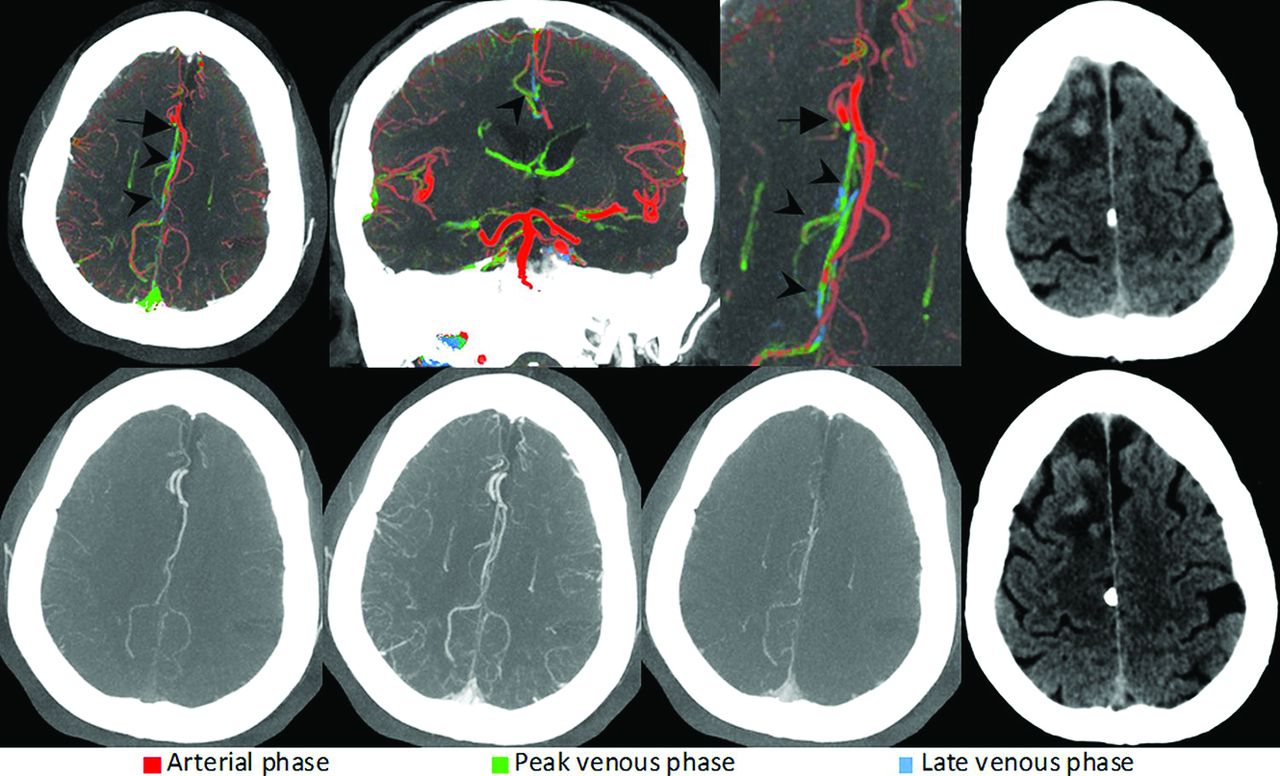
- FIG 6.
Right-sided fetal PCA occlusion (arrows). As opposed to the left PCA (lower middle image), the right distal PCA (lower left image) is blue in the color-coded summation images, suggestive of impaired filling of the distal vessel. The delayed arterial filling leads to a subsequent delay in venous filling in the posterior fossa on the affected side (arrowheads), a useful feature that is often encountered in posterior circulation occlusions and can help to identify the site of occlusion. Mechanical thrombectomy was performed. Follow-up MR imaging after 24 hours (lower right) reveals small right-sided occipital and thalamic infarct.
- FIG 7.
Bilateral M3 segment MCA occlusions (arrows) visualized through changes in the pial artery color compared with the surrounding pial arteries. Some cortical MCA branches of both hemispheres are depicted in green and blue (arrowheads), consistent with bilateral occlusions. Due to the symmetry of the occlusions, they are difficult to appreciate on conventional mCTA (lower row). CTP (lower right) shows prolonged time-to-maximum times in the affected territories, confirming the suspected occlusions.


Jump to section
Related Articles
Cited By...
- Interrater Agreement and Detection Accuracy for Medium-Vessel Occlusions Using Single-Phase and Multiphase CT Angiography
- A review of endovascular treatment for medium vessel occlusion stroke
- Multiphase CT Angiography: A Useful Technique in Acute Stroke Imaging--Collaterals and Beyond
- Missed Medium-Vessel Occlusions on CT Angiography: Make It Easier ... Easily!