Advertisement

Main menu

  • Home
  • Content
    • Current Issue
    • Accepted Manuscripts
    • Article Preview
    • Past Issue Archive
    • Video Articles
    • AJNR Case Collection
    • Case of the Week Archive
    • Case of the Month Archive
    • Classic Case Archive
  • Special Collections
    • AJNR Awards
    • Low-Field MRI
    • Alzheimer Disease
    • ASNR Foundation Special Collection
    • Photon-Counting CT
    • View All
  • Multimedia
    • AJNR Podcasts
    • AJNR SCANtastic
    • Trainee Corner
    • MRI Safety Corner
    • Imaging Protocols
  • For Authors
    • Submit a Manuscript
    • Submit a Video Article
    • Submit an eLetter to the Editor/Response
    • Manuscript Submission Guidelines
    • Statistical Tips
    • Fast Publishing of Accepted Manuscripts
    • Graphical Abstract Preparation
    • Imaging Protocol Submission
    • Author Policies
  • About Us
    • About AJNR
    • Editorial Board
    • Editorial Board Alumni
  • More
    • Become a Reviewer/Academy of Reviewers
    • Subscribers
    • Permissions
    • Alerts
    • Feedback
    • Advertisers
    • ASNR Home

User menu

  • Alerts
  • Log in

Search

  • Advanced search
American Journal of Neuroradiology

American Journal of Neuroradiology

ASHNR American Society of Functional Neuroradiology ASHNR American Society of Pediatric Neuroradiology ASSR
  • Alerts
  • Log in

Advanced Search

  • Home
  • Content
    • Current Issue
    • Accepted Manuscripts
    • Article Preview
    • Past Issue Archive
    • Video Articles
    • AJNR Case Collection
    • Case of the Week Archive
    • Case of the Month Archive
    • Classic Case Archive
  • Special Collections
    • AJNR Awards
    • Low-Field MRI
    • Alzheimer Disease
    • ASNR Foundation Special Collection
    • Photon-Counting CT
    • View All
  • Multimedia
    • AJNR Podcasts
    • AJNR SCANtastic
    • Trainee Corner
    • MRI Safety Corner
    • Imaging Protocols
  • For Authors
    • Submit a Manuscript
    • Submit a Video Article
    • Submit an eLetter to the Editor/Response
    • Manuscript Submission Guidelines
    • Statistical Tips
    • Fast Publishing of Accepted Manuscripts
    • Graphical Abstract Preparation
    • Imaging Protocol Submission
    • Author Policies
  • About Us
    • About AJNR
    • Editorial Board
    • Editorial Board Alumni
  • More
    • Become a Reviewer/Academy of Reviewers
    • Subscribers
    • Permissions
    • Alerts
    • Feedback
    • Advertisers
    • ASNR Home
  • Follow AJNR on Twitter
  • Visit AJNR on Facebook
  • Follow AJNR on Instagram
  • Join AJNR on LinkedIn
  • RSS Feeds

AJNR Awards, New Junior Editors, and more. Read the latest AJNR updates

  • Getting new auth cookie, if you see this message a lot, tell someone!
  • Getting new auth cookie, if you see this message a lot, tell someone!
Research ArticlePediatric Neuroimaging
Open Access

Longitudinal Assessment of Neuroradiologic Features in Wolfram Syndrome

A. Samara, H.M. Lugar, T. Hershey and J.S. Shimony
American Journal of Neuroradiology December 2020, 41 (12) 2364-2369; DOI: https://doi.org/10.3174/ajnr.A6831
A. Samara
aFrom the Department of Psychiatry (A.S., H.M.L.)
  • Find this author on Google Scholar
  • Find this author on PubMed
  • Search for this author on this site
  • ORCID record for A. Samara
H.M. Lugar
aFrom the Department of Psychiatry (A.S., H.M.L.)
  • Find this author on Google Scholar
  • Find this author on PubMed
  • Search for this author on this site
  • ORCID record for H.M. Lugar
T. Hershey
aFrom the Department of Psychiatry (A.S., H.M.L.)
bNeurology (T.H.)
cMallinckrodt Institute of Radiology (T.H., J.S.S.), Washington University School of Medicine, St. Louis, Missouri
  • Find this author on Google Scholar
  • Find this author on PubMed
  • Search for this author on this site
  • ORCID record for T. Hershey
J.S. Shimony
cMallinckrodt Institute of Radiology (T.H., J.S.S.), Washington University School of Medicine, St. Louis, Missouri
  • Find this author on Google Scholar
  • Find this author on PubMed
  • Search for this author on this site
  • ORCID record for J.S. Shimony
  • Article
  • Figures & Data
  • Info & Metrics
  • Responses
  • References
  • PDF
Loading

Article Figures & Data

Figures

  • Tables
  • FIG 1.
    • Download figure
    • Open in new tab
    • Download powerpoint
    FIG 1.

    Sagittal, coronal, and axial MR images show pons signal abnormalities as T1 hypointensity and T2 hyperintensity in a patient with Wolfram syndrome (white arrows). Brain stem atrophy is also evident on this MR image.
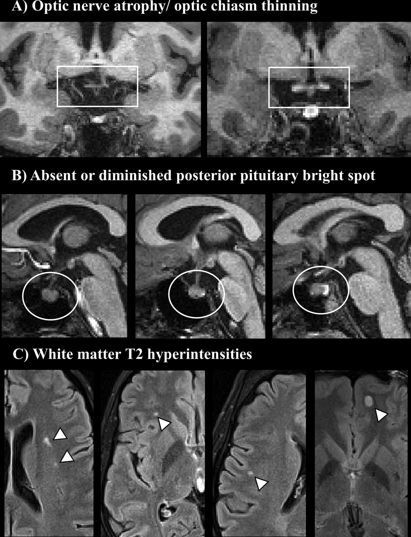
  • FIG 2.
    • Download figure
    • Open in new tab
    • Download powerpoint
    FIG 2.

    Examples of neuroradiologic findings in patients with Wolfram syndrome. A, Thinning of the optic chiasm (white box) as seen in coronal T1-weighted images (left, patient with Wolfram syndrome; right, healthy control). B, Abnormal PPBS signal (white circles) in midline sagittal T1-weighted images (from left to right, absent, diminished, and physiologic signal). C, White matter hyperintensities in FLAIR MR imaging (white arrowheads).
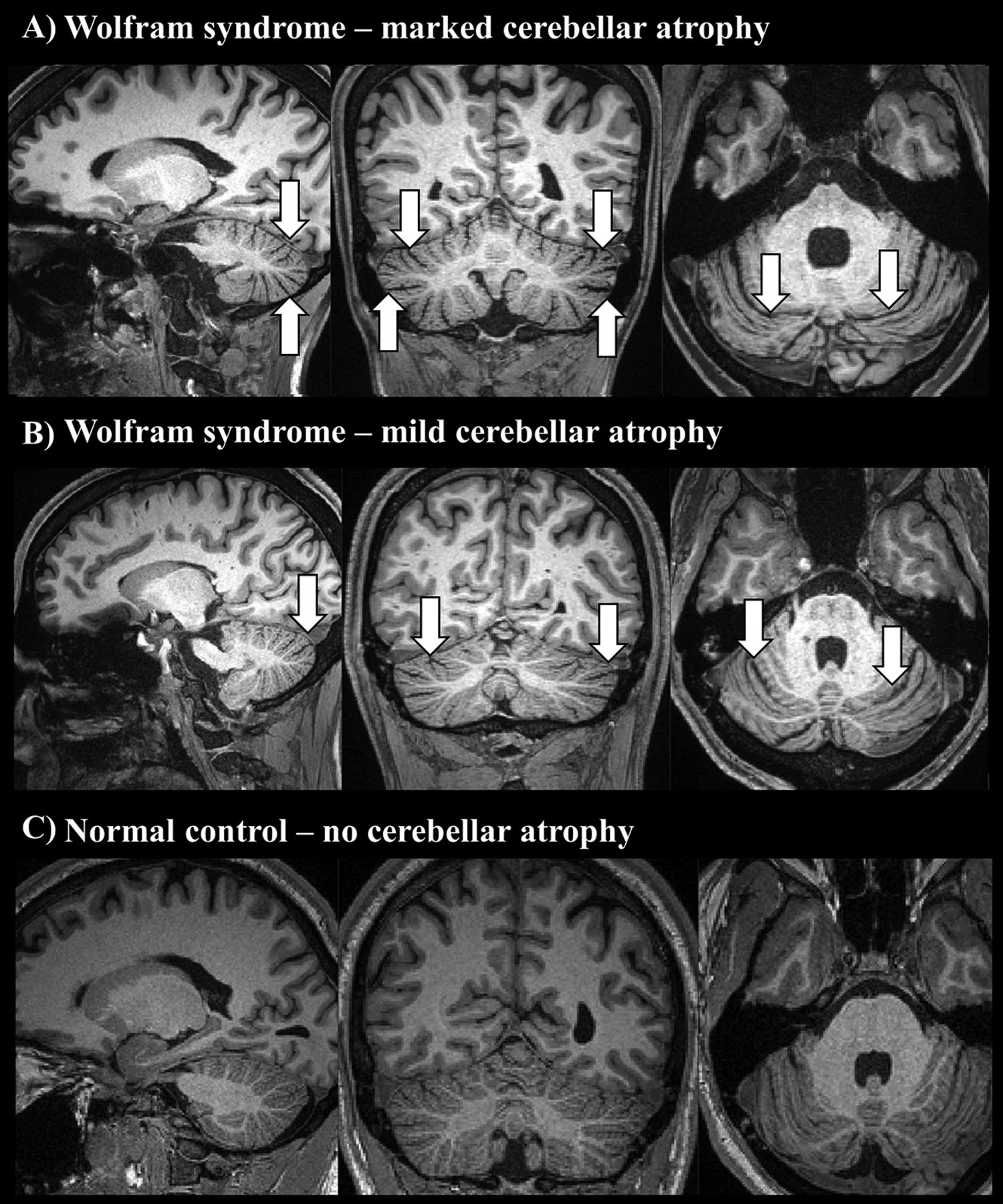
  • FIG 3.
    • Download figure
    • Open in new tab
    • Download powerpoint
    FIG 3.

    Marked (A) and mild (B) cerebellar atrophy in patients with Wolfram syndrome, as shown in sagittal, coronal, and axial T1-weighted MR images (white arrows), compared with a healthy control (C).

  • FIG 4.
    • Download figure
    • Open in new tab
    • Download powerpoint
    FIG 4.

    A, Frequency of neuroradiologic signs in patients with Wolfram syndrome at first and last visits. B, The relationship between age and the number of neuroradiologic signs. A line connecting a circle-shaped point (first visit) and a triangle-shaped point (last visit) represents each patient.

Tables

  • Figures
    • View popup
    Table 1:

    The demographic and clinical characteristics in patients with Wolfram syndrome at first and last follow-up visitsa

    First Visit (n = 30)Last Visit (n = 30)
    Age (yr)14 ± 619 ± 6
    Duration of disease (yr)3 ± 38 ± 4
    Diabetes mellitus29 (97) 30 (100)
    Vision impairment28 (93) 28 (93)
    Hearing loss 20 (67)23 (77)
    Diabetes insipidus15 (50)19 (63)
    Bladder dysfunction13 (43)26 (86)
    • ↵a For the age and duration of disease, means and SDs are reported. For comorbid conditions, numbers and percentages are reported.

    • View popup
    Table 2:

    Neuroradiologic findings in brain MR images in patients with Wolfram syndromea

    Radiologic SignFirst Visit (n = 30)Last Visit (n = 30)
    Negative PPBS signb16 (53)21 (70)
    T1/T2 pons signal abnormalities16 (53)20 (67)
    Optic nerve atrophy9 (30)24 (80)c
    White matter T2 hyperintensitiesd8 (27)10 (33)
    Cerebellum atrophy7 (23)21 (70)c
    • ↵a Numbers and percentages are reported.

    • ↵b Absent and diminished PPBS sign.

    • ↵c P value < .01 (χ2 test).

    • ↵d FLAIR images were not available to evaluate white matter T2 hyperintensity for 1 scan, and T2-weighted images were used instead.

PreviousNext
Back to top

In this issue

American Journal of Neuroradiology: 41 (12)
American Journal of Neuroradiology
Vol. 41, Issue 12
1 Dec 2020
  • Table of Contents
  • Index by author
  • Complete Issue (PDF)
Advertisement
Print
Download PDF
Email Article

Thank you for your interest in spreading the word on American Journal of Neuroradiology.

NOTE: We only request your email address so that the person you are recommending the page to knows that you wanted them to see it, and that it is not junk mail. We do not capture any email address.

Enter multiple addresses on separate lines or separate them with commas.
Longitudinal Assessment of Neuroradiologic Features in Wolfram Syndrome
(Your Name) has sent you a message from American Journal of Neuroradiology
(Your Name) thought you would like to see the American Journal of Neuroradiology web site.
CAPTCHA
This question is for testing whether or not you are a human visitor and to prevent automated spam submissions.
Cite this article
A. Samara, H.M. Lugar, T. Hershey, J.S. Shimony
Longitudinal Assessment of Neuroradiologic Features in Wolfram Syndrome
American Journal of Neuroradiology Dec 2020, 41 (12) 2364-2369; DOI: 10.3174/ajnr.A6831

Citation Manager Formats

  • BibTeX
  • Bookends
  • EasyBib
  • EndNote (tagged)
  • EndNote 8 (xml)
  • Medlars
  • Mendeley
  • Papers
  • RefWorks Tagged
  • Ref Manager
  • RIS
  • Zotero
0 Responses
Respond to this article
Share
Bookmark this article
Longitudinal Assessment of Neuroradiologic Features in Wolfram Syndrome
A. Samara, H.M. Lugar, T. Hershey, J.S. Shimony
American Journal of Neuroradiology Dec 2020, 41 (12) 2364-2369; DOI: 10.3174/ajnr.A6831
del.icio.us logo Twitter logo Facebook logo Mendeley logo
  • Tweet Widget
  • Facebook Like
  • Google Plus One
Purchase

Jump to section

  • Article
    • Abstract
    • ABBREVIATIONS:
    • MATERIALS AND METHODS
    • RESULTS
    • DISCUSSION
    • CONCLUSIONS
    • Acknowledgments
    • Footnotes
    • References
  • Figures & Data
  • Info & Metrics
  • Responses
  • References
  • PDF

Related Articles

  • No related articles found.
  • PubMed
  • Google Scholar

Cited By...

  • Expanding the spectrum of white matter abnormalities in Wolfram syndrome: A retrospective review
  • A deep phenotyping study in mouse and iPSC models to understand the role of oligodendroglia in optic neuropathy in Wolfram syndrome
  • Crossref
  • Google Scholar

This article has not yet been cited by articles in journals that are participating in Crossref Cited-by Linking.

More in this TOC Section

  • Frontal Paraventricular Cysts
  • Sodium MRI in Pediatric Brain Tumors
  • FRACTURE MR in Congenital Vertebral Anomalies
Show more Pediatric Neuroimaging

Similar Articles

Advertisement

Indexed Content

  • Current Issue
  • Accepted Manuscripts
  • Article Preview
  • Past Issues
  • Editorials
  • Editor's Choice
  • Fellows' Journal Club
  • Letters to the Editor
  • Video Articles

Cases

  • Case Collection
  • Archive - Case of the Week
  • Archive - Case of the Month
  • Archive - Classic Case

More from AJNR

  • Trainee Corner
  • Imaging Protocols
  • MRI Safety Corner
  • Book Reviews

Multimedia

  • AJNR Podcasts
  • AJNR Scantastics

Resources

  • Turnaround Time
  • Submit a Manuscript
  • Submit a Video Article
  • Submit an eLetter to the Editor/Response
  • Manuscript Submission Guidelines
  • Statistical Tips
  • Fast Publishing of Accepted Manuscripts
  • Graphical Abstract Preparation
  • Imaging Protocol Submission
  • Evidence-Based Medicine Level Guide
  • Publishing Checklists
  • Author Policies
  • Become a Reviewer/Academy of Reviewers
  • News and Updates

About Us

  • About AJNR
  • Editorial Board
  • Editorial Board Alumni
  • Alerts
  • Permissions
  • Not an AJNR Subscriber? Join Now
  • Advertise with Us
  • Librarian Resources
  • Feedback
  • Terms and Conditions
  • AJNR Editorial Board Alumni

American Society of Neuroradiology

  • Not an ASNR Member? Join Now

© 2025 by the American Society of Neuroradiology All rights, including for text and data mining, AI training, and similar technologies, are reserved.
Print ISSN: 0195-6108 Online ISSN: 1936-959X

Powered by HighWire