
Article Figures & Data
Figures
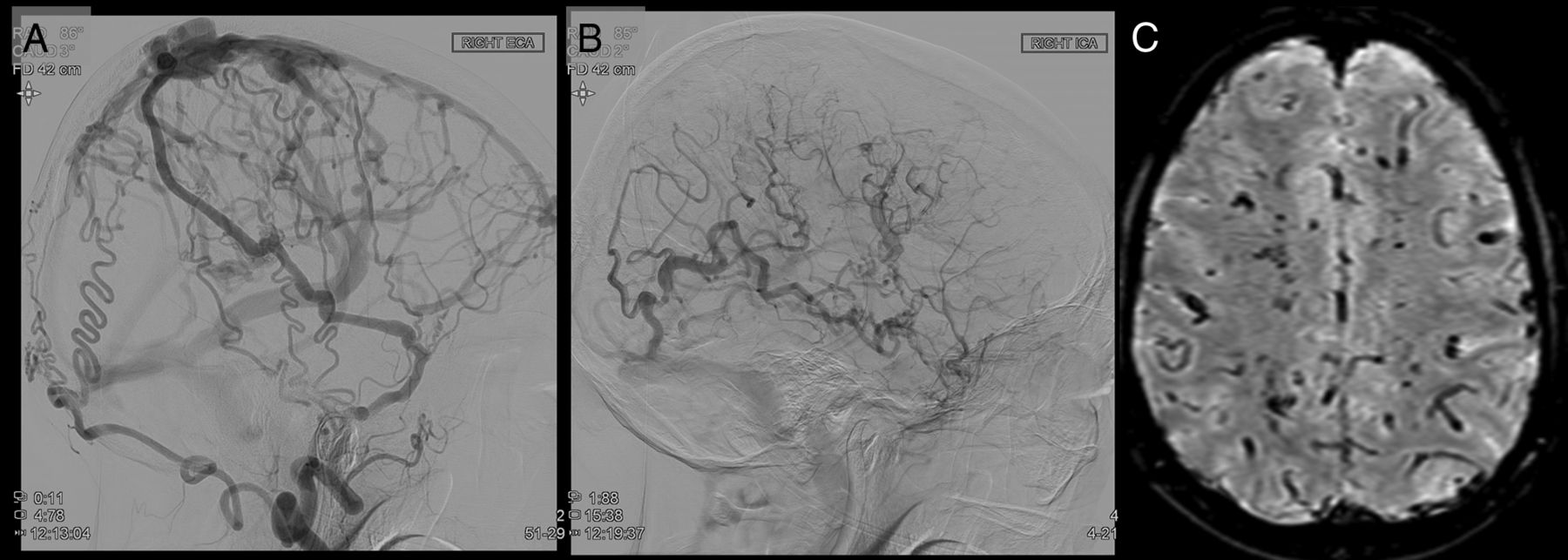
- FIG 1.
Pseudophlebitic pattern in a superior sagittal sinus fistula in a patient presenting with voice hoarseness. A, Right external carotid artery (ECA) injection shows the superior sagittal sinus fistula with reflux into the cortical veins. B, Right ICA cerebral angiogram shows dilated corkscrew veins in the right cerebral hemisphere during the parenchymal venous phase. There is stagnation of contrast due to the high venous pressure. C, SWI shows dilated T2-hypointense transmedullary veins reflective of venous hypertension.
- FIG 2.
A pseudophlebitic pattern in a patient with an extensive dural arteriovenous fistula presenting with cognitive decline. A, FLAIR imaging shows parenchymal FLAIR signal in the bilateral cerebral hemispheres, right greater than left. B, SWI shows a chronic hemorrhagic in the right parietal lobe and some dilated transmedullary veins in the left hemisphere. C, Left external carotid artery (ECA) injection shows the superior sagittal sinus and torcular fistulas with reflux into the deep venous system and parenchymal veins. D, Left ICA injection shows dilated corkscrew parenchymal cerebral veins, which have some stagnation consistent with a pseudophlelebitic pattern.
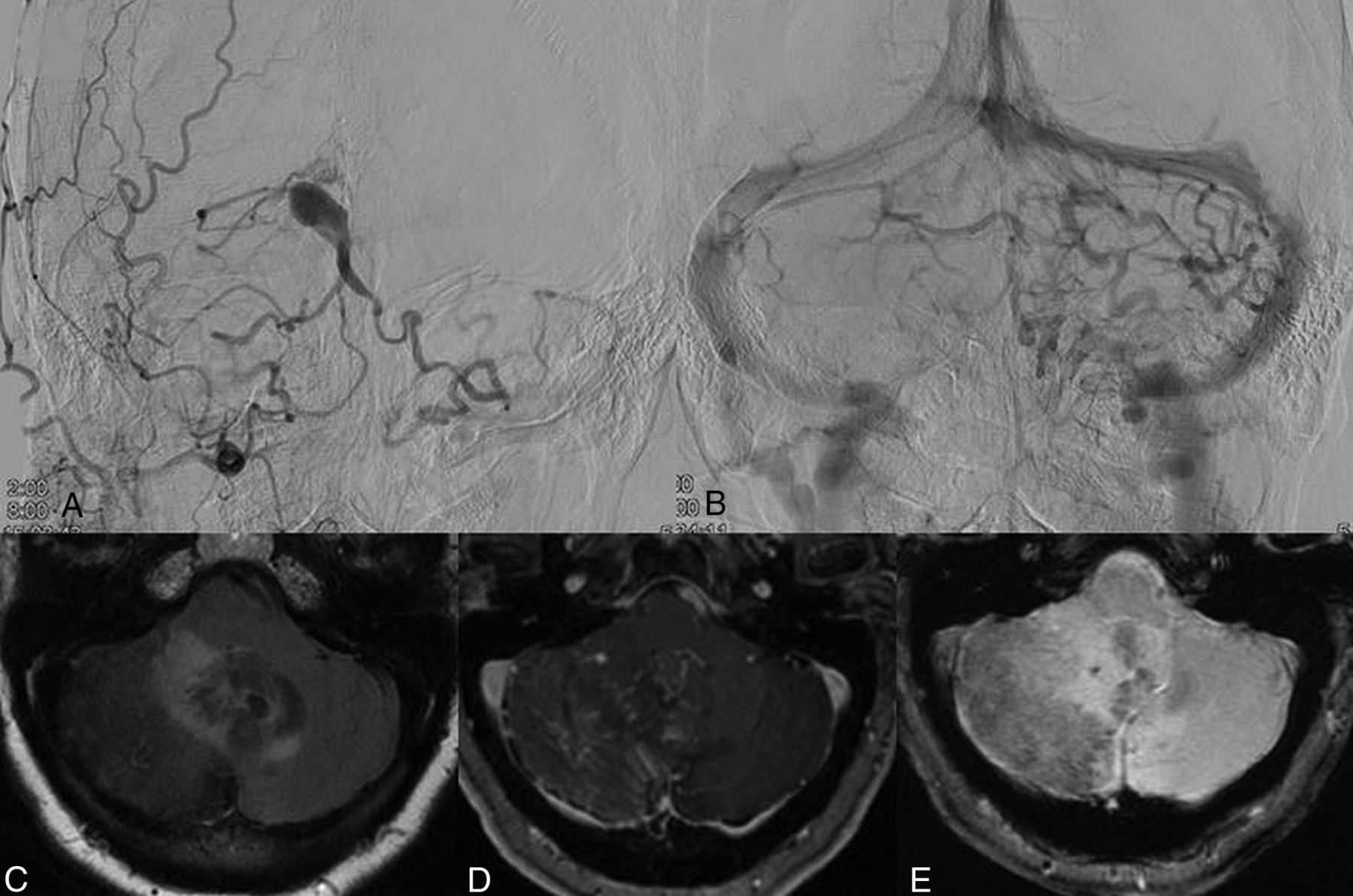
- FIG 3.
Pseudophlebitic pattern in a patient with a tentorial dural arteriovenous fistula presenting with ataxia. A, Right external carotid artery angiogram shows a tentorial dural arteriovenous fistula with reflux into the bilateral cerebellar hemispheres. B, Parenchymal venous phase of a left vertebral artery injection shows a pseudophlebitic pattern of the cerebellar parenchymal veins. C, FLAIR MR imaging shows edema in the bilateral cerebellar hemispheres and vermis. D, Postcontrast MR imaging shows dilated transmedullary veins in the right cerebellar hemisphere and vermis. E, Gradient recalled-echo MR imaging shows chronic hemosiderin deposition in the right cerebellar hemisphere.


Tables
Overall Nonpseudophlebitic Pseudophlebitic P No. 201 143 (71.1) 58 (28.9) – Mean age (SD (yr) 59.1 (13.7) 60.5 (13.5) 58.4 (13.8) .32 Sex Male (No.) (%) 104 (51.7) 64 (44.8) 40 (69.0) Female (No.) (%) 97 (48.3) 79 (55.2) 18 (31.0) .002 Clinical presentation (No.) (%) Hemorrhage 25 (12.5) 12 (8.4) 13 (22.8) .005 Incidental 22 (11.0) 18 (12.6) 4 (6.9) .24 Headache 23 (11.4) 15 (10.5) 8 (13.8) .51 Visual/ocular symptoms 37 (18.4) 29 (20.3) 8 (13.8) .28 Tinnitus/bruit 86 (42.8) 77 (53.9) 9 (15.5) <.0001 Cognitive changes 6 (3.0) 2 (1.4) 4 (6.9) .04 Gait changes/ataxia 4 (2.0) 0 (0.0) 4 (6.0) .002 Hydrocephalus 1 (0.5) 0 (0.0) 1 (1.7) .11 Seizure 8 (4.0) 3 (2.1) 5 (8.6) .03 Myelopathy 7 (3.5) 3 (2.1) 4 (6.0) .12 Other 17 (8.5) 6 (4.2) 11 (18.9) .0001 Note:—SD indicates standard deviation; yr, year; No., number; %, percentage.
Overall Nonpseudophlebitic Pseudophlebitic P Fistula location Transverse sigmoid 63 (31.3) 58 (40.6) 5 (8.6) <.0001 Cavernous sinus 27 (13.4) 23 (16.1) 4 (6.9) Tentorial 54 (26.9) 24 (16.8) 30 (51.7) Foramen magnum 7 (3.5) 4 (2.8) 3 (5.2) Superior sagittal sinus 6 (3.0) 1 (0.7) 5 (8.6) Anterior cranial fossa 5 (2.5) 5 (3.5) 0 (0.0) Extensive 1 (0.5) 1 (0.7) 0 (0.0) Multiple 9 (4.5) 4 (2.8) 5 (8.6) Hypoglossal/jugular foramen 16 (8.0) 13 (9.1) 3 (5.2) Convexity 7 (3.5) 5 (3.5) 2 (3.5) Middle cranial fossa 2 (1.0) 1 (0.7) 1 (1.7) Other 4 (2.0) 4 (2.8) 0 (0.0) Tentorial location Superior petrosal sinus 8 (14.6) 3 (12.5) 5 (16.1) .49 Straight sinus 1 (1.8) 0 (0.0) 1 (3.2) Galenic 11 (20.0) 4 (16.7) 7 (22.6) Tentorial sinus 25 (45.5) 14 (58.3) 11 (35.5) Torcular 10 (18.2) 3 (12.5) 7 (22.6) Borden classification (non-cavernous sinus) I 68 (33.8) 68 (56.7) 0 (0.0) <.0001 II 27 13 (10.8) 14 (25.9) III 79 39 (32.5) 40 (74.1) Cognard classification (non-cavernous sinus) I 52 52 (43.3) 0 (0.0) IIa 16 16 (13.3) 0 (0.0) IIb 6 5 (4.2) 1 (1.9) IIa+b 22 7 (5.8) 15 (27.8) <.0001 III 22 17 (14.2) 5 (9.3) IV 52 22 (18.3) 30 (55.6) V 4 1 (0.8) 3 (5.6) Venous dilation 76 (37.8) 34 (23.8) 42 (72.4) <.0001 Venous varix 57 (28.4) 20 (14.0) 37 (63.8) <.0001 Draining vein stenosis 48 (23.9) 22 (15.4) 26 (44.8) <.0001 Overall Nonpseudophlebitic Pseudophlebitic P Cerebral edema 43 (22.5) 4 (2.9) 39 (70.9) <.0001 Chronic hemosiderin deposition/microhemorrhage 12 (6.4) 3 (2.2) 9 (17.3) .0002 Venous infarct 2 (1.1) 1 (0.7) 1 (1.9) .49 Dilated transmedullary veins on SWI or CE T1 24 (12.9) 0 (0.0) 24 (47.1) <.0001 Sinus thrombosis 14 (7.4) 8 (5.9) 6 (11.1) .21 Cortical venous reflux on 3D-TOF 68 (38.9) 32 (24.6) 46 (80.0) <.0001 Note:—CE indicates contrast-enhanced.


