
Article Figures & Data
Figures
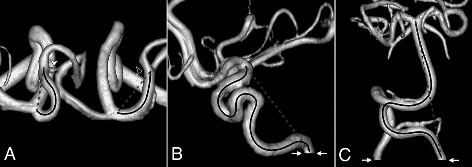
- FIG 1.
The TI measurement on 3D volume-rendered TOF-MRA. The solid black line indicates the centerline length, whereas the dashed gray line shows straight-line length. A, Superior view of A1 and M1 segments in a patient with LDS (all anterior cerebral artery segments distal to A2 have been removed; white arrowhead, anterior communicating artery; black arrowhead, right M1–M2 bifurcation). B, Lateral view of the right IICA in a patient with LDS (white arrows, level of the inferior opening of the carotid canal). C, Anterior view of VBS in a patient with Marfan syndrome (white arrows, level of the foramen magnum).
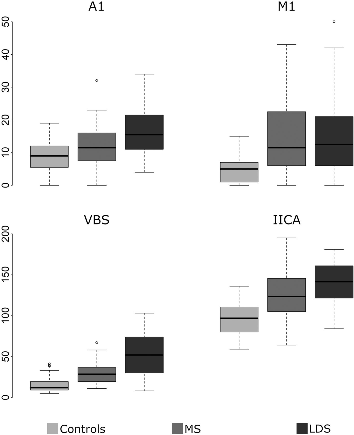
- FIG 2.
Boxplots of the TI of the 4 examined arterial segments for controls and patients with Marfan syndrome (MS) and LDS.
- FIG 3.
Examples of different degrees of tortuosity of IICA and VBS. A–C, Lateral view of the right IICA in a control (A), in a patient with Marfan syndrome (B), and in a patient with LDS (C). D–F, Anterior view of VBS in a control (D), in a patient with MS (E), and in a patient with LDS (F).
- FIG 4.
Receiver operating characteristic curves for the intracranial TI, with the outcome of LDS versus Marfan syndrome and CTD versus controls. Dashed gray line indicates the A1 TI; solid gray line, M1 TI; dashed black line, IICA TI; and solid black line, VBS TI.


Tables
Characteristic CTD (N = 68) Marfan Syndrome (n = 36) LDS (n = 32) P Age at MRA, median (IQR), y 38.5 (23–48) 32.5 (25–43.5) 42 (22–50) .21 Female, n (%) 33 (48.5) 15 (41.7) 18 (56.3) .33 Aortic root dilation, n (%) 59 (86.8) 33 (91.7) 26 (81.3) .29 Aortic dissection, n (%) 9 (13.2) 6 (16.7) 3 (9.4) .48 Arterial dissection, n (%) 4 (5.9) 0 (0.0) 4 (12.5) .04 Aortic surgery, n (%) 41 (60.3) 25 (69.4) 16 (50.0) .14 Age at surgery, median (IQR), y 28 (20–37) 26 (21–35) 33 (18–44) .47 Intracranial TI, median (IQR) P Controls (n = 52) Marfan Syndrome (n = 36) LDS (n = 32) Marfan Syndrome vs Controls LDS vs Controls LDS vs Marfan Syndrome A1 9 (6–12) 11.5 (8–16) 15.5 (11–21) .02 <.001 .02 M1 5 (1–7) 11.5 (6–22) 12.5 (6–21) <.001 <.001 .92 IICA 97 (80.5–110) 123.5 (105–144) 141.5 (123–160.5) <.001 <.001 .06 VBS 12 (9–19) 28.5 (20–36) 52 (32–73.5) <.001 <.001 <.001


