
Article Figures & Data
Figures
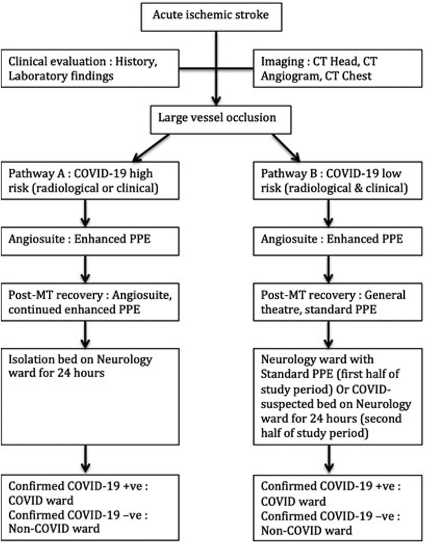
- FIG 1.
Comparison of the patient pathways according to the COVID-19 risk. High risk for COVID-19 on admission included typical or indeterminate pulmonary findings on the chest CT and/or any clinical suspicion. Low risk for COVID-19 included atypical or normal findings on the chest CT and no clinical suspicion. Enhanced PPE indicates filtering facepiece class 3 masks, visors, and long-sleeved fluid-repellent overalls. Standard PPE indicates fluid-resistant surgical masks, gloves, and aprons; −ve, negative; +ve, positive.
- FIG 2.
Typical features of COVID-19 pneumonia. Axial CT images at the level of the hilum show bilateral ground glass opacities (arrows) with areas of lobular sparing and sparing of the immediate subpleural area. This patient tested positive by RT-PCR analysis.
- FIG 3.
Indeterminate features of COVID-19 pneumonia. Axial CT images show unilateral, localized, peripheral ground glass opacities (arrows). This patient tested negative for COVID-19 by RT-PCR analysis.


Tables
COVID-19 Pneumonia Imaging Classification Rationale CT Findings Typical appearance Commonly reported imaging features of greater specificity for COVID-19 pneumonia Peripheral, bilateral GGOs with or without consolidation or visible intralobular lines (crazy-paving) Multifocal GGOs of rounded morphology with or without consolidation or visible intralobular lines (crazy-paving) Reverse halo sign or other findings of organizing pneumonia (seen later in the disease) Indeterminate Nonspecific imaging features of COVID-19 pneumonia Absence of typical features AND Presence of multifocal, diffuse, perihilar, or unilateral GGO with or without consolidation lacking a specific distribution; they are nonrounded or nonperipheral Few very small GGOs with a nonrounded and nonperipheral distribution Negative Uncommonly or not reported features of COVID-19 pneumonia Absence of typical or indeterminate features AND Presence of isolated lobar or segmental consolidation without GGO Discrete small nodules (centrilobular, tree-in-bud sign) Lung cavitation Smooth interlobular septal thickening with pleural effusion OR no abnormal findings Note:—GGO indicates ground glass opacity.
↵a Adapted from the proposed Radiological Society of North America and British Society of Thoracic Imaging chest CT classification for reporting of COVID-19 pneumonia.15,16
- Table 3:
Comparison of classification of COVID-19 pulmonary findings between the primary reporter (general radiologist at primary stroke centers) and secondary reviewer (cardiothoracic radiologist) according to the adapted RSNA and BSTI guidance
COVID-19 Pneumonia Imaging Classification Primary Reporter (n = Patients) Secondary Reviewer (n = Patients) Typical 2 3 Indeterminate 7 7 Negative (atypical or normal) 29 28 Note:—BSTI indicates British Society of Thoracic Imaging; RSNA, Radiological Society of North America.
- Table 4:
List of incidental findings identified on review of the thoracic imaging in this cohort, summarized according to their clinical relevance
Incidental Pulmonary/CVS Findings of Substantial Clinical Relevance (n = Patients) Incidental Pulmonary/CVS Findings of Indeterminate Clinical Relevance (n = Patients) Lung malignancy/suspicious nodule2 Emphysema4 Pulmonary edema4 Lung fibrosis/pneumoconiosis3 Left ventricular thrombus/aneurysm2 Ascending aortic dilation2 Left atrial dilation: anterior-posterior diameter >45 mm16 Note:—CVS indicates cardiovascular.


Jump to section
Related Articles
Cited By...
- No citing articles found.