
Article Figures & Data
Figures
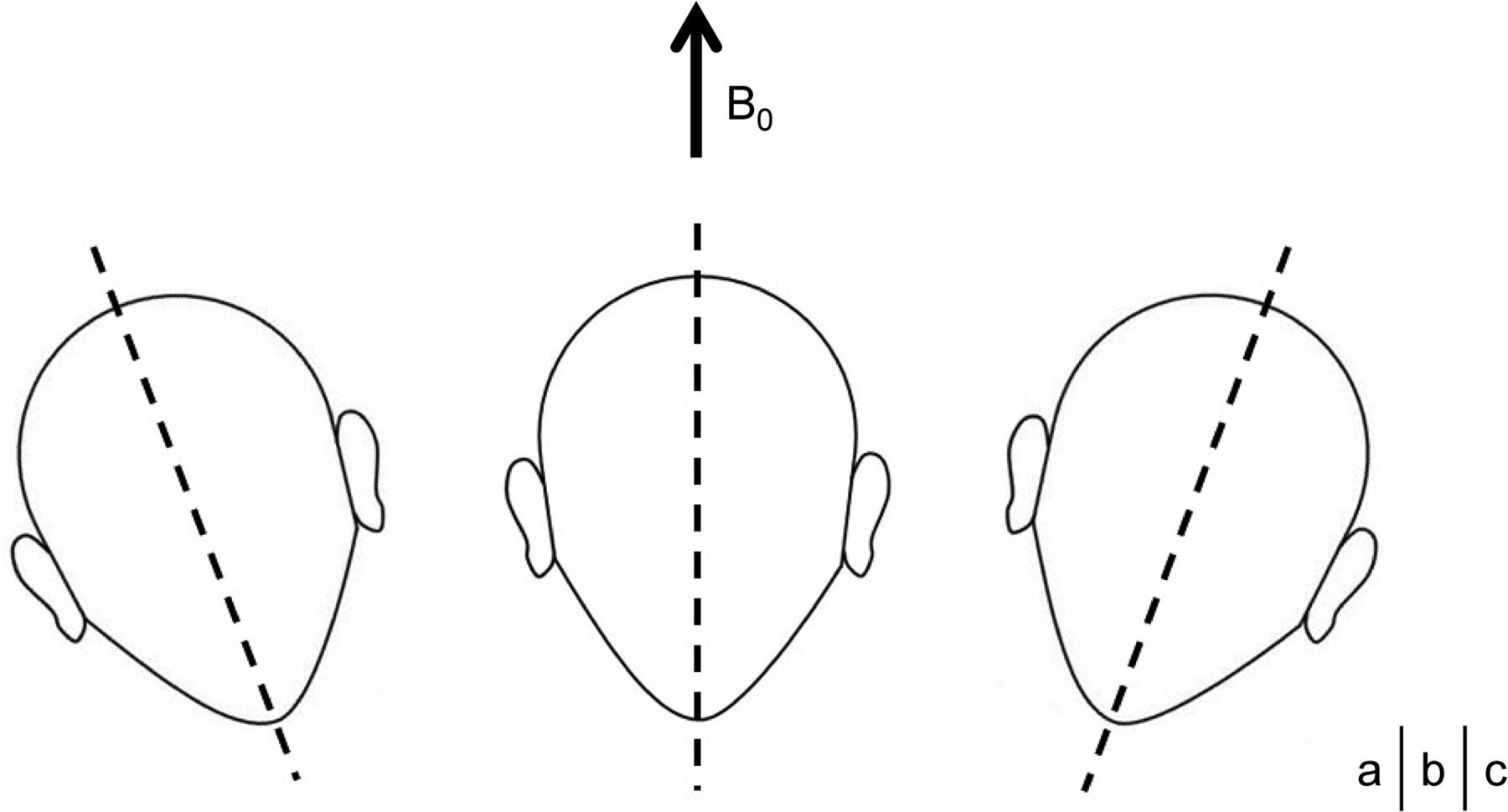
- Fig 1.
Volunteers’ heads were held in 1 of 3 ways: tilted to the right (A), not tilted (B), and tilted to the left (C) in the B0 direction.
- Fig 2.
The 3D spoiled gradient-echo sequences with multiecho acquisitions are oriented orthogonally to the anterior midbrain (A), and nigrosome 1 is visualized in the caudal and mediolateral part of the substantia nigra pars compacta (B). When the subjects’ heads were tilted to the right or left, the MR imaging signal intensities measured on the basis of the depicted ROI (C) differentiated nigrosome 1 and SNc.
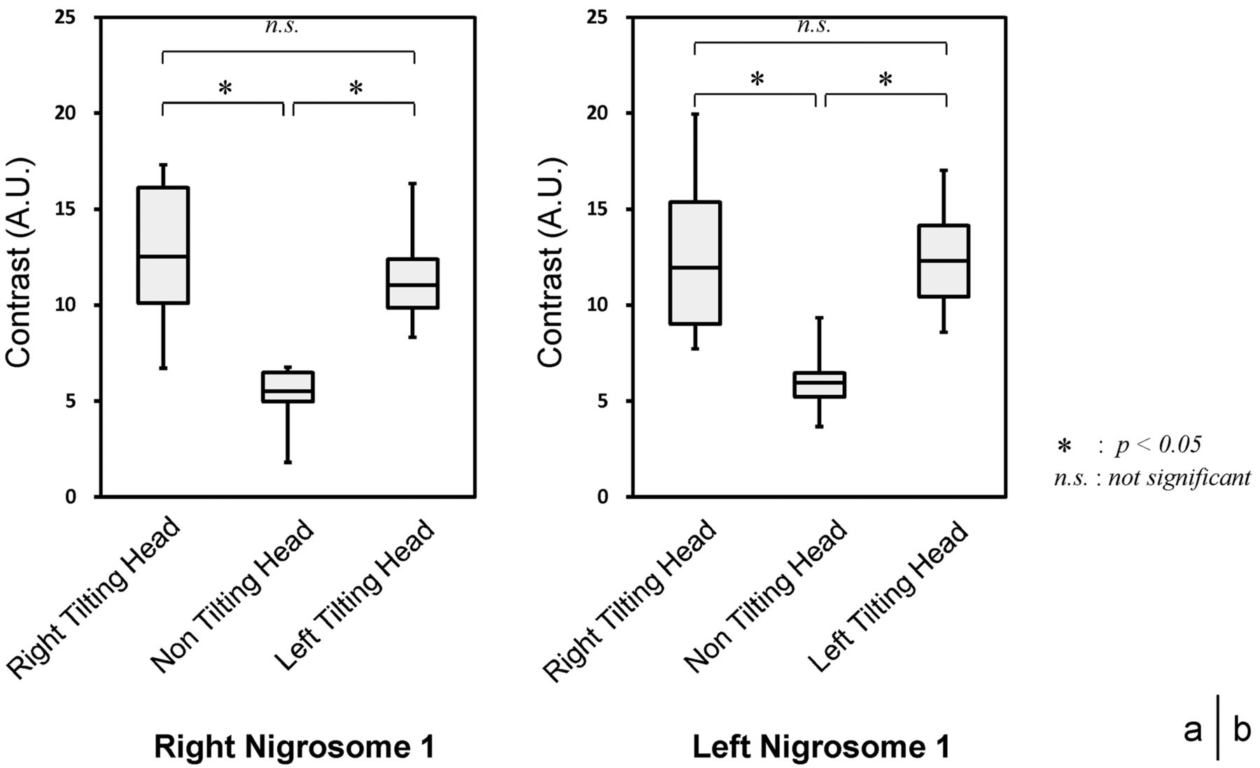
- Fig 3.
Visualization of the right (A) and left (B) nigrosome 1, in which the volunteers’ heads were tilted to the right or left in the B0 direction or not at all.
- Fig 4.
Examples of visualization of nigrosome 1 with a right head tilt (A), without a head tilt (B), and with a left head tilt (C) in the B0 direction.
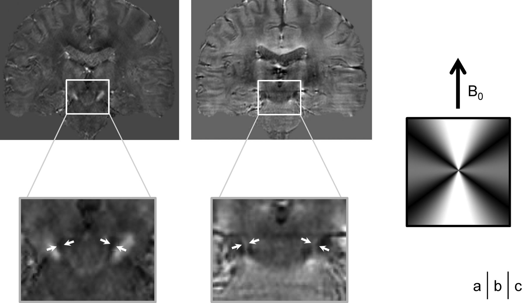
- Fig 5.
Coronal sections of the 3D susceptibility map (A) and the local field map (B). The anatomic slant structure of nigrosome 1 is coherent between the axis of nigrosome 1 (arrows) and the magic angle that occurs at approximately 54.7° in the B0 direction by dipolar interaction (C).


Tables
- Table 1:
Number of visualizations of discriminable nigrosome 1 when the subjects’ heads were tilted in the B0 direction (n = 14)
Tilting Head in B0 Direction Right None Left Nigrosome 1 Right Left Right Left Right Left Discriminable nigrosome 1 (No.) 9 9 6 5 10 11 - Table 2:
Results of contrast measurement repeated 6 times in 1 volunteer with right head tilting and left head tilting
Tilting Head in B0 Direction Right Left Nigrosome 1 and SNc Right Left Right Left Times 1 12.0 11.8 10.6 12.6 2 12.1 11.4 11.0 12.3 3 12.3 11.4 10.6 13.0 4 11.9 11.9 10.4 12.4 5 12.5 11.0 10.5 12.6 6 12.3 11.4 10.9 12.4 SD 0.20 0.32 0.21 0.26


